

Abstract
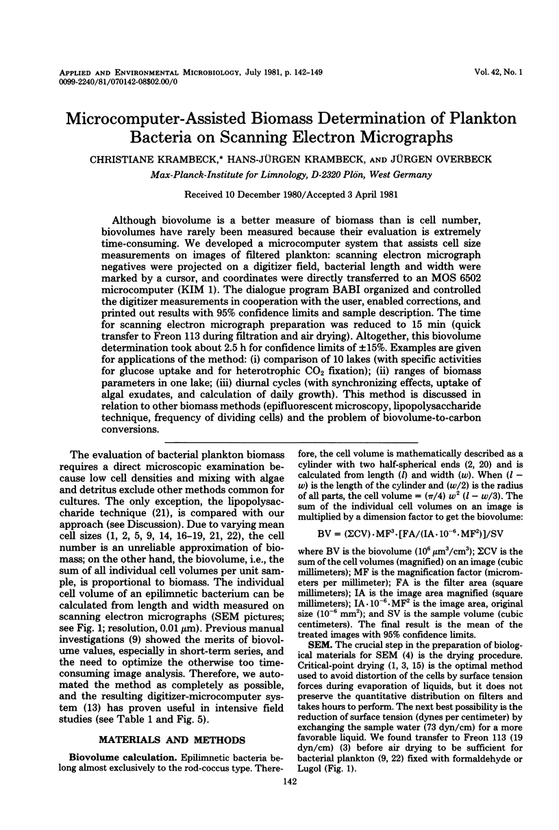
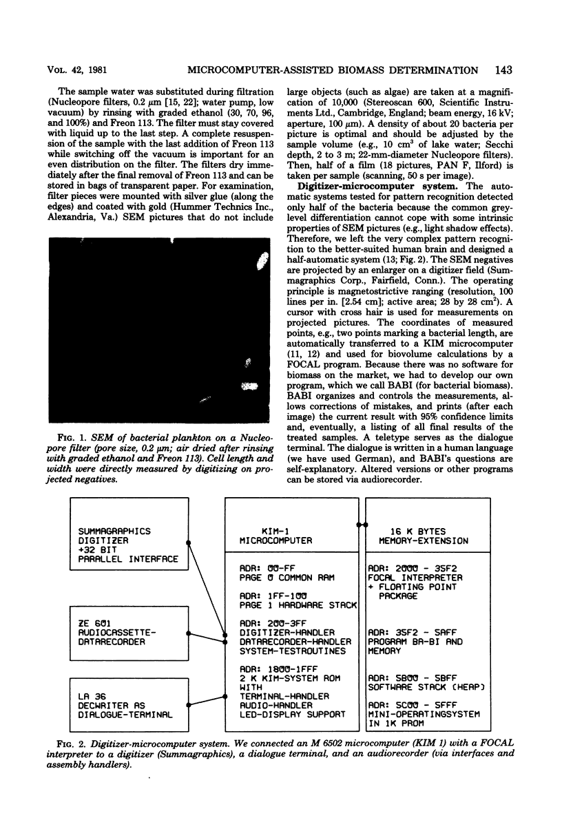
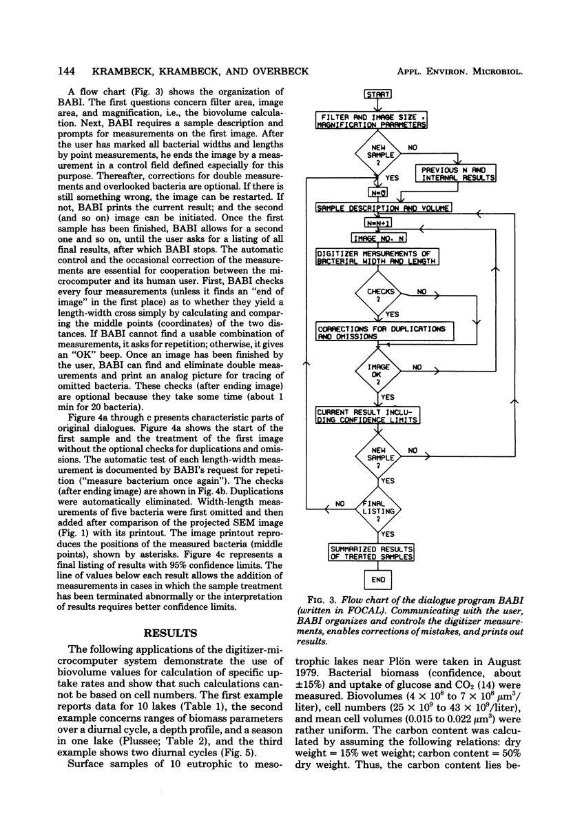
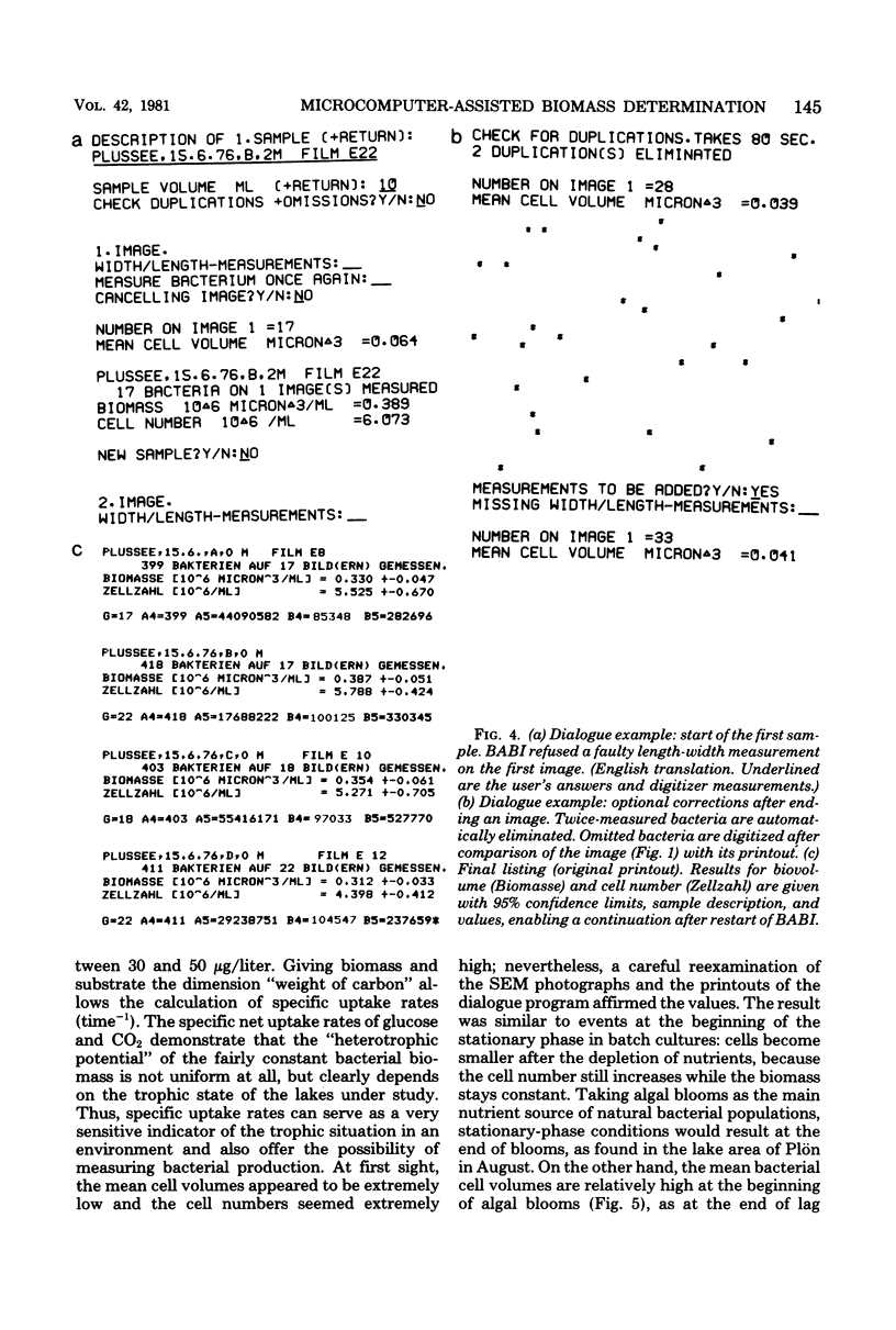
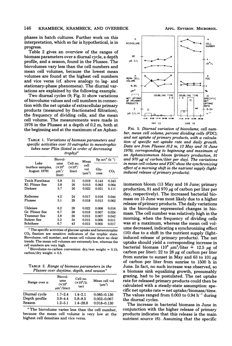
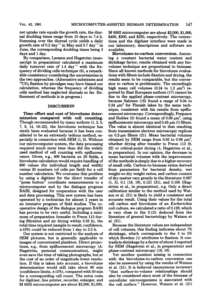
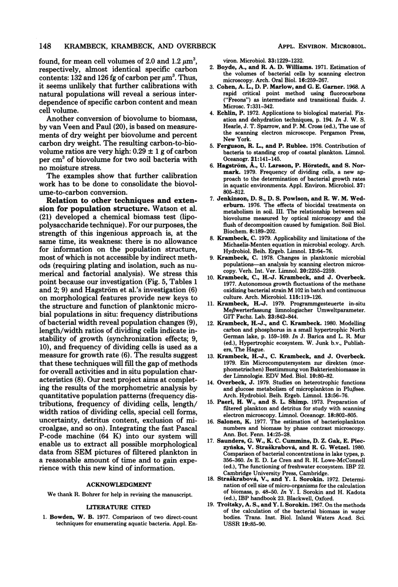
Although biovolume is a better measure of biomass than is cell number, biovolumes have rarely been measured because their evaluation is extremely time-consuming. We developed a microcomputer system that assists cell size measurements on images of filtered plankton: scanning electron micrograph negatives were projected on a digitizer field, bacterial length and width were marked by a cursor, and coordinates were directly transferred to an MOS 6502 microcomputer (KIM 1). The dialogue program BABI organized and controlled the digitizer measurements in cooperation with the user, enabled corrections, and printed out results with 95% confidence limits and sample description. The time for scanning electron micrograph preparation was reduced to 15 min (quick transfer to Freon 113 during filtration and air drying). Altogether, this biovolume determination took about 2.5 h for confidence limits of ±15%. Examples are given for applications of the method: (i) comparison of 10 lakes (with specific activities for glucose uptake and for heterotrophic CO2 fixation); (ii) ranges of biomass parameters in one lake; (iii) diurnal cycles (with synchronizing effects, uptake of algal exudates, and calculation of daily growth). This method is discussed in relation to other biomass methods (epifluorescent microscopy, lipopolysaccharide technique, frequency of dividing cells) and the problem of biovolume-to-carbon conversions.
Full text
PDF
Images in this article
Selected References
These references are in PubMed. This may not be the complete list of references from this article.
- Bowden W. B. Comparison of two direct-count techniques for enumerating aquatic bacteria. Appl Environ Microbiol. 1977 May;33(5):1229–1232. doi: 10.1128/aem.33.5.1229-1232.1977. [DOI] [PMC free article] [PubMed] [Google Scholar]
- Boyde A., Williams R. A. Estimation of the volumes of bacterial cells by scanning electron microscopy. Arch Oral Biol. 1971 Mar;16(3):259–267. doi: 10.1016/0003-9969(71)90019-7. [DOI] [PubMed] [Google Scholar]
- Hagström A., Larsson U., Hörstedt P., Normark S. Frequency of dividing cells, a new approach to the determination of bacterial growth rates in aquatic environments. Appl Environ Microbiol. 1979 May;37(5):805–812. doi: 10.1128/aem.37.5.805-812.1979. [DOI] [PMC free article] [PubMed] [Google Scholar]
- Krambeck C., Krambeck H. J., Overbeck J. Autonomous growth fluctuations of the methane oxidizing bacterial strain M 102 in batch and continuous culture. Arch Microbiol. 1977 Nov 18;115(2):119–126. doi: 10.1007/BF00406364. [DOI] [PubMed] [Google Scholar]
- Watson S. W., Novitsky T. J., Quinby H. L., Valois F. W. Determination of bacterial number and biomass in the marine environment. Appl Environ Microbiol. 1977 Apr;33(4):940–946. doi: 10.1128/aem.33.4.940-946.1977. [DOI] [PMC free article] [PubMed] [Google Scholar]
- van Veen J. A., Paul E. A. Conversion of biovolume measurements of soil organisms, grown under various moisture tensions, to biomass and their nutrient content. Appl Environ Microbiol. 1979 Apr;37(4):686–692. doi: 10.1128/aem.37.4.686-692.1979. [DOI] [PMC free article] [PubMed] [Google Scholar]