Abstract
The recent identification of a second mesodermal region as a source of cardiomyocytes has challenged the views on the formation of the heart. This second source of cardiomyocytes is localized centrally on the embryonic disc relative to the remainder of the classic cardiac crescent, a region also called the pharyngeal mesoderm. In this review, we discuss the concept of the primary and secondary cardiogenic fields in the context of folding of the embryo, and the subsequent temporal events involved in formation of the heart. We suggest that, during evolution, the heart developed initially only with the components required for a systemic circulation, namely a sinus venosus, a common atrium, a ‘left’ ventricle and an arterial cone, the latter being the myocardial outflow tract as seen in the heart of primitive fishes. These components developed in their entirety from the classic cardiac crescent. Only later in the course of evolution did the appearance of novel signalling pathways permit the central part of the cardiac crescent, and possibly the contiguous pharyngeal mesoderm, to develop into the cardiac components required for the pulmonary circulation. These latter components comprise the right ventricle, and that part of the left atrium that derives from the mediastinal myocardium, namely the dorsal atrial wall and the atrial septum. It is these elements which are now recognized as developing from the second field of pharyngeal mesoderm. We suggest that, rather than representing development from separate fields, the cardiac components required for both the systemic and pulmonary circulations are derived by patterning from a single cardiac field, albeit with temporal delay in the process of formation.
Keywords: cardiovascular development, cardiogenesis, second heart field, anterior heart field
1. Introduction
The new concept that there is a second heart-forming field, which provides a second source of cardiomyocytes for the definitive chambers of the developing heart, has been exceedingly productive in terms of our appreciation of the building plan of the heart (Kelly et al. 2001; Mjaatvedt et al. 2001; Waldo et al. 2001). It is probably due to this acquisition of new knowledge that the time-honoured concept of a pre-existing cardiac tube, containing from the outset all the components of the definitive heart (Davis 1927; Srivastava & Olson 2000), has rapidly been superseded. Owing to this change, it has proved possible to retrieve from oblivion previous morphological studies that had long since demonstrated that cardiac muscle was added during development at both extremities of the heart tube (Virágh & Challice 1973; Arguello et al. 1975). The new insights are derived from modern techniques of transgenesis, combined with analysis of genetic lineages, which have provided additional and unambiguous molecular evidence concerning those cardiac components which are already represented in the primary heart tube, as opposed to those that are added in later phases of development (Cai et al. 2003; Zaffran et al. 2004).
The new concept of two heart fields, therefore, has provided a great impetus for the understanding of cardiac morphogenesis; but it has also raised additional questions. These relate in part to the failure of those introducing the novel concepts to provide precise definitions of their own understanding of the expression ‘cardiac field’. In this light, the interested reader should consult the elegant methodological discussions of the embryologic meaning of ‘field’ provided by Slack (1983) and Minelli (2003). Taking into account their suggestions, we might question whether there are indeed two sources of myocardial cells that form discrete components of the formed heart. Why should there be two fields, as opposed to just one, three, or perhaps more? How are the alleged fields distinguished from the surrounding tissues and from each other? Do the fields represent territories of gene expression, or of morphogenetic signalling? Most importantly, do the two fields emphasized thus far represent distinct and unbridgeable cellular lineages, the descendants of which form distinct compartments within the definitive heart?
To provide answers to these questions, we need to understand the mechanics of formation of the cardiac tube itself. This occurs following a complex morphogenetic process of folding, which is part of a more general process of folding by which the embryo itself is formed from the embryonic germ layers. By focusing on this embryonic process of formation of the heart, as opposed to the adjacent but contiguous ‘non-cardiac’ regions, we will address these questions, and at the same time seek to highlight some general principles of development.
2. Frames of reference: the embryonic disc and the axes of the developing body
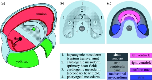
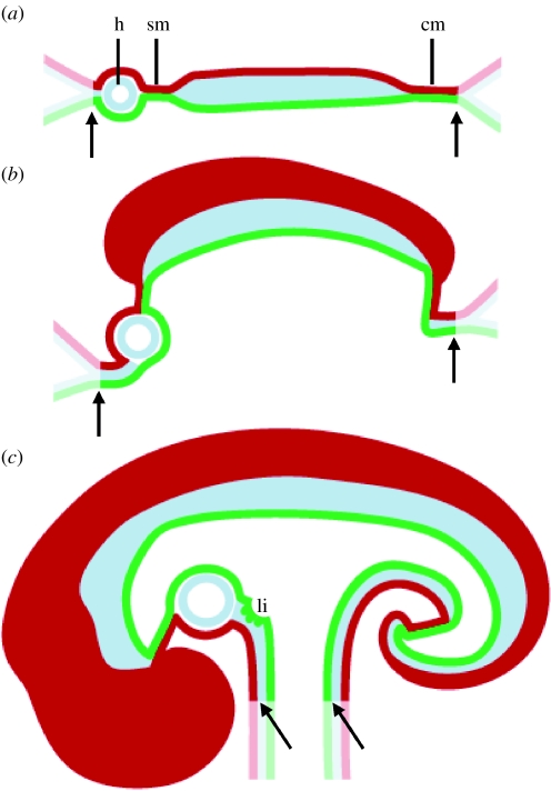
Subsequent to gastrulation, the embryo is composed of three germ layers, the ectoderm facing the amniotic cavity, the endoderm facing the yolk sac and the mesoderm located between these layers (figure 1a). The periphery of the embryonic disc is marked by the boundary between the embryo proper and the extra-embryonic tissues. Owing to subsequent extensive growth of the embryonic disc relative to the peripheral boundary of amnion and yolk sac, the process of folding produces the recognized shape of the embryo. After this process, the original peripheral boundary of the embryonic disc itself becomes no more than the navel of the embryo (figure 2a–c). Although this process of folding is well described, and accounts are included in all medical textbooks of embryology, we are unaware of any account in which the peripheral boundary of the disc has been associated with the navel of the embryo. Perhaps in keeping with this, it seems to us that it has not always been appreciated how the components of the embryonic disc achieve their positions in the formed body. Subsequent to folding, the peripheral parts of the disc become positioned ventrally, or towards the belly of the embryo. We explain the terminology of the axes of the body in Box 1. If we then focus on that part of the flat mesodermal germ layer closest to the site of formation of the head, namely the stomatopharyngeal membrane, which closes the orifice of the developing mouth, then we find that the membrane is flanked by pharyngeal mesoderm. This pharyngeal mesoderm, in turn, is bordered at its periphery by the cardiogenic mesoderm. This, again in turn, is contiguous with the mesoderm of the transverse septum, in which the liver will develop. This so-called septum is the most peripheral part of the mesodermal layer of the embryonic disc. After completion of the process of folding, it is situated cranially relative to the navel (figure 2). These topographic relationships are subsequently maintained during further development, although their position may alter relative to the axes of the body as the embryo continues to fold.
Figure 1.
(a) The trilaminar embryonic disc in the context of the amnion and the yolk sac. Red, ectoderm; blue, mesoderm; green, endoderm; sm, stomatopharyngeal membrane; Hn, Hensen's node and ps, primitive streak. The ectoderm has been removed in the region of the stomatopharyngeal membrane to show the cardiac crescent. The adjacent pharyngeal mesoderm is central relative to the crescent, while the mesoderm of the septum transversum is peripheral; these relationships are shown in detail in (b). In panel (c), we provide a detail of the cardiogenic region, and suggest a hypothetical fate for the different cardiac regions, largely based on the previous work of Stalsberg & de Haan (1969).
Figure 2.
(a–c) The cartoon shows folding of the embryonic disc, and is based on Tuchmann et al. (1971). Red, ectoderm; blue, mesoderm; green, endoderm; sm, stomatopharyngeal membrane; cm, cloacal membrane; h, heart; li, liver and arrows, edge of the embryonic disc, or ‘navel’.
Box 1.
The three body planes, frontal, sagittal and transversal, are used in the description of the location and relation of organs and structures in both human anatomy and biology. But, owing to the unique upright position of humans, the usual terminology for the body axes is different in the two fields. Cranial and caudal directions are dubbed superior and inferior in the medical sciences, but are called anterior and posterior in the biological world. Dorsal and ventral are dubbed anterior and posterior in human, and superior and inferior in the mouse. To prevent confusion, throughout this review, we use the terms cranial and caudal, and dorsal and ventral, to describe these coordinates. For the flat embryonic disc, however, it is inappropriate to employ any of these terms, and hence we try to avoid their use when describing the disc itself.
3. Morphological analyses: formation of the heart tube
It has long been recognized that, during the process of gastrulation, cardiogenic precursor cells leave the anterior part of the primitive streak and give rise to two heart-forming regions. These areas are located in the anterior mesodermal germ layer and are positioned on either side of the midline (Rawles 1943; Rosenquist & de Haan 1966; Rosenquist 1970; Garcia-Martinez & Schoenwolf 1993; Tam et al. 1997). The two heart-forming regions then fuse across the midline to form a crescent-shaped area of epithelium, which subsequently becomes the heart tube. As yet, no definitive patterning has been recognized in the primitive streak and early heart-forming regions (Redkar et al. 2001). Only at later stages, after formation of the cardiac crescent, is it possible to recognize patterning (Stalsberg & de Haan 1969; Redkar et al. 2001; Yutzey & Kirby 2002) sufficient to permit a morphological description of the subsequent remodelling required to form the cardiac tube (see below).
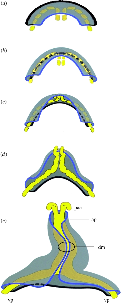
We have sought to encapsulate the morphogenetic movements of the anterior mesodermal germ layer, along with the different steps involved in the process of folding, in the cartoons and animation shown in figures 2 and 3. In figure 3d,e, we show that the myocardial tube is initially a trough, which starts to close dorsally along the margins closest to the developing spine. The closure starts at the level of the left ventricle, and then proceeds by ‘zipping’ so that the trough is closed initially in caudal, and later also in cranial direction (Arguello et al. 1975; Moreno-Rodriguez et al. 2006).
Figure 3.
(a–e) Formation of the cardiac tube from the primary cardiac crescent, represented in five consecutive phases based on de Jong et al. (1997). See online data supplement for a link to an animation to visualize this process. The cardiac crescent is shown from the amniotic cavity (dorsal view). The other germ layers, and the rest of the mesoderm, are not shown. The myocardium is shown in transparent grey; the endocardium is shown in yellow as it forms between the endoderm and the mesodermal myocardium. The blue line marks the central border of the cardiac crescent, which is contiguous with the pharyngeal mesoderm. The dark grey line marks the peripheral border, which is contiguous with the mesoderm of the septum transversum, in which develops the systemic venous tributaries. Note that central ‘blue’ region becomes positioned dorsally, explaining the relationship of outflow components with the mediastinal component at the venous pole. paa, pharyngeal arch artery; ap, arterial pole; vp, venous pole and dm, dorsal mesocardium.
Subsequent to the completion of folding, the initial central part of the cardiac crescent (figure 3, blue line) has retained its location, and hence is located medio-dorsally in the formed body. Concomitant with the folding, when considered relative to the axes of the body, the peripheral region (figure 3, dark grey line) is transformed into the ventral part of the heart tube. If the heart-forming region initially positioned centro-medially (in other words, the region that has become known as the second heart field) contributes to the definitive cardiac structures located dorsally, then this central region is able to contribute to structures developing at the arterial pole, such as the right ventricle and outflow tract, and also at the venous pole. As we will show, it is at the dorsal part of the venous pole that the mediastinal myocardium is formed (Franco et al. 2000), this being the area where the pulmonary veins enter the heart (Soufan et al. 2004; Anderson et al. 2006). The peripheral region of the initial cardiac crescent also flanks the future transverse septum, the site of formation of the liver, pro-epicardium and the systemic venous tributaries.
For didactic reasons, our cartoons lack the three-dimensional context of the surrounding tissues, but we have made them topologically correct. Thus, irrespective of the model system used, be it avian or mammalian, the peripheral border of the cardiogenic mesoderm faces the mesoderm of the transverse septum, while the central border of the cardiogenic mesoderm is contiguous with the pharyngeal mesoderm, this being the area of formation of the outflow tract and right ventricle, the mediastinal, or pulmonary, myocardium, and the site of the persisting dorsal mesocardium. As far as we know, it has never been possible thus far to determine directly the precise size of the ‘classic’ cardiac crescent, or ‘heart-forming region’, this rather being determined by retrospective fate mapping, with the drawback that the estimated size of the precursor field depends on the size of the heart at the stage of analysis. And, because the stage of analysis in all experiments conducted thus far was prior to completion of the incorporation of all cells into the definitive heart, the size of the classic crescent has certainly been underestimated. It coincides roughly with the precursors of the left ventricle, these being the only cardiac cells present at the time of analysis (Ehrman & Yutzey 1999; Redkar et al. 2001; Moorman & Christoffels 2003). Recent (Kelly et al. 2001; Mjaatvedt et al. 2001; Waldo et al. 2001; Cai et al. 2003) and classic experiments (Virágh & Challice 1973; Arguello et al. 1975), nonetheless, suggest that the potential cardiogenic component of the mesoderm is larger than generally thought. More importantly, we should note that the central, or medial, tissues adjacent to the classical cardiac crescent are made up of pharyngeal mesoderm (figure 1b). Thus, from the outset of development, the tissues destined to give rise to the right ventricle and outflow tract are directly confluent with the site of formation of the pharyngeal arches. It is this area of conjunction between the classic cardiac crescent and the pharyngeal mesoderm that is now identified as the second heart field. This pharyngeal mesoderm contributes not only to the myocardial components within the right ventricle and outflow tract, but also to the non-myocardial intra-pericardial portion of the arterial pole (Waldo et al. 2005). From a morphological stance, if we argue that the primary heart field gives rise only to the left ventricle, it is axiomatic that we accept that cells need to be added at both extremities of the developing heart. The cells added at the arterial and venous poles, however, are derived fundamentally from different regions of the mesoderm of the trilaminar embryonic disc, specifically the central pharyngeal mesoderm as opposed to the peripheral mesoderm. As we have discussed, nonetheless, the transverse septum and the systemic venous tributaries, including the smooth-walled component of the right atrium, or the sinus venarum, are also formed from the peripheral mesoderm. It should now be possible to determine the precise morphological boundary between these two different regions at the venous pole of the heart by following the fate of cells which express Islet1.
4. Classic lineage analyses
A fate map demonstrating that the separate regions of the cardiogenic mesoderm in the avian embryo was established in 1969 by Stalsberg & de Haan, who transplanted fragments of tissue labelled with 3H-thymidine and tracked their fate. Experiments using DiI labelling have now confirmed and extended these observations (Redkar et al. 2001). These, and other lineage experiments, have been discussed in detail by Eisenberg (2002). The meticulous and precise fate map produced by Stalsberg and de Haan already showed that the peripheral part of the cardiogenic mesoderm formed the venous pole of the heart, and was contiguous with the mesenchyme of the transverse septum. The centrally localized explants, in contrast, were shown to contribute to the inner curvature, or dorsal side, of the forming heart tube, along with the arterial pole. The chicken embryos, however, were analysed only up to embryonic stage 12. Of necessity, this means that the extent of the cardiogenic mesoderm was underestimated, because, as pointed out previously (Moorman & Christoffels 2003), the myocardium of the outflow tract, and that clothing the systemic venous tributaries, has not, at this stage, been added to the heart tube.
Stalsberg & de Haan (1969) also performed marking experiments using injections of particles of iron oxide. By following the fate of these particles in successive developmental stages, they were able to demonstrate that the early heart tube continues to elongate by progressive recruitment of cardiac cells to the venous pole. Arguello et al. (1975), using similar techniques, demonstrated similar recruitment at the arterial pole. The same group performed ablation experiments, in which the heart tube was cut at the cranial side of the fusion of the two cardiac primordia. Formation of cardiac tissues was observed from the remaining cranial part, demonstrating unequivocal recruitment of cardiac cells to the arterial pole of the heart. Furthermore, Virágh & Challice (1973) had also described differentiation of cardiac muscle cells in the mouse in the flanking extra-cardiac mesenchyme. It is the region of origin of these additional cells that is now designated as the second heart field, thus distinguishing it from the primary heart field, which gives rise only to the initial linear heart tube. Different authors have used a variety of terminologies to describe this region, or parts of it, as discussed in several reviews (Abu-Issa et al. 2004; van den Hoff et al. 2004; Buckingham et al. 2005; Kelly 2005). Here, we concentrate on the conceptual framework regarding the origin and location of the second source of cells.
5. Molecular lineage analyses
Recent molecular lineage analyses also demonstrate in unambiguous fashion that myocardium is added to the already functioning myocardial heart tube, the techniques used currently probably being more convincing than ever was possible with conventional techniques. It was the groups of Markwald (Mjaatvedt et al. 2001) and Kirby (Waldo et al. 2001, 2005) that first demonstrated the addition of myocardium at the arterial pole of the chicken heart. Waldo and co-workers then demonstrated that the formation of this new myocardium recapitulates a similar programme as seen in the differentiation of the primary heart field, including the involvement of the transcription factors Nkx2-5, Gata4 and Mef2c, as well as fibroblast growth factors and bone morphogenetic proteins. Accounts of the extent of addition of myocardium, however, differed between these research groups, with the former arguing only for addition of the outflow tract, and the latter claiming that, as well as the outflow tract, the second field also gave rise to the non-myocardial but intra-pericardial portion of the arterial pole. This difference in interpretation may be more apparent than real, since from the stance of lineages, boundaries between morphological regions are not necessarily made up at subsequent stages by the same cells as were present at that site initially. This possibility was well demonstrated by the classic labelling experiments of Stalsberg & de Haan (1969). The failure as yet to resolve this issue has hampered discussions on the topic of lineage. A second confounding factor is the assumption that, in avian hearts, the right ventricular precursors make up a large part of the initial linear heart tube, whereas in mammalian hearts, the linear heart tube contains the left ventricular precursors (Abu-Issa et al. 2004; van den Hoff et al. 2004). If this is correct, then it impacts markedly on any conclusions drawn concerning the potential contribution from the second field at the arterial pole of the heart. The assumption is based on lineage analyses made by De la Cruz and co-workers (De la Cruz et al. 1989; De la Cruz & Sanchez-Gomez 1998). Having restudied their account in depth, we agree with their general conclusion that there are no primitive cavities in the linear heart tube that correspond with the definitive cardiac cavities. Thus far, however, we are unable to find unambiguous evidence to demonstrate the presence of the primordium of the trabeculated apical portion of the right ventricle within the chicken linear heart tube. When DiI was used to label the straight heart tube at the pericardial reflection of the arterial pole, the experiments showed that the entirety of the right ventricle is added to the linear heart tube later in development (Rana et al. 2007), this being consistent with the data derived by Kelly and co-workers in the mouse (Kelly et al. 2001; Zaffran et al. 2004; Kelly 2005). This latter group has demonstrated, both by DiI-labelling experiments, and using a LacZ transgene integrated in the Fgf10 locus, that both the outflow tract and the right ventricle are derived from pharyngeal mesoderm. Cai et al. (2003) have now extended these observations, demonstrating that a larger population of cardiac precursor cells, which includes the precursors of the pharyngeal mesoderm, contributes not only to the right ventricle and outflow tract at the arterial pole, but also form parts of the atrial chambers at the venous pole. This population of cardiac precursor cells is characterized by the expression of the LIM homeodomain transcription factor Islet1. Null mutants of Islet1 display complete truncation of the above-mentioned cardiac domains, indicating the requirement of this transcription factor for the differentiation of the second pool of cardiac precursor cells. Another Cre-directed lineage study (Verzi et al. 2005), however, using a so-called mef2c anterior heart field promoter, does not identify this atrial component, but identifies at the arterial pole an even larger area, also including the ventricular septum and parts of the left ventricle. These data demonstrate the difficulties in identifying the primordia for cardiac components using patterns of gene expression and do not support the notion that the cardiac components are built from two precisely defined sources of progenitors.
6. The heart-forming fields: one or multiple?
In a recent review entitled: ‘Heart fields: one, two or more’, Abu-Issa et al. (2004), conclude that ‘current evidence points to complex patterning in the primary heart field rather than the existence of multiple fields’. The comparability of the transcriptional programme in both of the alleged fields underscores this conclusion (Waldo et al. 2001). Expression of Islet1 seems not to be a distinguishing feature, because it is expressed in the alleged primary heart field both in chicken (Yuan & Schoenwolf 2000) and in mouse (Prall et al. 2007). Based on our own evaluation of the gradual processes of folding that lead to the formation of the linear heart tube, as shown by our cartoons in figure 3, we endorse this notion, as extensively discussed previously (van den Hoff et al. 2004). Acceptance of this notion, nonetheless, carries the implication that all cells within this solitary field have the capacity to form different cardiac compartments, depending on their position in the field. Such a view is entirely in keeping with current views on the development of cellular diversity (Gurdon et al. 1999; Ashe & Briscoe 2006). Differences in the concentration of a diffusing morphogen will eventually create a number of different cell fates, leading to a progressive restriction of developmental potency, and progressive generation of diversity within a field that was initially homogeneous.
Although our own bias is to endorse the view of a solitary field, we must also consider the possibility of additional cardiac fields, and whether these fields constitute unbridgeable lineages. Meilhac et al. (2004) sought to address this question by performing retrospective clonal analyses. This genetic approach depends on the visualization of rare spontaneous events of intragenic recombination, this then permitting clonal analysis. Clones spanning two or more adjacent cardiac compartments were consistently observed for all cardiac compartments, as well as clones exclusively present in the compartments at the extremities of the tube. But clones were also found exclusively in the middle part of the cardiac tube, as well as clones spanning all cardiac components. These data are not consistent with the idea that the heart is built from two precisely defined sources of precursors, namely one for segments such as right ventricle and outflow regions, the second heart field, and another for the left ventricle and the inflow areas, the classic cardiac crescent (Buckingham et al. 2005). Meilhac et al. (2004), nonetheless, did conclude that two distinct lineages would contribute to the heart. The first lineage was proposed to contribute to both ventricles, to the atrioventricular canal and to both atrial chambers. The second lineage was held not to contribute to the left ventricle, albeit that they found large clones which did involve the left ventricle. An alternative interpretation is that the observed distribution of clones over the heart reflects the gradual and progressive formation of the heart tube, as outlined in figure 3. Thus, it is possible that early recombination events give rise to clones only in the precursors of the left ventricle, and in contiguous compartments which are formed first. It is the later recombination events that give rise to labelling of the compartments at the venous and arterial extremities of the heart tube. Irrespective of such differences in interpretation, the data certainly demonstrate that none of the cardiac compartments of the formed heart derive from a single lineage, but rather support gradual formation of the heart. These meticulous lineage analyses, nonetheless, cannot be taken as unequivocal evidence for the existence of only two cardiac lineages.
In this light, we should also ponder on the temporal order of differentiation of the two alleged heart fields. In our view, this order may simply reflect the evolutionary development of the cardiovascular system. Thus, the heart that initially developed during evolution contained only the components for a systemic circulation, namely a sinus venosus, a common atrium, the left ventricle and the myocardial outflow tract. Only later in the evolutionary tree do we see the development of the cardiac components of the pulmonary circulation, specifically the right ventricle, and most, but not all, of the left atrium. The primitive bony fishes (Teleostomi) possessed a gas bladder that may have served both as a primitive lung and a buoyancy-controlling device (Foxon 1955; Farmer 1999; Kardong 2006). This structure evolved into the swim bladder in the ray-finned bony fishes (Actinopterygii) and into the lungs in lungfish (Dipnoi) and tetrapods, both belonging to the branch of the lobe-finned fishes (Sarcopterygii). In the great majority of these fishes, the veins from the ‘gas bladder’ drain to the systemic venous tributaries. It is only in the lungfish that the veins unite and drain directly to the left atrium, this setting the scene for a separate pulmonary circulation. The hearts of the other fishes possess a left wall of the common atrial chamber, but they lack a definitive left atrium because there is no atrial septum. It is only with the appearance, during the evolutionary sequence, of the atrial septum, first seen in the lungfish, that we are able to define the presence of a separate left atrium. Significantly, the process coincides with the formation of the dorsal atrial wall, which incorporates the site of entrance of the pulmonary veins. In mouse, the atrial septum, along with this dorsal atrial wall, is formed from mediastinal myocardium, and both display a unique molecular phenotype (Franco et al. 2000; Kruithof et al. 2003; Soufan et al. 2004). This mediastinal myocardium, along with the right ventricle, is added relatively late to the heart during its development (Mjaatvedt et al. 2001; Cai et al. 2003; Zaffran et al. 2004). In figure 1c, we offer a hypothetical view of the location of the precursor cells for the distinct cardiac components within the cardiogenic mesoderm. This scheme relies heavily upon the meticulous data of Stalsberg & de Haan (1969). We emphasize that this scheme should be considered as a pictogram, showing only the topographical relationships between the different cardiac components, rather than real dimensions. The pictogram clearly shows the spatial relationship of the mediastinal myocardium, from which the atrial septum takes origin, in the central part of the crescent and the cranial part of this region where the right ventricle and outflow tract develop. The myocardial outflow tract of the primitive fishes, the conus arteriosus, is homologous with the amphibian bulbus cordis, and with the embryonic outlet component of the heart tube of the mammal, which gives rise eventually to the right ventricle (Holmes 1975; Romer & Parsons 1986). Thus, important elements representing potential components of the pulmonary circulation are to be found in the primitive fish heart. These evolutionary considerations fail to provide support for the necessity of defining an additional heart field. Instead, they suggest novel patterning within a single heart field to accommodate the construction of the four-chambered hearts of birds and mammals.
It is also clear that the addition of myocardium to the heart from the peripheral mesoderm, specifically the transverse septum, is old in evolutionary terms, since we know that the fish heart possesses a specific ‘sinus venosus’. This systemic venous sinus persists in the embryonic stages of avian cardiac development (deRuiter et al. 1995; Webb et al. 2000), while its remnants can still be recognized as the myocardial sleeves of the systemic venous tributaries, produced during formation of the venous pole of the heart in mammals (Soufan et al. 2004; van den Hoff et al. 2004; Anderson et al. 2006). Recent work from our laboratory has shown that it is expression of Tbx18, rather than Islet1, that marks these precursors (Christoffels et al. 2006), highlighting the distinction between the precursors of the second heart field found in the central pharyngeal mesoderm, and the precursors of the systemic venous sinus located in the peripheral mesoderm.
Although we do not imply any preferred place for initial closure of the ‘cardiac zipper’ within the morphogenetic principles of formation of the cardiac tube that we have outlined in figure 3, it is known that the heart tube closes first at the level of the left ventricle. It is possible, of course, that any other part of the heart could have been formed first. However, we would argue that the evolutionary history of the development of the heart, with differentiation first of the cardiac components of the systemic circulation from the classic primary heart field, followed by the differentiation of the cardiac components of the pulmonary circulation from the second heart field, militates strongly against this possibility.
7. Conclusions
In our opinion, all the evidence thus far available, be it culled from morphological, molecular, lineage or evolutionary studies, supports the view that there is but a single heart field, albeit that the patterning of the field has changed during evolution to accommodate the construction of a four-chambered heart. Although recent investigations have clarified many molecular aspects of early cardiac development (Cripps & Olson 2002; Harvey 2002; Brand 2003; Kelly 2005), we still do not know why, either in birds or mammals, the so-called primary heart field should be first to differentiate. Equally enigmatic is the mechanism by which the so-called second heart field remains initially undifferentiated, although fibroblast growth factor 8 and Islet1 may play a crucial role in these processes (Alsan & Schultheiss 2002; Laugwitz et al. 2005). In this review, we have discussed the possibility that the temporal differences in differentiation of the distinct cardiac components relate to their evolutionary origin. The right ventricle and outflow tract are derived from cardiomyocytes that are centrally localized on the embryonic disc, contiguous with the pharyngeal mesenchyme. This mesenchyme is initially localized centrally within the embryonic disc, being contiguous caudally with the mediastinal mesenchyme. As far as we are aware, the formation of myocardium from this area has yet to be studied in sufficient morphogenetic detail unambiguously to determine its contribution to the venous pole of the heart. It is our opinion, nonetheless, that the mediastinal myocardium (Franco et al. 2000; Soufan et al. 2004), including the atrial septum, is formed from this region. And it is within this myocardium that the pulmonary veins drain to the atrial component of the heart at the left side of the forming atrial septum. This hypothesis, of course, remains to be tested.
Acknowledgments
We greatly appreciate the help provided by Martin Kruijff in producing our illustrations, and thank Didi Berkhof for secretarial help, Dr André Linnenbank for the animation of figure 3 and Dr Jan Ruijter for critical reading the manuscript. Research of A.F.M.M., V.M.C. and M.J.B.vdH. is supported by The Netherlands Heart Foundation grant no. 1996M002. R.H.A. is supported by the British Heart Foundation together with the Joseph Levy Foundation.
Footnotes
One contribution of 21 to a Theme Issue ‘Bioengineering the heart’.
References
- Abu-Issa R, Waldo K, Kirby M.L. Heart fields: one, two or more? Dev. Biol. 2004;272:281–285. doi: 10.1016/j.ydbio.2004.05.016. doi:10.1016/j.ydbio.2004.05.016 [DOI] [PubMed] [Google Scholar]
- Alsan B.H, Schultheiss T.M. Regulation of avian cardiogenesis by Fgf8 signaling. Development. 2002;129:1935–1943. doi: 10.1242/dev.129.8.1935. [DOI] [PubMed] [Google Scholar]
- Anderson R.H, Brown N.A, Moorman A.F.M. Development and structures of the venous pole of the heart. Dev. Dyn. 2006;235:2–9. doi: 10.1002/dvdy.20578. doi:10.1002/dvdy.20578 [DOI] [PubMed] [Google Scholar]
- Arguello C, De la Cruz M.V, Gomez C.S. Experimental study of the formation of the heart tube in the chick embryo. J. Embryol. Exp. Morphol. 1975;33:1–11. [PubMed] [Google Scholar]
- Ashe H.L, Briscoe J. The interpretation of morphogen gradients. Development. 2006;133:385–394. doi: 10.1242/dev.02238. doi:10.1242/dev.02238 [DOI] [PubMed] [Google Scholar]
- Brand T. Heart development: molecular insights into cardiac specification and early morphogenesis. Dev. Biol. 2003;258:1–19. doi: 10.1016/s0012-1606(03)00112-x. doi:10.1016/S0012-1606(03)00112-X [DOI] [PubMed] [Google Scholar]
- Buckingham M, Meilhac S, Zaffran S. Building the mammalian heart from two sources of myocardial cells. Nat. Rev. Genet. 2005;6:826–837. doi: 10.1038/nrg1710. doi:10.1038/nrg1710 [DOI] [PubMed] [Google Scholar]
- Cai C.L, Liang X, Shi Y, Chu P.H, Pfaff S.L, Chen J, Evans S. Isl1 identifies a cardiac progenitor population that proliferates prior to differentiation and contributes a majority of cells to the heart. Dev. Cell. 2003;5:877–889. doi: 10.1016/s1534-5807(03)00363-0. doi:10.1016/S1534-5807(03)00363-0 [DOI] [PMC free article] [PubMed] [Google Scholar]
- Christoffels V.M, et al. Formation of the venous pole of the heart from an Nkx2–5–negative precursor population requires Tbx18. Circ. Res. 2006;98:1555–1563. doi: 10.1161/01.RES.0000227571.84189.65. doi:10.1161/01.RES.0000227571.84189.65 [DOI] [PubMed] [Google Scholar]
- Cripps R.M, Olson E.N. Control of cardiac development by an evolutionarily conserved transcriptional network. Dev. Biol. 2002;246:14–28. doi: 10.1006/dbio.2002.0666. doi:10.1006/dbio.2002.0666 [DOI] [PubMed] [Google Scholar]
- Davis C.L. Development of the human heart from its first appearance to the stage found in embryos of twenty paired somites. Contrib. Embryol. 1927;19:245–284. [Google Scholar]
- de Jong F, Virágh Sz, Moorman A.F.M. Cardiac development: a morphologically integrated molecular approach. Card. Young. 1997;7:131–146. [Google Scholar]
- De la Cruz M.V, Sanchez-Gomez C. Straight tube heart. Primitive cardiac cavities vs. primitive cardiac segments. In: De la Cruz M, Markwald R.R, editors. Living morphogenesis of the heart. Birkhäuser; Boston, MA: 1998. pp. 85–98. ch. 3. [Google Scholar]
- De la Cruz M.V, Sánchez-Gómez C, Palomino M. The primitive cardiac regions in the straight tube heart (stage 9) and their anatomical expression in the mature heart: an experimental study in the chick embryo. J. Anat. 1989;165:121–131. [PMC free article] [PubMed] [Google Scholar]
- deRuiter M.C, Gittenberger-de Groot A.C, Wenink A.C.G, Poelmann R.E, Mentink M.M.T. In normal development pulmonary veins are connected to the sinus venosus segment in the left atrium. Anat. Rec. 1995;243:84–92. doi: 10.1002/ar.1092430110. doi:10.1002/ar.1092430110 [DOI] [PubMed] [Google Scholar]
- Ehrman L.A, Yutzey K.E. Lack of regulation in the heart forming region of avian embryos. Dev. Biol. 1999;207:163–175. doi: 10.1006/dbio.1998.9167. doi:10.1006/dbio.1998.9167 [DOI] [PubMed] [Google Scholar]
- Eisenberg L.M. Belief vs. scientific observation: the curious story of the precardiac mesoderm. Anat. Rec. 2002;266:194–197. doi: 10.1002/ar.10066. doi:10.1002/ar.10066 [DOI] [PubMed] [Google Scholar]
- Farmer C.G. Evolution of the vertebrate cardio-pulmonary system. Annu. Rev. Physiol. 1999;61:573–592. doi: 10.1146/annurev.physiol.61.1.573. doi:10.1146/annurev.physiol.61.1.573 [DOI] [PubMed] [Google Scholar]
- Foxon G.E.H. Problems of the double circulation in vertebrates. Biol. Rev. Camb. Phil. Soc. 1955;30:196–228. [Google Scholar]
- Franco D, Campione M, Kelly R, Zammit P.S, Buckingham M, Lamers W.H, Moorman A.F.M. Multiple transcriptional domains, with distinct left and right components, in the atrial chambers of the developing heart. Circ. Res. 2000;87:984–991. doi: 10.1161/01.res.87.11.984. [DOI] [PubMed] [Google Scholar]
- Garcia-Martinez V, Schoenwolf G.C. Primitive streak origin of the cardiovascular system in avian embryos. Dev. Biol. 1993;159:706–719. doi: 10.1006/dbio.1993.1276. doi:10.1006/dbio.1993.1276 [DOI] [PubMed] [Google Scholar]
- Gurdon J.B, Standley H, Dyson S, Butler K, Langon T, Ryan K, Stennard F, Shimizu K, Zorn A. Single cells can sense their position in a morphogen gradient. Development. 1999;126:5309–5317. doi: 10.1242/dev.126.23.5309. [DOI] [PubMed] [Google Scholar]
- Harvey R.P. Patterning the vertebrate heart. Nat. Rev. Genet. 2002;3:544–556. doi: 10.1038/nrg843. doi:10.1038/nrg843 [DOI] [PubMed] [Google Scholar]
- Holmes E.B. A reconsideration of the phylogeny of the tetrapod heart. J. Morphol. 1975;147:209–228. doi: 10.1002/jmor.1051470207. doi:10.1002/jmor.1051470207 [DOI] [PubMed] [Google Scholar]
- Kardong K.V. McGraw-Hill; New York, NY: 2006. Vertebrates: comparative anatomy, function, evolution. pp. 1–782. [Google Scholar]
- Kelly R.G. Molecular inroads into the anterior heart field. Trends Cardiovasc. Med. 2005;15:51–56. doi: 10.1016/j.tcm.2005.02.001. doi:10.1016/j.tcm.2005.02.001 [DOI] [PubMed] [Google Scholar]
- Kelly R.G, Brown N.A, Buckingham M.E. The arterial pole of the mouse heart forms from Fgf10-expressing cells in pharyngeal mesoderm. Dev. Cell. 2001;1:435–440. doi: 10.1016/s1534-5807(01)00040-5. doi:10.1016/S1534-5807(01)00040-5 [DOI] [PubMed] [Google Scholar]
- Kruithof B.P.T, van den Hoff M.J.B, Tesink-Taekema S, Moorman A.F.M. Recruitment of intra- and extra-cardiac cells into the myocardial lineage during mouse development. Anat. Rec. A: Discov. Mol. Cell Evol. Biol. 2003;271:303–314. doi: 10.1002/ar.a.10033. doi:10.1002/ar.a.10033 [DOI] [PubMed] [Google Scholar]
- Laugwitz K.L, et al. Postnatal isl1+ cardioblasts enter fully differentiated cardiomyocyte lineages. Nature. 2005;433:647–653. doi: 10.1038/nature03215. doi:10.1038/nature03215 [DOI] [PMC free article] [PubMed] [Google Scholar]
- Meilhac S.M, Esner M, Kelly R.G, Nicolas J.F, Buckingham M.E. The clonal origin of myocardial cells in different regions of the embryonic mouse heart. Dev. Cell. 2004;6:685–698. doi: 10.1016/s1534-5807(04)00133-9. doi:10.1016/S1534-5807(04)00133-9 [DOI] [PubMed] [Google Scholar]
- Minelli A. Cambridge University Press; Cambridge, UK: 2003. The development of animal form: ontogeny, morphology and evolution. pp. 1–323. [Google Scholar]
- Mjaatvedt C.H, Nakaoka T, Moreno-Rodriguez R, Norris R.A, Kern M.J, Eisenberg C.A, Turner D, Markwald R.R. The outflow tract of the heart is recruited from a novel heart-forming field. Dev. Biol. 2001;238:97–109. doi: 10.1006/dbio.2001.0409. doi:10.1006/dbio.2001.0409 [DOI] [PubMed] [Google Scholar]
- Moorman A.F.M, Christoffels V.M. Cardiac chamber formation: development, genes and evolution. Physiol. Rev. 2003;83:1223–1267. doi: 10.1152/physrev.00006.2003. [DOI] [PubMed] [Google Scholar]
- Moreno-Rodriguez R.A, Krug E.L, Reyes L, Villavicencio L, Mjaatvedt C.H, Markwald R.R. Bidirectional fusion of the heart-forming fields in the developing chick embryo. Dev. Dyn. 2006;235:191–202. doi: 10.1002/dvdy.20601. doi:10.1002/dvdy.20601 [DOI] [PMC free article] [PubMed] [Google Scholar]
- Prall O.W, et al. An Nkx2-5/Bmp2/Smad1 negative feedback loop controls heart progenitor specification and proliferation. Cell. 2007;128:947–959. doi: 10.1016/j.cell.2007.01.042. doi:10.1016/j.cell.2007.01.042 [DOI] [PMC free article] [PubMed] [Google Scholar]
- Rana M.S, Horsten N.C.A, Tesink-Taekema S, Lamers W.H, Moorman A.F.M, van den Hoff M.J.B. Trabeculated right ventricular free wall in the chicken forms by ventricularization of the myocardium initially forming the outflow tract. Circ. Res. 2007;100:1000–1007. doi: 10.1161/01.RES.0000262688.14288.b8. [DOI] [PubMed] [Google Scholar]
- Rawles M.E. The heart-forming areas of the early chick blastoderm. Physiol. Zool. 1943;16:22–42. [Google Scholar]
- Redkar A, Montgomery M, Litvin J. Fate map of early avian cardiac progenitor cells. Development. 2001;128:2269–2279. doi: 10.1242/dev.128.12.2269. [DOI] [PubMed] [Google Scholar]
- Romer A.S, Parsons T.S. Saunders College Publications; Philadelphia, PA: 1986. The vertebrate body. pp. 1–679. [Google Scholar]
- Rosenquist G.C. Location and movements of cardiogenic cells in the chick embryo: the heart forming portion of the primitive streak. Dev. Biol. 1970;22:461–475. doi: 10.1016/0012-1606(70)90163-6. doi:10.1016/0012-1606(70)90163-6 [DOI] [PubMed] [Google Scholar]
- Rosenquist, G. C. & de Haan, R. L. 1966 Migration of precardiac cells in the chick embryo: a radioautographic study. In Carnegie Inst. Washington Publ 625, 111–121.
- Slack J.M.W. From egg to embryo. In: Barlow P.W, Green P.B, Wylie C.C, editors. Determinative events in early development. Cambridge University Press; Cambridge, UK: 1983. pp. 3–241. [Google Scholar]
- Soufan A.T, van den Hoff M.J.B, Ruijter J.M, de Boer P.A.J, Hagoort J, Webb S, Anderson R.H, Moorman A.F.M. Reconstruction of the patterns of gene expression in the developing mouse heart reveals an architectural arrangement that facilitates the understanding of atrial malformations and arrhythmias. Circ. Res. 2004;95:1207–1215. doi: 10.1161/01.RES.0000150852.04747.e1. doi:10.1161/01.RES.0000150852.04747.e1 [DOI] [PubMed] [Google Scholar]
- Srivastava D, Olson E.N. A genetic blueprint for cardiac development. Nature. 2000;407:221–226. doi: 10.1038/35025190. doi:10.1038/35025190 [DOI] [PubMed] [Google Scholar]
- Stalsberg H, de Haan R.L. The precardiac areas and formation of the tubular heart in the chick embryo. Dev. Biol. 1969;19:128–159. doi: 10.1016/0012-1606(69)90052-9. doi:10.1016/0012-1606(69)90052-9 [DOI] [PubMed] [Google Scholar]
- Tam P.P, Parameswaran M, Kinder S.J, Weinberger R.P. The allocation of epiblast cells to the embryonic heart and other mesodermal lineages: the role of ingression and tissue movement during gastrulation. Development. 1997;124:1631–1642. doi: 10.1242/dev.124.9.1631. [DOI] [PubMed] [Google Scholar]
- Tuchmann H, David G, Haegel P. Embryogenesis. vol. I. Masson and Company; New York, NY: 1971. Illustrated human embryology. pp. 1–110. [Google Scholar]
- van den Hoff M.J.B, Kruithof B.P.T, Moorman A.F.M. Making more heart muscle. Bioessays. 2004;26:248–261. doi: 10.1002/bies.20006. doi:10.1002/bies.20006 [DOI] [PubMed] [Google Scholar]
- Verzi M.P, McCulley D.J, De V.S, Dodou E, Black B.L. The right ventricle, outflow tract, and ventricular septum comprise a restricted expression domain within the secondary/anterior heart field. Dev. Biol. 2005;287:134–145. doi: 10.1016/j.ydbio.2005.08.041. doi:10.1016/j.ydbio.2005.08.041 [DOI] [PubMed] [Google Scholar]
- Virágh Sz, Challice C.E. Origin and differentiation of cardiac muscle cells in the mouse. J. Ultrastruct. Res. 1973;42:1–24. doi: 10.1016/s0022-5320(73)80002-4. doi:10.1016/S0022-5320(73)80002-4 [DOI] [PubMed] [Google Scholar]
- Waldo K.L, Kumiski D.H, Wallis K.T, Stadt H.A, Hutson M.R, Platt D.H, Kirby M.L. Conotruncal myocardium arises from a secondary heart field. Development. 2001;128:3179–3188. doi: 10.1242/dev.128.16.3179. [DOI] [PubMed] [Google Scholar]
- Waldo K.L, Hutson M.R, Ward C.C, Zdanowicz M, Stadt H.A, Kumiski D, Abu-Issa R, Kirby M.L. Secondary heart field contributes myocardium and smooth muscle to the arterial pole of the developing heart. Dev. Biol. 2005;281:78–90. doi: 10.1016/j.ydbio.2005.02.012. doi:10.1016/j.ydbio.2005.02.012 [DOI] [PubMed] [Google Scholar]
- Webb S, Brown N.A, Anderson R.H, Richardson M.K. Relationship in the chick of the developing pulmonary vein to the embryonic systemic venous sinus. Anat. Rec. 2000;259:67–75. doi: 10.1002/(SICI)1097-0185(20000501)259:1<67::AID-AR8>3.0.CO;2-5. doi:10.1002/(SICI)1097-0185(20000501)259:1<67::AID-AR8>3.0.CO;2-5 [DOI] [PubMed] [Google Scholar]
- Yuan S, Schoenwolf G.C. Islet-1 marks the early heart rudiments and is asymmetrically expressed during early rotation of the foregut in the chick embryo. Anat. Rec. 2000;260:204–207. doi: 10.1002/1097-0185(20001001)260:2<204::AID-AR90>3.0.CO;2-5. doi:10.1002/1097-0185(20001001)260:2<204::AID-AR90>3.0.CO;2-5 [DOI] [PubMed] [Google Scholar]
- Yutzey K.E, Kirby M.L. Wherefore heart thou? Embryonic origins of cardiogenic mesoderm. Dev. Dyn. 2002;223:307–320. doi: 10.1002/dvdy.10068. doi:10.1002/dvdy.10068 [DOI] [PubMed] [Google Scholar]
- Zaffran S, Kelly R.G, Meilhac S.M, Buckingham M.E, Brown N.A. Right ventricular myocardium derives from the anterior heart field. Circ. Res. 2004;95:261–268. doi: 10.1161/01.RES.0000136815.73623.BE. doi:10.1161/01.RES.0000136815.73623.BE [DOI] [PubMed] [Google Scholar]


