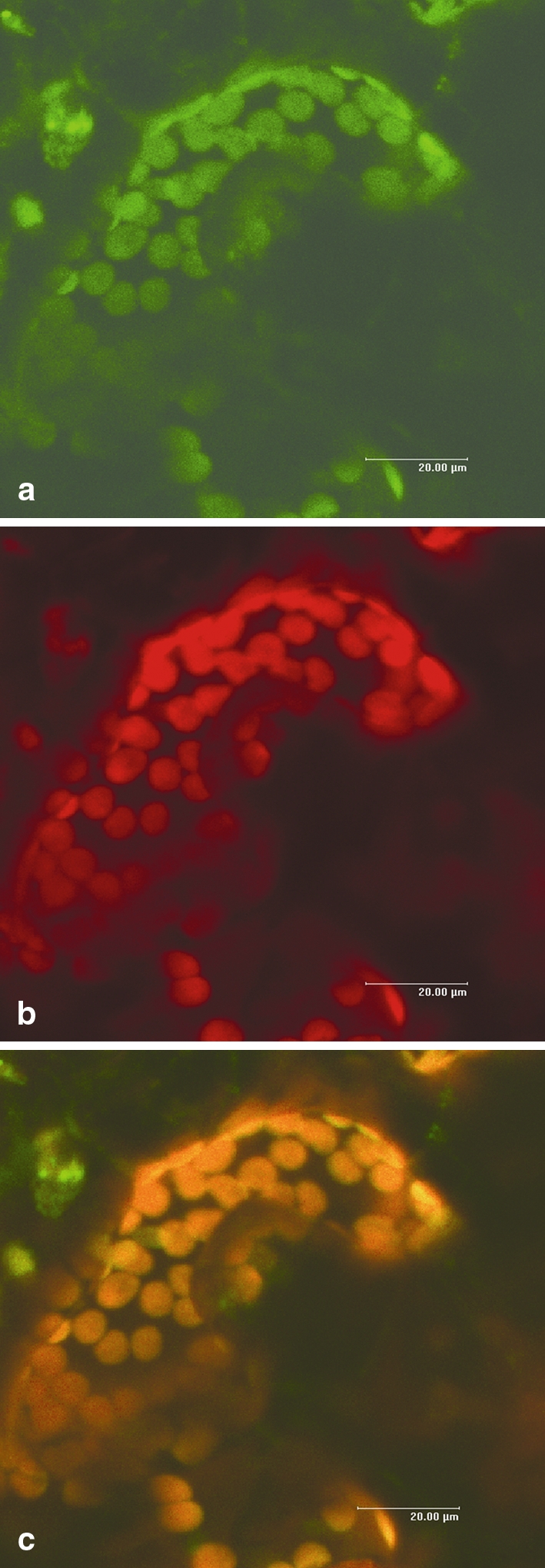
Fig. 2.

Subcellular localization of StAOS2. Confocal laser scanning microscope photographs of a N. benthamiana leaf epidermal guard cell transformed with the fusion construct 2x35S::StAOS2-GFP. a GFP fluorescence (emission spectrum 490–560 nm). b Chlorophyll auto-fluorescence (emission spectrum 650–700 nm). c Overlay of a and b, demonstrating perfect co-localization of StAOS2-GFP with chloroplasts
