Abstract
Bone scans are widely utilized for detection of metastases from bone sarcomas. Technetium methylene diphosphonate scan ([99mTc]MDP) is one of the most popular radiotracers used for that purpose. Lymphatic spread of bone sarcomas is unusual and often difficult to diagnose. Unfortunately, bone scans are not as sensitive in demonstrating lymphatic spread of sarcomas as they are at demonstrating hematogenous spread. A bone scan will often fail to demonstrate lymph nodes metastases until there is mineralization at the affected node. In this report, we highlight an interesting case of a patient with secondary osteogenic sarcoma (OS) from Paget's disease in the distal femur with non-ossified inguinal nodal metastasis diagnosed with [99mTc]MDP. Lymph node involvement was not appreciated on plain radiographs or computed tomography (CT).
Keywords: Metastatic sarcoma, lymph node metastasis, bone scan, Paget's sarcoma
Case report
A 72-year-old male presented to our clinic with a 3-month history of progressive right knee pain associated with swelling and stiffness; this pain was exacerbated by activity and alleviated with narcotics. The past medical history was significant for Paget's disease of several years duration. Physical examination showed a decreased range of motion of the knee (5–120 degrees), mild swelling and elevated temperature, tenderness on palpation of the distal femur, no signs of ligamentous or meniscal injuries. No lymph nodes were palpated throughout the lower extremities and were not enlarged.
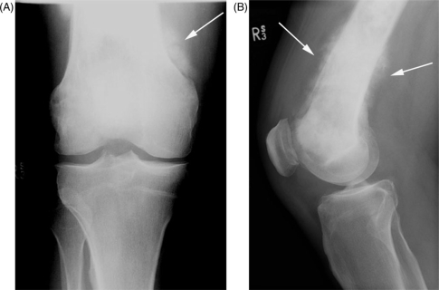
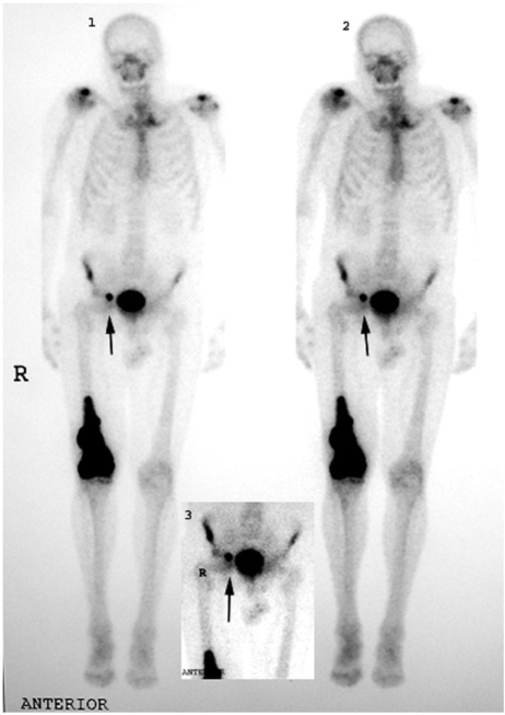
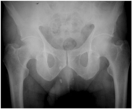
The plain radiographs (Fig. 1) demonstrated an aggressive bone-forming lesion of the right distal femur. Magnetic resonance imaging and computerized scan confirmed the extent of the soft tissue mass. [99mTc]MDP scintigraphy showed increased activity at the lesion site (Fig. 2) and identified an increased uptake of radiotracer localized in the lymph nodes of the right inguinal region. The plain film of the pelvis (Fig. 3), along with other imaging exams, did not show any abnormalities in the inguinal region.
Figure 1.
Anterior–posterior (A) and lateral (B) plain radiographs of the right knee demonstrate an ill-defined, bone-forming, aggressive looking lesion of the distal femur (arrows).
Figure 2.
Three-phase [99mTc]MDP bone scan shows increased uptake in the right distal femur lesion and also an increased uptake of radiotracer in the right inguinal area (arrows).
Figure 3.
Anterior–posterior (A) radiograph of the pelvis does not demonstrate the inguinal lymph node that was identified on the bone scan.
An incisional biopsy of the bony lesion and excisional biopsy of the lymph nodes confirmed the diagnosis of metastatic osteogenic sarcoma (OS). Following a wide resection of the right femoral mass and endoprosthetic reconstruction, chemotherapy was administered. Despite all efforts the patient developed widespread metastatic disease and died 6 months post-operatively, due to pulmonary complications.
Discussion
Approximately 15–20% of patients with OS will present with metastases at time of diagnosis[1]. OS generally spreads through the blood stream and as a result, the most common site for metastases is the lung, followed by bones and soft tissues[1,2].
Lymphatic spread of sarcomas, particularly OS is rare and likely represents less than 3% of all cases[3,4]. Weingrad reported a 2.6% incidence (3/374 patients) of lymphatic spread with OS5. Ceceres et al. reported an incidence of regional lymph nodes metastases in OS of 10.4%. This series included 182 consecutive patients with operable OS that underwent radical surgery and lymph node dissection6.
The estimated incidence of sarcomas in patients with Paget's disease is less than 1%7 however some have reported rates as high as 10%[8,9]. In a review of 1649 OS, only 54 (3.3%) arose in pagetic bone10.
Radiographic features of OS vary depending on the level of osteoblastic activity. This is especially true for bone scans. Non-osseous lesions are better identified when osteoblastic activity produces ossification. Although rare, mineralized lymph nodes are more readily detected on bone scan than non-mineralized nodes2. Bone scans are a cost-effective and sensitive method for detection of lung, osseous and soft-tissue metastases associated with OS11. Bone scans also usually reveal areas of intense tracer activity with or without extension into the soft tissue adjacent to the bone[12,13]. One drawback to bone scans is that other bones in the extremity bearing the tumor may show mild to moderate diffusely increased uptake. This uptake may be more intense around the joints, a finding known as ‘extended pattern’14. Bone scans are also useful in the detection of multicentric OS13. Bone scans with [99mTc]MDP have been widely used for detection of lung and skeletal metastases from OS, often detecting disease before other imaging modalities15.
Paget's disease usually presents with increased activity of radiotracer uptake16; however OS secondary to Paget's disease often has a photon-deficient pattern17. In one previous study, conducted at our institution, 85 patients with bone sarcoma associated with Paget's disease were reviewed, [99mTc]MDP was cold (negative), while gallium uptake was often observed17. Gallium scans were also shown to be more useful in defining the likelihood of malignant degeneration, and the disease extent7.
Heyman, the first to report the detection of lymphatic spread of OS by [99mTc]MDP18, concluded that bone scans are a valuable tool for following the progression of the disease. Caner et al.19 reported a case of metastatic lymph node from an OS of the tibia using hexakis technetium (Tc-MIBI) following a negative three-phase [99mTc]MDP. Positive bone scans have been described in patients with calcified lymph nodes associated with primary OS, however in these cases it is usually possible to make the diagnosis concomitantly with plain films, computed tomography (CT) or magnetic resonance imaging (MRI)[4,20]. Tobias reported lymphatic metastases of OS in 4 of 176 patients, however one other patient presented with calcified lymph nodes in the absence of metastatic disease. It did not seem that patients with lymphatic spread had a different prognosis than those with hematogenous spread4.
Bone scans are not routinely used for the detection of lymph node metastases, however, in the present case, it was useful in identifying an uncommon site of metastasis. For staging purposes gallium scans seem to be more reliable when the diagnosis of lymphatic spread is suspected.
References
- 1.Meyers PA, Heller G, Healey JH, et al. Osteogenic sarcoma with clinically detectable metastasis at initial presentation. J Clin Oncol. 1993;11:449–53. doi: 10.1200/JCO.1993.11.3.449. [DOI] [PubMed] [Google Scholar]
- 2.Kim SJ, Choi JA, Lee SH, et al. Imaging findings of extrapulmonary metastases of osteosarcoma. Clin Imaging. 2004;28:291–300. doi: 10.1016/S0899-7071(03)00206-7. [DOI] [PubMed] [Google Scholar]
- 3.Jeffree GM, Price CH, Sissons HA. The metastatic patterns of osteosarcoma. Br J Cancer. 1975;32:87–107. doi: 10.1038/bjc.1975.136. [DOI] [PMC free article] [PubMed] [Google Scholar]
- 4.Tobias JD, Pratt CB, Parham DM, Green AA, Rao B. The significance of calcified regional lymph nodes at the time of diagnosis of osteosarcoma. Orthopedics. 1985;8:49–52. doi: 10.3928/0147-7447-19850101-08. [DOI] [PubMed] [Google Scholar]
- 5.Weingrad DN, Rosenberg SA. Early lymphatic spread of osteogenic and soft-tissue sarcomas. Surgery. 1978;84:231–40. [PubMed] [Google Scholar]
- 6.Caceres E, Zaharia M, Calderon R. Incidence of regional lymph node metastasis in operable osteogenic sarcoma. Semin Surg Oncol. 1990;6:231–3. doi: 10.1002/ssu.2980060408. [DOI] [PubMed] [Google Scholar]
- 7.Healey JH, Buss D. Radiation and pagetic osteogenic sarcomas. Clin Orthop Relat Res. 1991;270:128–34. [PubMed] [Google Scholar]
- 8.Dieckmann C, Bruns J, Maas R. [Secondary osteosarcoma in Paget disease] Aktuelle Radiol. 1996;6:191–3. [PubMed] [Google Scholar]
- 9.Schajowicz F, Santini Araujo E, Berenstein M. Sarcoma complicating Paget's disease of bone. A clinicopathological study of 62 cases. J Bone Joint Surg Br. 1983;65:299–307. doi: 10.1302/0301-620X.65B3.6573330. [DOI] [PubMed] [Google Scholar]
- 10.Unni KK. Dahlin's bone tumors: general aspects and data on 11087 cases. 5th. Philadelphia: Lippincott-Raven; 1996. [Google Scholar]
- 11.Brown ML. Bone scintigraphy in benign and malignant tumors. Radiol Clin North Am. 1993;31:731–8. [PubMed] [Google Scholar]
- 12.Brown ML. Significance of the solitary lesion in pediatric bone scanning: concise communication. J Nucl Med. 1983;24:114–5. [PubMed] [Google Scholar]
- 13.Wang K, Allen L, Fung E, Chan CC, Chan JC, Griffith JF. Bone scintigraphy in common tumors with osteolytic components. Clin Nucl Med. 2005;30:655–71. doi: 10.1097/01.rlu.0000178027.20780.95. [DOI] [PubMed] [Google Scholar]
- 14.Chew FS, Hudson TM. Radionuclide bone scanning of osteosarcoma: falsely extended uptake patterns. AJR Am J Roentgenol. 1982;139:49–54. doi: 10.2214/ajr.139.1.49. [DOI] [PubMed] [Google Scholar]
- 15.Hoefnagel CA, Bruning PF, Cohen P, Marcuse HR, van der Schoot JB. Detection of lung metastases from osteosarcoma by scintigraphy using 99mTc-methylene diphosphonate. Diagn Imaging. 1981;50:277–84. [PubMed] [Google Scholar]
- 16.Wellman HN, Schauwecker D, Robb JA, Khairi MR, Johnston CC. Skeletal scintimaging and radiography in the diagnosis and management of Paget's disease. Clin Orthop Relat Res. 1977;127:55–62. [PubMed] [Google Scholar]
- 17.Smith J, Botet JF, Yeh SD. Bone sarcomas in Paget disease: a study of 85 patients. Radiology. 1984;152:583–90. doi: 10.1148/radiology.152.3.6235535. [DOI] [PubMed] [Google Scholar]
- 18.Heyman S. The lymphatic spread of osteosarcoma shown by Tc-99m-MDP scintigraphy. Clin Nucl Med. 1980;5:543–5. doi: 10.1097/00003072-198012000-00003. [DOI] [PubMed] [Google Scholar]
- 19.Caner B, Kitapci M, Aras T, Erbengi G, Ugur O, Bekdik C. Increased accumulation of hexakis (2-methoxyisobutylisonitrile)technetium(I) in osteosarcoma and its metastatic lymph nodes. J Nucl Med. 1991;32:1977–8. [PubMed] [Google Scholar]
- 20.Nouyrigat P, Berdah JF, Roullet B, Olivier JP. Osteosarcoma with calcified regional lymph nodes. Pediatr Radiol. 1993;23:74–5. doi: 10.1007/BF02020235. [DOI] [PubMed] [Google Scholar]


