Abstract
Defined candidate human vaccines for treating infection by Yersinia pestis, the agent of plague, have been developed. To facilitate evaluation of the vaccines' efficacy, the in vitro caspase-3 assay for cytotoxicity-neutralizing activity was modified and reevaluated. Immune serum-mediated caspase-3 neutralizing activity correlated with protection against infection in a nonhuman primate vaccine model of plague immunity.
Yersinia pestis, the etiologic agent of plague, has a significant potential to be utilized as a biothreat or a bioweapon (6, 7). Countermeasures, especially those that are effective against pneumonic plague, are critically needed. New candidate vaccines consisting of the F1 capsule antigen and the LcrV, or V, virulence antigen have been developed (5, 10). Both the F1 and the V antigens are highly immunogenic, and they stimulate antibodies that confer passive protection. As the efficacy of these new vaccines cannot be tested in humans, it is essential to develop in vitro surrogate assays that are valid predictors of immunity. We recently described the development of macrophage (MΦ)-based correlate assays of immunity to infection by Y. pestis (1). These assays included a microtiter fluorometric assay of the apoptosis-specific enzyme caspase-3 in J774.A1 MΦs, and a fluorescence-activated cell sorter (FACS)-based live-dead staining assay of terminal necrosis of human-derived HL60 cells (1). Sera from mice and nonhuman primates (NHP) vaccinated with the F1-V fusion protein vaccine (5) were evaluated for the levels at which they neutralized the cytotoxicity induced by infection of MΦs with Y. pestis or the Y. pseudotuberculosis strain Yptb (pTrcV) (1). The results of both the FACS live-dead assay and the caspase-3 assay for serum cytotoxicity neutralization activity (CNA) correlated well with immunity to plague in mice. However, the caspase-3 assay results did not correlate as well as those of the FACS assay with survival in vaccinated NHP, the species that appears to best model human responses to plague, and the FACS assay was proposed as a candidate in vitro correlate assay. Nonetheless, compared to the FACS assay, the caspase-3 assay is easier to perform and analyze, it is faster and requires less expensive equipment, and it is more amenable to large-scale testing of sera from vaccinees. In addition, NIAID- and DOD-sponsored research of new biodefense countermeasures stipulates the development of a validated in vitro correlate assay(s) of immunity (4). Such validated assays are also required, as part of the advanced development, to secure FDA licensure of the F1-V human plague vaccine; and phase 1 and 2 clinical trials of the F1-V fusion vaccine have been initiated (2). Results of prevalidation studies using the FACS and caspase-3 assays (data not shown) indicated that the latter would more readily fulfill the validation requirements that include demonstration of assay reproducibility, repeatability, and robustness (3, 8). Thus, the purposes of this study were to modify and optimize the performance of the caspase-3 procedure and to test this modified assay as a potential in vitro predictor of immunity in vaccinated NHP. Sera from a large, recently completed study done with cynomolgus macaques to test F1-V vaccine efficacy were available for use, as described below.
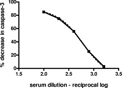
Numerous changes were made to the caspase-3 microtiter assay, and a complete protocol is shown in Table 1. Changes were made in the following: the cell and bacterial culture media, the final concentration of bacteria in the assay, the amounts of antibody used in pretreatment and titration steps, and the assay incubation periods. Also, greater emphasis was given to important aspects of bacterial propagation, and more details were provided for statistical analyses of data (Table 1). The modified procedure was evaluated initially by titrating the CNA of a rabbit anti-V immunoglobulin G (IgG) preparation, which was used as the positive control in the original MΦ assay (1). A good dose response was observed in twofold titration assays as determined by regression analysis. Whereas rabbit anti-V IgG diluted 1/100 protected 85% of the cells, the highest dilution of 1/1,600 protected only 3% of the MΦs against Yersinia infection-induced apoptosis (Fig. 1), and a 50% reciprocal neutralization titer value of 457 was determined, as described in the legend to Fig. 1.
TABLE 1.
Modified caspase-3 assay for serum neutralization of macrophage cytotoxicity
| Assay stage | Step | Descriptiona | Comment |
|---|---|---|---|
| Cell and bacterial culture | 1 | Seed J774.A1 MΦs into 96-well trays (200 μl/well) of a 5 × 10⁵ cell/ml suspension. | Use freshly prepared EMEM with Earle's salts (Invitrogen) supplemented with 2 mM nonessential amino acids (Sigma), 2 mM l-glutamine (HyClone), and 10% FBS (Gibco).b |
| The J774.A1 MΦs performed optimally in the assay when used at a passage less than 25; cells passaged at <22 were used. | |||
| 2 | Inoculate an overnight culture of Yersinia pseudotuberculosis strain Yptb pTrcV (1) in BHI broth supplemented with ampicillin, kanamycin, EDTA, and MgCl2, as described previously (1). | Use fresh growth from a BHI agar slant consisting of BHI agar with ampicillin and kanamycin. | |
| 3 | Dilute the overnight culture 1/20 into 25 ml of BHI broth and incubate the flask at 37°C for 2 h with shaking at 225 to 250 rpm. | ||
| Preparation of bacterial inoculum | 4a | After the culture has incubated for 2 h, transfer the culture to a 50-ml centrifuge tube and centrifuge the culture for 10 min at 2,600 rpm to sediment the bacteria. | Vigorous growth of the bacteria during the 2-h preculture step is important; cultures that do not increase at an OD620 of at least six- to eightfold |
| 4b | After centrifugation is complete, resuspend the pellet in 3 ml of EMEM-FBS and determine the bacterial concn (with, e.g., an Ultrospec UV spectrophotometer; Amersham Pharmacia) at OD620. Dilute and adjust the bacterial inoculum in warm EMEM-FBS to an OD620 of 0.25 in the required vol (see step 6). | (3-4 doublings) are often not sufficiently cytotoxic. | |
| Pretreatment | 5 | Prepare the J774 tray. Just before treating the bacteria with anti-V Ab/serum, remove the medium and immediately add prewarmed EMEM-FBS to the 96-well trays (200 μl of EMEM-FBS in uninfected control wells and 100 μl in the well to be infected). | |
| 6a | Pretreat the bacteria and add the sample to the tray. For each sample, combine 0.5 ml of adjusted bacteria in a microtube with Ab/serum (in amts of 10-40 μl) diluted to twice that of the final desired dilution in the tray. Use medium instead of Ab for the negative control sample. For titration experiments, the Ab/serum is prediluted in EMEM-FBS in a microtiter tray, and 20 μl of each is added to a tube of bacteria. | ||
| 6b | Incubate the microtubes at 38°C for 5 min with shaking (1, 9) and add 100 μl of the pretreated bacteria to the wells that are to be infected. Test all samples in quadruplicate. | ||
| Incubation and development | 7 | Centrifuge the tray for 5 min at 800 rpm in a centrifuge that accommodates 96-well-plate carriers. | |
| 8 | Incubate the tray for 2 h at 37°C in 5% CO2. Dilute gentamicin (Invitrogen) into EMEM-FBS and add 20 μl of the dilution to each well, for a final concn of 50 μg/ml gentamicin. | ||
| 9 | Incubate the tray once more for 1.5 h, remove the supernatants from the wells, and wash the samples once, gently, in 10-20 mM PBS without magnesium or calcium. | ||
| 10 | Remove the medium and measure caspase-3 levels as described in the EnzChek caspase-3 II kit instructions (Molecular Probes). | ||
| After adding the lysis buffer, place the tray in a −70°C freezer until the medium is frozen, and then thaw the medium completely by placing the tray in a 37°C incubator. | |||
| Centrifuge the tray and transfer the supernatants to a black, clear bottom tray (Costar). Add an equal volume of reaction solution and, after incubating the sample for 30 min, read the fluorescence with, e.g., Softmax Pro software on a Gemini or M2e spectrometer (Molecular Devices) with settings of 485 nm em and 530 nm ex. Export the data to an Excel spreadsheet for regression analyses (see text). |
Abbreviations: EMEM, Eagle's minimal essential medium; FBS, fetal bovine serum; BHI, brain heart infusion; OD620, optical density measured at 620 nm; Ab, antibody; Ab/serum, antibody or serum; em, emission; ex, excitation.
The complete medium, containing the components listed here, is referred to herein as EMEM-FBS.
FIG. 1.
Titration of the CNA of the rabbit anti-V IgG positive control. Twofold dilutions of the IgG were assayed in the caspase-3 microtiter fluorescence assay. Caspase-3 levels represent a marker for MΦ cytotoxicity (apoptotic cell death) associated with the Yersinia infection. The data shown (dashed line with symbols) are the mean percentages of decrease in caspase-3 values for each dilution tested (in quadruplicate). These values were analyzed by four-parameter nonlinear regression (solid line) using GraphPad Prism5 software. The dilution interpolated to represent 50% neutralizing activity in this analysis was 1/457 (log 2.66); r2 value, 0.9998.
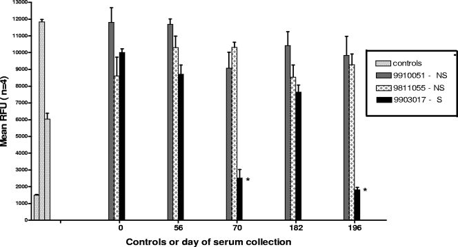
Sera from a recent study of the efficacy of the F1-V fusion vaccine in cynomolgus macaques were tested with the caspase-3 assay; the results of that study will be reported separately. That study, which involved four cohorts of 16 animals each, assessed the association between vaccine dose and protection against lethal aerosol challenge with the virulent Y. pestis strain C092 (cohorts 2 to 4). It also analyzed the long-term immunity of the vaccine (cohort 1). The NHP in all four groups were vaccinated three times with F1-V by the intramuscular route on days 0, 56, and 182 and were challenged by aerosol exposure to lethal doses of strain C092 at 1 month (cohorts 2 to 4) or 1 year (day 547; cohort 1) after the third vaccine dose. Sera were collected at various times over the course of the prechallenge vaccination period for in vitro evaluation. Further details of the vaccination doses and schedule are described in the figure legends and by Adamovicz et al. (manuscript in preparation). Figure 2 illustrates the caspase-3 results of tests done with sera collected on five separate days from three representative F1-V-vaccinated NHP of cohort 1. These results, as well as those of assays of additional sets of sera and those including sera collected on days 0, 14, 56, 70, 182, 196, and 469 (cohort 4 only) of the vaccination period, confirmed a finding reported previously for the live-dead assays (1). For all four animal groups, the day 70 and day 196 sera, which were collected 2 weeks after the second and third doses of vaccine, respectively, exhibited the greatest extent of MΦ protection compared to that associated with sera collected at other times during vaccination (Fig. 2 and data not shown).
FIG. 2.
Neutralization of Yersinia-mediated J774.A1 macrophage cytotoxicity by immune sera from F1-V-vaccinated cynomolgus macaques, as detected in the caspase-3 fluorescence assay. The caspase-3 data are shown as mean relative fluorescence units (RFU) (± standard deviations) of four replicates per serum sample (1). Tests were done with sera from three representative F1-V-vaccinated NHP of cohort 1 of this vaccine trial, and the results shown are those using sera collected on vaccination days 0, 56, 70, 182, and 196. Data are grouped by the day of serum collection. The three controls were (left to right) uninfected MΦs, infected untreated MΦs, and MΦs infected with bacteria pretreated with the positive control rabbit anti-V IgG. NS, nonsurvivor; S, survivor. *, P ≤ 0.0001, compared to the mean value for the day 0 serum-treated samples (analysis of variance and t test).
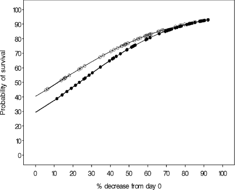
The relationship between the caspase-3 levels of MΦs infected with serum-pretreated Yersinia and the survival of the vaccinated NHP was then evaluated by logistic regression analysis of the pooled cohort data. As illustrated in Fig. 3, the extent to which caspase-3 levels decreased was significantly associated with survival for both the day 70 and the day 196 results (P = 0.0038 and P = 0.0024, respectively). The serum CNA was also highly correlated with the anti-V and anti-F1 enzyme-linked immunosorbent assay titers for both the day 70 and the day 196 sera (P ≤ 0.001, data not shown). The significance was observed when the caspase-3 levels measured in MΦs infected with the pretreated bacteria were compared to either the experimental negative control level (infected, nontreated MΦs; P = 0.0020 to 0.0001; data not shown) or to the caspase-3 levels measured in infected MΦs pretreated with day 0 (preimmune) sera from each animal (P < 0.0001) (Fig. 3).
FIG. 3.
The relationship between the level of caspase-3 in infected MΦs and the probability of survival after challenge in F1-V-immunized NHP. J774.A1 MΦs were infected with bacteria [Yptb (pTrcV)] that had been pretreated with serum collected from F1-V-vaccinated (or nonvaccinated control) cynomolgus macaques on vaccination days 70 (open symbols) and 196 (filled symbols). The effects of the treatment on apoptotic cell death of the MΦs were determined with the caspase-3 assay, and the results were expressed as the percent of decrease in caspase-3 levels (%); the latter were calculated based on the day 0 value for each NHP sera set. The significances of the association between the probability of survival after challenge with virulent Y. pestis and the in vitro cytotoxicity-neutralizing activity (percent of decrease in caspase-3) were evaluated by logistic regression. Data were analyzed with SAS/STAT software, version 9.1.3 (SAS system for PC; SAS Institute Inc.).
Thus, the results of the modified caspase-3 microtiter assay for serum-mediated CNA were highly correlated with survival status of F1-V-vaccinated NHP after they were challenged with virulent Y. pestis.
Acknowledgments
The research described herein was supported by JSTO-CBD/DTRA, project no. 5.10047-05-RDB.
The views expressed in this paper are those of the authors and do not reflect the official policy or position of the Department of the Army, Department of Defense, or the United States Government.
Footnotes
Published ahead of print on 14 May 2008.
REFERENCES
- 1.Bashaw, J., S. Norris, S. Trevino, J. Adamovicz, and S. Welkos. 2007. In vitro correlate assays of immunity to infection with Yersinia pestis. Clin. Vaccine Immunol. 14:605-616. [DOI] [PMC free article] [PubMed] [Google Scholar]
- 2.Computer Sciences Corporation (CSC). April 2008, posting date. Case studies for government. Department of Defense Joint Vaccine Acquisition program (U.S.): CSC's DVC edges one step closer to plague vaccine. http://www.csc.com/industries/government/casestudies/1710.shtml.
- 3.DHHS-FDA, CDR, and CVM. Posting date, May 2001. Guidance for industry: bioanalytical method validation. Center for Drug Evaluation and Research (CDER), Rockville, MD.
- 4.DHHS-NIH-NIAID. Posting date, September 2005. RFP-NIH-NIAID-DMID-07-06. Development of animal models and assays for plague vaccines. http://www.fedbizopps.gov.
- 5.Heath, D. G., G. W. Anderson, Jr., J. M. Mauro, S. L. Welkos, G. P. Andrews, J. Adamovicz, and A. M. Friedlander. 1998. Protection against experimental bubonic and pneumonic plague by a recombinant capsular F1-V antigen fusion protein vaccine. Vaccine 16:1131-1137. [DOI] [PubMed] [Google Scholar]
- 6.Henderson, D. A. 1999. The looming threat of bioterrorism. Science 283:279-282. [DOI] [PubMed] [Google Scholar]
- 7.Inglesby, T. V., D. T. Dennis, D. A. Henderson, J. G. Bartlett, M. S. Ascher, E. Eitzen, A. D. Fine, A. M. Friedlander, J. Hauer, J. F. Koerner, M. Layton, J. McDade, M. T. Osterholm, T. O'Toole, G. Parker, T. M. Perl, P. K. Russell, M. Schoch-Spana, and K. Tonat. 2000. Plague as a biological weapon: medical and public heath management. JAMA 283:2281-2290. [DOI] [PubMed] [Google Scholar]
- 8.International Conference on Harmonisation (ICH). 1997. Guideline on the validation of analytical procedures: methodology, availability, notice. Fed. Regist. 62:27463-27467. [Google Scholar]
- 9.Weeks, S., J. Hill, A. Friedlander, and S. Welkos. 2002. Anti-V antigen antibody protects macrophages from Yersinia pestis-induced cell death and promotes phagocytosis. Microb. Pathog. 32:227-237. [DOI] [PubMed] [Google Scholar]
- 10.Williamson, E. D., S. M. Eley, K. F. Griffin, M. Green, P. Russell, S. E. C. Leary, P. C. F. Oyston, T. Easterbrook, K. M. Reddin, A. Robinson, and R. W. Titball. 1995. A new improved sub-unit vaccine for plague: the basis of protection. FEMS Immunol. Med. Microbiol. 12:223-230. [DOI] [PubMed] [Google Scholar]


