Abstract
Bacterial endospore dormancy and resistance properties depend on the relative dehydration of the spore core, which is maintained by the spore membrane and its surrounding cortex peptidoglycan wall. During spore germination, the cortex peptidoglycan is rapidly hydrolyzed by lytic enzymes packaged into the dormant spore. The peptidoglycan structures in both dormant and germinating Bacillus anthracis Sterne spores were analyzed. The B. anthracis dormant spore peptidoglycan was similar to that found in other species. During germination, B. anthracis released peptidoglycan fragments into the surrounding medium more quickly than some other species. A major lytic enzymatic activity was a glucosaminidase, probably YaaH, that cleaved between N-acetylglucosamine and muramic-δ-lactam. An epimerase activity previously proposed to function on spore peptidoglycan was not detected, and it is proposed that glucosaminidase products were previously misidentified as epimerase products. Spore cortex lytic enzymes and their regulators are attractive targets for development of germination inhibitors to kill spores and for development of activators to cause loss of resistance properties for decontamination of spore release sites.
Bacterial endospores are metabolically dormant and are highly resistant to many treatments that rapidly kill vegetative cells. The resistance properties are dependent on the relative dehydration of the spore core or cytoplasm and the high concentrations of calcium dipicolinic acid (DPA) and other core solutes (26). When exposed to a nutrient-rich environment, spores initiate germination and outgrowth into vegetative cells. Nutrient germinant molecules bind to Ger protein family receptors in the inner forespore membrane surrounding the spore core and trigger biophysical changes (20). The spore becomes partially rehydrated as water enters the spore core, and the calcium DPA that was in the core is released into the environment. To complete the germination process and resume metabolism (11, 24), the spore must go through a second stage, which is more biochemical (20). This involves activation of spore cortex peptidoglycan (PG) lytic enzymes, which are present in an inactive state within the dormant spore. As the cortex is degraded, the cell expands to its full size and regains full metabolic activity. Due to the key role that cortex lytic enzymes play in germination, they are potential targets for spore decontamination methods that could involve either irreversible inactivation or gratuitous activation of the enzymes. The latter would lead to premature spore germination into vegetative forms that are much more easily killed.
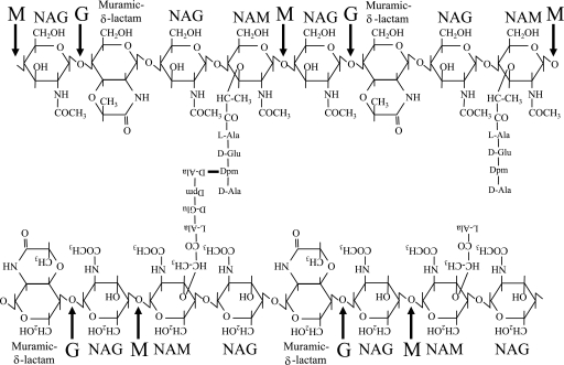
PG consists of glycan strands with alternating residues of N-acetylglucosamine (NAG) and N-acetylmuramic acid (NAM) that are cross-linked by peptide side chains located on the NAM residues (Fig. 1). While the PG structure in vegetative cells can vary between species, primarily in the amino acid composition of the peptide side chains, spore PG structure has been found to be highly conserved across species (3, 30). The peptide side chains contain l-alanine, d-glutamate, meso-diaminopimelic acid (Dpm), and d-alanine. The cross-linking occurs between the Dpm on one strand and the d-alanine of a tetrapeptide on an adjacent strand. The spore PG is composed of the inner, germ cell wall layer (10 to 20% of the PG), which resembles the PG of vegetative cells (6, 19), and the cortex, which has important and unique chemical modifications (2, 3, 5, 23, 28, 30). In the cortex, approximately 50% of the muramic acid residues (the residues on every alternate disaccharide) have their peptide side chains completely removed, and these NAM residues are converted to muramic-δ-lactam. In the Bacillus species previously examined, ∼25% of the cortex muramic acid residues have tetrapeptide side chains, while the majority of the remaining residues have their peptides cleaved to single l-alanine residues. Both the shortening and the removal of peptide side chains cause a decrease in cross-linking between the PG strands. The muramic-δ-lactam serves as the specificity determinant for germination lytic enzymes (5, 8, 9, 18, 24). This allows specific hydrolysis of the cortex during germination, while the germ cell wall remains intact as the initial wall of the outgrowing spore.
FIG. 1.
Spore PG structure and hydrolysis. Spore PG structure is well conserved across bacilli and clostridia (2, 3, 5, 23, 28, 30). The glycan strands contain alternating NAG and NAM residues in which approximately every second muramic acid residue is converted to muramic-δ-lactam. Approximately 26% of muramic acid residues have tetrapeptide side chains, 20% have l-alanine side chains, and a small percentage carry tripeptides. The peptides are utilized in cross-linking the glycan strands. Arrows indicate the cleavage sites of the muramidase mutanolysin (M) and of a glucosaminidase (G) active during germination. The actions of these enzymes singly produce tetrasaccharide and together produce trisaccharide muropeptides. The presence of adjacent muramic-δ-lactam residues results in the production of hexasaccharide and pentasaccharide muropeptides.
Hydrolysis of the cortex PG during germination of spores of several different species has been studied previously. Structural characterization of PG fragments allowed prediction of enzymatic activities present during germination. In Clostridium perfringens, muramidase, lytic transglycosylase, and amidase enzymatic products were found (9, 18). In Bacillus subtilis, lytic transglycosylase, glucosaminidase, and putative muramic-δ-lactam epimerase products were identified (4, 6), while in Bacillus megaterium, glucosaminidase and putative muramic-δ-lactam epimerase products were found (2). Studies with B. subtilis mutant strains allowed correlation of lytic transglycosylase activity with an intact sleB gene (7), while putative epimerase activity was associated with the sleL (yaaH) gene (10). The cwlJ product plays a clear role in cortex lysis (15), but no enzymatic activity has yet been associated with this protein. Separately, enzymatic activities have been directly demonstrated for a few proteins. SleL (YaaH) of Bacillus cereus was shown to be a glucosaminidase (8). SleC and SleM of C. perfringens, which do not appear to have orthologs in Bacillus species, were shown to be a bifunctional lytic transglycosylase/amidase and a muramidase, respectively (9, 18).
Germination-specific cortex lysis of B. anthracis spores is of great interest for two reasons. First, spores are the infectious agent in anthrax, and thus germination is an essential step in the progression of the infection. Germination within the host body results in release of cortex PG fragments that may serve as novel stimulators of the host immune system (14, 16). Second, efficient stimulation of spore germination in a contaminated site renders the spores susceptible to simpler, less destructive decontamination methods. We analyzed the structure of the PG in both dormant and germinating B. anthracis spores. While these spores progressed through germination and outgrowth with kinetics similar to those of spores of other species, they released PG fragments into the surrounding medium much more quickly. The major cortex lytic products were those produced by a glucosaminidase activity. We present evidence and an argument against the presence of a previously postulated PG epimerase activity. Further characterization of cortex lytic enzymes and their regulation should contribute to development of better spore decontamination methods.
MATERIALS AND METHODS
Strains, growth conditions, and spore preparation.
B. anthracis Sterne strain 34F2, obtained from P. Hanna, was used throughout this study. Cultures in DSM (modified Schaeffer's sporulation medium) (21) or modified G medium (17) were shaken at 37°C for at least 48 h or until ≥ 90% of the visible spores were released from sporangia. Spores were collected by centrifugation, washed several times at 4°C in 0.1 M NaCl containing 0.1% Triton X-100, heated at 70°C for 10 min, washed several additional times, and stored at 4°C with gentle shaking. Washing in Triton X-100 did not affect the rate or efficiency of germination in the presence of l-alanine plus inosine or the pattern of muropeptide peaks detected, but it did affect the efficiency of germination in the presence of other nutrient germinants (data not shown). Additional washing over several days resulted in the production of spore preparations that were more than 90% free of contaminating cellular debris, which were further cleaned by centrifugation through a metrizoic acid gradient as previously described (21). The resulting spores were >95% free of contaminating material, and they were washed several times and stored at 4°C in 0.1 M NaCl containing 0.1% Triton X-100.
PG purification and analysis.
Cortex PG was purified from dormant spores and analyzed as previously described (23). Briefly, spores were suspended at an optical density at 600 nm (OD600) of 60 in 1 ml of 50 mM Tris-HCl (pH 7.5), 1% sodium dodecyl sulfate, 50 mM dithiothreitol and boiled for 20 min to permeabilize and extract the coat protein layers. The spores were washed, treated with 5% trichloroacetic acid, digested with trypsin (Worthington TRTPCK), and finally digested with mutanolysin (Sigma) at 37°C for 16 h. The supernatant, containing the muropeptides, was lyophilized and stored at −20°C. The terminal sugar residues were reduced to the corresponding alcohols using NaBH4, and the pH was adjusted to 2 with H3PO4. Muropeptides were separated on a Hypersil octyldecyl silane column (250 by 4.6 mm; 3 μm; Thermo Scientific) using a sodium phosphate buffer and a methanol gradient. A second, volatile buffer system, containing trifluoroacetic acid and acetonitrile, was used for a second verification of cochromatography with previously identified muropeptides and to remove phosphate buffer prior to amino acid analysis and mass spectrometry (23).
Purified muropeptides were subjected to amino acid and amino sugar analysis with comparison to purified standards of individual amino acids and sugars (13). Matrix-assisted laser desorption ionization-time of flight mass spectrometry of muropeptides (23) was performed at the Washington University Mass Spectrometry Facility, and electrospray ionization mass spectrometry was performed as previously described (22).
Spore germination.
Prior to initiation of germination, spores were suspended in 8.2 ml of H2O at an OD600 of 24, heat activated for 30 min at 70°C, cooled on ice for 10 min, and then added to a 125-ml flask containing 0.8 ml of 0.5 M NaPO4 (pH 7.0). The resulting spore suspension was incubated at 37°C for 5 min, and the OD600 was determined before the germinant was added. To initiate germination, 1.0 ml of 1 M l-alanine, 10 mM inosine was added to the spore suspension, and the preparation was incubated at 37°C; during incubation samples were removed for measurement of the OD600, for an assay of the release of DPA and cortex fragments, and for purification of PG.
For analysis of DPA release, 100-μl samples were centrifuged for 45 s at 13,000 × g. The supernatant was discarded, and the pellet was stored at −20°C. Pellets were resuspended in 10 mM Tris-HCl (pH 8.0) and boiled for 20 min to release DPA from the spores. The DPA in each sample was assayed colorimetrically as previously described (21). Purified DPA (Sigma) was used to prepare a standard curve.
For analysis of cortex fragment release, 10-μl aliquots were added to 90 μl of water, the samples were immediately centrifuged for 45 s at 13,000 × g, and the exudate was transferred to a separate microcentrifuge tube. The pellet and supernatant samples were stored at −20°C prior to analysis as previously described (13, 19).
For PG purification, samples corresponding to 60 OD600 units of dormant spores were centrifuged for 45 s at 13,000 × g, and the germination exudate was removed, boiled for 20 min, and stored at −80°C until analysis. The germinated spore pellet was resuspended in 1 ml of 50 mM Tris-HCl (pH 7.5), 1% SDS, 50 mM dithiothreitol and boiled for 30 min. The PG was then purified as described above for dormant spores. The exudate samples were lyophilized, resuspended in 100 μl of H2O, and split in half. One half was digested with mutanolysin as described above. Muropeptides in the exudate samples were reduced and analyzed as described above.
RESULTS
Structure of the PG in dormant B. anthracis spores.
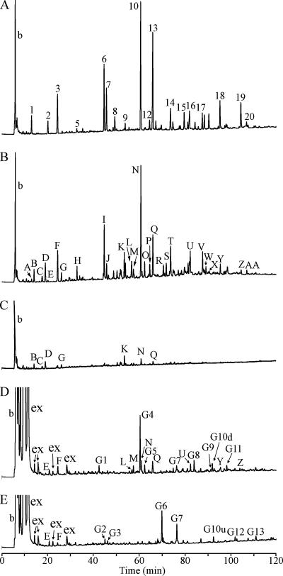
In order to examine the action of lytic enzymes on the B. anthracis Sterne 34F2 spore cortex during germination, we first determined the structure of the PG in dormant spores. PG was extracted from purified spores and then was digested with muramidase, reduced, and separated using reverse-phase high-performance liquid chromatography (HPLC) as previously described for B. subtilis (23). The process was performed in parallel with B. subtilis spore PG for comparison. The pattern of muropeptide peaks derived from B. anthracis spores was very similar to that derived from B. subtilis spores, except that there were several additional peaks (Fig. 2A and 2B). The identities of the muropeptides were determined by cochromatography with previously identified compounds (1, 23) using two different buffer systems (23), with amino acid analysis, and with mass spectrometry (Table 1). The major muropeptide species were found to be identical to those derived from B. subtilis spores, while the novel minor muropeptides resulted from deacetylation of some of the amino sugars, as has been observed previously for B. cereus and B. megaterium spores (2, 3). A small percentage (<9%) of the muropeptides contained amidated Dpm residues, which are found normally in Bacillus vegetative cell walls (25, 29). These residues may have represented contamination of the spore preparation with a small amount of vegetative cell wall PG, or there may be some Dpm amidation in the B. anthracis spores. Significant quantities of these muropeptides were not released into the germination exudate, indicating that if the muropeptides are within the spore, then they are associated with the germ cell wall. During the reduction of terminal sugars required to achieve good HPLC separation, some of the muramic-δ-lactam residues become reduced (30). A number of these reduced muropeptides were identified. Quantification of the muropeptides allowed calculation of some structural parameters of the spore PG (Table 2). Overall, the values and the PG structure in B. anthracis spores were similar to those determined for spores of multiple other species (2, 3, 5, 23, 30).
FIG. 2.
HPLC separation of B. anthracis spore muropeptides. Unless otherwise noted, PG preparations were digested with muramidase, reduced, and separated as previously described (19). (A) Muropeptides derived from dormant B. subtilis spores. Peaks are numbered as in reference 19. Early-eluting peaks labeled “b” are buffer components present in blank samples. (B) Muropeptides derived from dormant B. anthracis spores. Peaks are designated as in Table 1. (C) Muropeptides derived from B. anthracis spores 15 min after the initiation of germination. (D) Muropeptides derived from the exudate of B. anthracis spores 15 min after the initiation of germination. Peaks labeled “ex” are spore exudate components that, based upon amino acid analysis, are not derived from PG. Peaks are designated as in Table 1; however, the “a” in germination-specific peak designations is omitted due to space limitations. (E) Muropeptides derived from the exudate of B. anthracis spores 15 min after the initiation of germination, but with no muramidase digestion.
TABLE 1.
Muropeptide peak identification
| B. subtilis muropeptidea | B. anthracis muropeptide | Structureb | Dormant sporec | Germinated spore pelletc | Germinated spore exudate after mutanolysin digestionc | Germinated spore exudate before mutanolysin digestionc | Cochromatography | m/z expectedd | m/z observedd | Amino acid/amino sugar analysise
|
||||||
|---|---|---|---|---|---|---|---|---|---|---|---|---|---|---|---|---|
| Muramic acid derived from NAM or muramic-δ-lactam | Glucosamine | Muramitol | Glucosaminitol | Ala | Glu | Dpm | ||||||||||
| Muropeptides produced from dormant spore PG | ||||||||||||||||
| 1 | A | DS-TriP | + | * | + | + | + | 1 | 1 | 1 | ||||||
| B | DS-Ac-TriP | + | + | 827.4 | 827.3 | + | + | 1 | 1 | 1 | ||||||
| C | DS-TriP+Am | + | + | 868.7 | 869.7 | + | + | 1 | 1 | 1 | ||||||
| D | DS-Ac-TriP+Am | + | + | 826.4 | 826.3 | + | + | 1 | 1 | 1 | ||||||
| 2 | E | DS-Ala | + | * | + | + | + | + | + | 1 | ||||||
| 3 | F | DS-TP | + | * | + | + | + | + | + | 2 | 1 | 1 | ||||
| G | DS-Ac-TP | + | + | 898.4 | 898.3 | + | + | 2 | 1 | 1 | ||||||
| 5 | H | TS-TP open lactam | + | * | + | + | + | + | 2 | 1 | 1 | |||||
| 6 | I | TSred-TP | + | * | + | + | + | 2 | 1 | 1 | ||||||
| 7 | J | TSred-Ala | + | * | + | + | + | 1 | ||||||||
| K | DS-Ac-TP x TriP+Am-DS-Ac | + | + | 1707.8 | 1707.7 | + | + | 3 | 2 | 2 | ||||||
| L | TS-TP-Ac | + | * | + | 1316.5 | 1317.8 | + | + | + | 2 | 1 | 1 | ||||
| M | DS-Ac-TP+Am x TriP+Am-DS-Ac | + | * | + | + | |||||||||||
| 10 | N | TS-TP | + | + | + | + | 1358.6 | 1358.5 | + | + | + | 2 | 1 | 1 | ||
| 11 | O | TS-TP x TP | + | * | + | + | + | + | 2 | 1 | 1 | |||||
| 12 | P | DS-TP x TP-TSred | + | * | + | + | + | 2 | 1 | 1 | ||||||
| 13 | Q | TS-Ala | + | + | + | + | 986.6 | 986.4 | + | + | + | 1 | ||||
| R | HSred-Ac-TP (right lactam reducedh) | + | * | 1720.7 | 1721.6 | |||||||||||
| S | HSred-Ac-TP (left lactam reducedh) | + | * | 1720.7 | 1721.7 | |||||||||||
| 14 | T | DS-TP x TP-TS | + | * | + | + | 2283.3 | 2283.4 | + | + | + | 2 | 1 | 1 | ||
| U | HS-TP-Ac | + | * | + | 1734.7 | 1734.6 | + | + | + | 2 | 1 | 1 | ||||
| 17 | V | TS-TP x TP-TS | + | * | + | + | + | + | + | 2 | 1 | 1 | ||||
| W | HS-Ala-Ac | + | * | 1361.8 | 1361.7 | |||||||||||
| X | HS-Ala-Ac | + | * | 1361.8 | 1361.7 | |||||||||||
| 18 | Y | HS-TP | + | * | + | + | + | + | + | 2 | 1 | 1 | ||||
| 19 | Z | HS-Ala | + | * | + | + | + | + | + | 1 | ||||||
| 20 | AA | TS-TP x TP-HS | + | * | + | + | + | + | 2 | 1 | 1 | |||||
| Muropeptides produced from germinated spore exudate following mutanolysin digestion | ||||||||||||||||
| aG1 | TriS-TP Red | + | 1141.5 | 1141.4 | + | + | 2 | 1 | 1 | |||||||
| G8f | aG4 | TriS-TP | + | 1155.5 | 1155.4 | + | + | + | 2 | 1 | 1 | |||||
| G8Ag | aG5 | TriS-Ala | + | 782.5 | 782.4 | + | + | + | 1 | |||||||
| aG8 | PS-TP-Ac | + | 1531.6 | 1531.5 | + | + | + | 2 | 1 | 1 | ||||||
| aG9 | PS-Ala-Ac | + | 1160.1 | 1159.9 | + | + | + | 1 | ||||||||
| aG10d | TriS-TP x TP-TriS | + | 2293.8 | 2293.8 | + | + | + | 2 | 1 | 1 | ||||||
| G8Bg | aG11 | PS-TP | + | 1573.7 | 1573.6 | + | + | + | 2 | 1 | 1 | |||||
| Muropeptides produced from germinated spore exudate with no mutanolysin digestion | ||||||||||||||||
| G2f | aG2 | TS-TP NAGr Red | + | 1344.8 | 1344.6 | + | + | + | 2 | 1 | 1 | |||||
| G1f | aG3 | TS-Ala NAGr Red | + | 972.7 | 972.6 | + | + | + | 2 | 1 | 1 | |||||
| G3f | aG6 | TS-TP NAGr | + | 1358.6 | 1358.5 | + | + | + | 2 | 1 | 1 | |||||
| G4f | aG7 | TS-Ala NAGr | + | + | 986.6 | 986.4 | + | + | + | 1 | ||||||
| aG10u | HS-TP-Ac NAGr | + | 1734.7 | 1735.2 | + | + | + | 2 | 1 | 1 | ||||||
| aG12 | HS-Ala-Ac NAGr | + | 1361.8 | 1361.8 | + | + | + | 1 | ||||||||
| G6f | aG13 | HS-TP NAGr | + | 1776.8 | 1777.2 | + | + | + | 2 | 1 | 1 | |||||
Numbering as in reference 23.
DS, disaccharide (NAG-NAM); TS, tetrasaccharide (NAG-lactam-NAG-NAM); HS, hexasaccharide (NAG-lactam-NAG-lactam-NAG-NAM); TriS, trisaccharide (lactam-NAG-NAM); PS, pentasaccharide (lactam-NAG-lactam-NAG-NAM); TriP, tripeptide (Ala-Glu-Dpm); TP, tetrapeptide (Ala-Glu-Dpm-Ala); Ac, deacetylated glucosamine; +Am, amidated Dpm; Red, reduced lactam (an artifact of sample preparation [30])); NAGr, NAG at the reducing end; x, cross-link between two peptides.
A plus sign indicates that the muropeptide was detected in the sample, was integrated for structural parameter determinations (Table 2), and is indicated in Fig. 2. An asterisk indicates that the muropeptide was detected in the sample and was integrated for structural parameter determinations (Table 2) but is very small and not indicated in Fig. 2.
m/z expected is the mass-to-charge ratio predicted for the deprotonated ion of the muropeptide structure indicated. m/z observed is the value determined using matrix-assisted laser desorption ionization-time of flight mass spectrometry or electrospray ionization mass spectrometry.
The analysis method used does not produce reliable quantitative results for amino sugars. A plus sign indicates that the compound was clearly detected. For amino acids, the values are the molar ratios, which are rounded to the nearest whole number.
Numbering as in reference 6.
Numbering as in reference 7.
The position of lactam reduction was determined by fragmentation of the muropeptide during tandem mass spectrometry analysis.
TABLE 2.
Structural parameters of B. anthracis spore PG
| Sample | % Muramic acid witha:
|
|||||
|---|---|---|---|---|---|---|
| Lactam | l-Ala | Tripeptide | Tetrapeptide | Cross-link | Deacetylation of NAG | |
| Dormant | 48.5 | 13.9 | 4.2 | 34.4 | 5.6 | 11.3 |
| Germinated pellet | 35.9 | 11.6 | 21.8 | 31.2 | 7.7 | 30.0 |
| Exudate after mutanolysin digestion | 52.0 | 16.2 | 0.9 | 30.9 | 2.0 | 7.3 |
| Exudate before mutanolysin digestion | 51.7 | 24.5 | 0.3 | 23.4 | 0.2 | 3.8 |
All values are averages of three analyses of independent spore preparations. The standard deviation of each value was ≤4%.
Germination kinetics of B. anthracis spores.
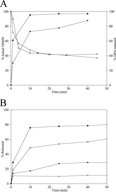
B. anthracis spores were heat activated and then stimulated to germinate in a buffered suspension by addition of l-alanine and inosine. The progress and efficiency of germination were monitored by using the change in optical density of the suspension and the release of DPA (Fig. 3A). Spores released >90% of their DPA during the first 10 min following addition of germinants and lost >50% of their optical density during the first 15 min. This indicates that highly efficient and synchronous germination was achieved.
FIG. 3.
Kinetics of B. anthracis and B. subtilis spore germination. Purified spores were heat activated and stimulated to germinate by addition of l-alanine and inosine at time zero. Samples were removed for measurement of the optical density, DPA release, and PG release at the indicated times. (A) Decrease in optical density (open symbols) and release of DPA (filled symbols) were determined for B. anthracis (squares) and B. subtilis (circles). (B) Release of Dpm (open symbols) and muramic acid (filled symbols) was determined for B. anthracis (squares) and B. subtilis (circles). Data from a single experiment are shown; however, similar results were obtained in two additional experiments.
The release of cortex PG fragments from the spores was assayed by amino acid/amino sugar analysis of the muramic acid and Dpm remaining in the spores collected by centrifugation. During the first 10 min following germinant addition, >75% of the muramic acid and >50% of the Dpm were released into the medium (Fig. 3B). We compared these germination kinetics to those obtained with B. subtilis spores purified in an identical manner and stimulated to germinate with the same germinant mixture (although B. subtilis was likely responding solely to the l-alanine and not to the inosine [20]). Loss of optical density occurred at the same rate in the two species, and the levels of DPA release were similar, although they were slightly less in B. subtilis (Fig. 3A). The two species differed significantly in the release of cortex fragments from the spores. While B. anthracis released >75% of the muramic acid in 10 min, B. subtilis released only 20% in this time and only 30% after 60 min (Fig. 3B). A similar pattern was observed for the release of Dpm. These differences do not necessarily indicate a difference in the germination rates. When spores of the two species were germinated in rich medium, they progressed through outgrowth and reached the first cell division at essentially the same time (data not shown). A similar slow release of muramic acid and Dpm was observed with Bacillus sphaericus spores (data not shown).
Structure of the PG in germinating B. anthracis spores.
PG was recovered from germinating spore suspensions at 5, 15, and 30 min after addition of germinants. Spore suspensions were centrifuged, and muropeptides were derived both from the germinating spore pellet and from the spore exudate (Fig. 2C to E). Larger amounts of material were present in the exudate at 15 min than at 5 min, but the numbers and identities of the muropeptide peaks observed in the samples did not differ (data not shown). There was no clear difference between the samples obtained 15 and 30 min after the addition of germinants (data not shown). The 15-min samples were analyzed in detail (Fig. 2C to E).
Muropeptide peaks were collected, and a total of 38 compounds from germinated spore pellets and germination exudates both with and without mutanolysin treatment were identified using a variety of methods (Table 1). Twenty-seven muropeptides were identical to muropeptides found in dormant spores. Six muropeptides were tentatively identified based on their appearance in germination samples and relative elution times in comparison to peaks identified for B. subtilis (6). All of the germination-specific muropeptides were subjected to amino acid/amino sugar analysis and to mass spectrometry (Table 1). The muropeptides recovered from the germinated spore pellet (Fig. 2C) were reminiscent of those previously associated with germ cell wall PG (1, 19). The structural parameters of this PG include a lower level of muramic-δ-lactam and relatively high levels of tripeptide side chains, peptide cross-linking, and deacetylation compared to the total dormant spore PG (Table 2).
Novel muropeptides associated with germination-specific cortex hydrolysis were found in the germination exudate. These muropeptides were classified into groups that were present either before or after mutanolysin digestion (Table 1). Germination-specific muropeptides were designated consecutively based on the order of elution by using the designation aG (for “anthracis germination”) to differentiate them from muropeptides with similar designations derived from other species. Muropeptides aG10d and aG10u eluted in the same position in mutanolysin-digested and undigested samples, respectively. Muropeptides produced by digestion with mutanolysin, which cleaves on the reducing side of NAM, have NAM at their reducing ends and thus have muramitol in the reduced form that is resolved by HPLC. Muropeptides aG1, aG4, aG5, aG8, aG9, aG10d, and aG11 were found to be tri- and pentasaccharides with various peptide side chains and the expected modifications of deacetylation and lactam reduction (Table 1). This indicates that they were produced as a result of a glucosaminidase activity that cleaved the glycan strands between NAG and muramic-δ-lactam, followed by mutanolysin digestion (Fig. 1).
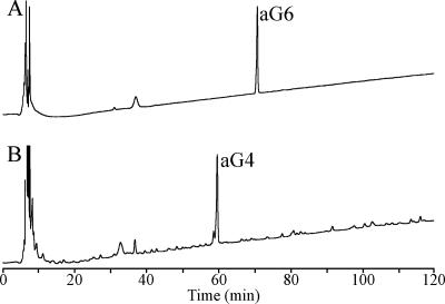
Several muropeptides were detected in the exudate in the absence of mutanolysin digestion (Fig. 2E) and thus must be the direct products of spore lytic enzymes. These muropeptides, including aG6, aG7, aG10u, aG12, and aG13, were all found to contain NAG as their reducing terminal sugar. The amino acid/amino sugar analysis of these muropeptides showed that in the reduced form that was separated by HPLC, they contained glucosaminitol (Table 1) rather than the muramitol that is found in the muropeptides produced by mutanolysin digestion. Furthermore, some of these muropeptides were sensitive to mutanolysin digestion and were converted to other muropeptides that were detected in the exudate following this digestion. Purified muropeptide aG6 was quantitatively converted to aG4 by mutanolysin (Fig. 4). The same relationship was observed for conversion of aG7 to aG5, conversion of aG10u to aG8, conversion of aG12 to aG9, and conversion of aG13 to aG11 (data not shown).
FIG. 4.
Muramidase cleaves the glucosaminidase product muropeptide aG6 to produce muropeptide aG4. Muropeptide aG6 was purified using HPLC separation as shown in Fig. 2 and was then desalted and further purified by HPLC using an acetonitrile/trifluoroacetic acid buffer system. The purified muropeptide was incubated in 12.5 mM NaPO4 (pH 5.5) without (A) and with (B) mutanolysin, followed by reduction and HPLC separation as shown in Fig. 2.
Structural parameters determined for PG detected in the exudate following mutanolysin digestion are more representative of the exudate fraction than the structural parameters of PG detected prior to digestion (Fig. 2), as the undigested material certainly contained some larger fragments that were not resolved or identified during HPLC analysis. PG in the exudate was enriched in muramic-δ-lactam and had few tripeptide side chains, as expected for cortex PG (Table 2). This PG also had a lower level of deacetylation than the total dormant spore PG. The 36% of the muramic acid in the germinated spore pellet that was in the muramic-δ-lactam form (Table 2) probably represented cortex fragments that were too large to escape through the spore coats only 15 min into germination. If the germ cell wall PG is 10% of the total spore PG and the 75% of the PG released after 15 min is cortex PG, then the remaining 15% in the spore pellet would be residual cortex PG. The 10% germ cell wall PG with no muramic-δ-lactam plus the 15% cortex PG with 50% muramic-δ-lactam would result in a total of approximately 30% muramic-δ-lactam in the pellet.
DISCUSSION
The PG structure present in B. anthracis Sterne spores is not significantly different from the PG structures found in the spores of a number of other species (3, 23, 28). Almost 50% of the muramic acid residues are in the muramic-δ-lactam form, and the majority of the remaining muramic acid residues carry tetrapeptide or single-alanine side chains. A small percentage of the side chains are tripeptides. Tripeptides are expected to be predominantly in the germ cell wall, consistent with their retention in the germinated spore pellet. The overall level of peptide cross-linking is low, as in the spores of other species (3, 23, 28). Approximately 10% of the glucosamine residues were deacetylated in B. anthracis. This modification has previously been found in B. cereus, B. megaterium, Clostridium botulinum (3), and C. perfringens (22).
Rapid, synchronous germination of B. anthracis spores upon addition of nutrient germinants resulted in very rapid release of the majority of the spore PG into the medium. In this regard, B. anthracis differs from some other sporulating species, which release PG fragments more slowly. Our data showed that B. subtilis spores released <30% of their PG 40 min into germination, while B. anthracis released >75% after only 10 min. Despite different rates of PG release, spores of these species progressed through germination and outgrowth with similar kinetics. They both released >70% of their DPA within 10 min, and when germinated in rich medium, they progressed through outgrowth and reached their first cell division within 120 min. The rapid release of PG fragments by B. anthracis spores may be due to higher levels of lytic enzymes that more quickly produce smaller PG fragments and thus more rapid diffusion through the spore coat layers, or it may be due to thinner (12), more permeable spore coat layers. Whatever the reason, the rapid release of PG fragments may have implications for recognition of germinating spores by a host innate immune system (14, 16). Retention of highly deacetylated PG within the germinated spore pellet indicates that this modification is concentrated in the germ cell wall relative to the cortex. The higher level of deacetylation may be important for protecting the germinating spore from lytic enzymes, such as lysozyme (27), during germination within a host.
Analysis of muropeptide structures produced during germination revealed the products of a glucosaminidase that cleaves between NAG and muramic-δ-lactam. Germination-specific glucosaminidase products were previously found in B. subtilis (6) and B. megaterium (2). Chen et al. demonstrated that the SleL protein of B. cereus possessed glucosaminidase activity that was specific for spore cortex PG fragments (8). All of these Bacillus species, including B. anthracis, possess sleL orthologs. When a B. subtilis mutant lacking this ortholog (yaaH) was examined, it was found to retain glucosaminidase activity during germination, but it did not contain the products of a putative epimerase (10).
Germination-specific epimerase products were previously postulated for B. subtilis and B. megaterium (2, 6). These products were said to have amino acid contents and molecular masses indistinguishable from those of similar tetrasaccharide and hexasaccharide muropeptides produced by muramidase digestion of dormant spore PG, and nuclear magnetic resonance spectra revealed no clear structural modifications (6). Importantly, in the previous studies, amino sugar contents were estimated only from mass spectrometry data (5). It was proposed that the germination-specific muropeptides were altered in the stereochemistry of a muramic acid residue. We did not identify any of these putative epimerase products. Rather, we found muropeptides eluting in the same positions as the putative epimerase products, which we demonstrated to be glucosaminidase products. These muropeptides had the same masses as corresponding muramidase products derived from dormant spore PG, but there was a subtle difference in the amino sugar analysis. Following the reduction required prior to HPLC analysis, these muropeptides contained glucosaminitol at their reducing termini rather than the muramitol found in the muramidase products. We further demonstrated that the tetrasaccharides and hexasaccharides with NAG at their reducing termini could be converted to trisaccharides and pentasaccharides by muramidase treatment. These trisaccharides and pentasaccharides are the same glucosaminidase products (which had also been muramidase digested) that were previously found by the researchers who postulated the epimerase products (2, 6, 7). We suggest that the tetrasaccharide and hexasaccharide muropeptides that we identified as the direct products of a glucosaminidase are the same molecules that were previously postulated to be epimerase products. We are then left to explain two discrepancies with the results of previous researchers. First, the previous researchers observed only minimal loss of these muropeptides and production of trisaccharides and pentasaccharides upon muramidase digestion. We suggest that the muramidase is less active on the small glucosaminidase products than on the glycan strands that are the normal substrates in PG analyses and that we may have used excess muramidase that allowed full digestion. Alternatively, the mutanolysin preparation that we used may have been more active with these small substrates than the Cellosyl used in previous studies (2, 6, 7). Second, the finding that an sleL (yaaH) mutant lacked putative epimerase products but contained glucosaminidase products (10) is not consistent with the hypothesis that both classes of products are actually glucosaminidase products. We suggest that either there was a glucosaminidase in addition to the YaaH present or that the mutant studied was leaky and produced a reduced amount of glucosaminidase. A small amount of glucosaminidase may cleave glycan strands, leaving them with NAG at their reducing termini, but glucosaminidase may only extremely rarely cut twice in close proximity to produce the smaller tetrasaccharide and hexasaccharide products. The longer glycan strands would be excellent muramidase substrates, and complete digestion of them would produce only the small amounts of trisaccharide and pentasaccharide glucosaminidase products that were observed (10). Conversely, in the wild-type strain, high levels of glucosaminidase would produce an abundance of tetrasaccharide and hexasaccharide products, which may be poor substrates for the muramidase. Clearly, further examination of the genes and proteins having glucosaminidase activity is required.
Previous studies identified lytic transglycosylase activity in germinating B. subtilis and B. megaterium spores (2, 6), and this activity was associated with the sleB gene (4, 7). We did not identify any lytic transglycosylase products in our analysis, although B. anthracis does possess a sleB ortholog. Lytic transglycosylase activity may be present, but the level of the products may be below the limit of detection of our analysis. As lytic transglycosylase products are potent inducers of the host immune system (14, 16), perhaps there is selection against these products in this pathogenic species. Studies of B. anthracis mutants lacking orthologs of the germination-specific lytic enzymes CwlJ, SleB, and YaaH should help to clarify the enzymatic and phenotypic roles of these genes and proteins. Lytic enzymes and their regulatory systems may be attractive targets for methods of inactivating spores.
Acknowledgments
This work was supported by Public Health Service grant AI060726 from the National Institute of Allergy and Infectious Diseases. Mass spectrometry was provided by the Washington University Mass Spectrometry Resource with support from the NIH National Center for Research Resources (grant P41RR0954). ABI mass spectrometers were donated to Virginia Tech by PPD, Inc., Richmond, VA.
Footnotes
Published ahead of print on 2 May 2008.
REFERENCES
- 1.Atrih, A., G. Bacher, G. Allmaier, M. P. Williamson, and S. J. Foster. 1999. Analysis of peptidoglycan structure from vegetative cells of Bacillus subtilis 168 and role of PBP 5 in peptidoglycan maturation. J. Bacteriol. 1813956-3966. [DOI] [PMC free article] [PubMed] [Google Scholar]
- 2.Atrih, A., G. Bacher, R. Korner, G. Allmaier, and S. J. Foster. 1999. Structural analysis of Bacillus megaterium KM spore peptidoglycan and its dynamics during germination. Microbiology 1451033-1041. [DOI] [PubMed] [Google Scholar]
- 3.Atrih, A., and S. J. Foster. 2001. Analysis of the role of bacterial endospore cortex structure in resistance properties and demonstration of its conservation amongst species. J. Appl. Microbiol. 91364-372. [DOI] [PubMed] [Google Scholar]
- 4.Atrih, A., and S. J. Foster. 2001. In vivo roles of the germination-specific lytic enzymes of Bacillus subtilis 168. Microbiology 1472925-2932. [DOI] [PubMed] [Google Scholar]
- 5.Atrih, A., P. Zollner, G. Allmaier, and S. J. Foster. 1996. Structural analysis of Bacillus subtilis 168 endospore peptidoglycan and its role during differentiation. J. Bacteriol. 1786173-6183. [DOI] [PMC free article] [PubMed] [Google Scholar]
- 6.Atrih, A., P. Zollner, G. Allmaier, M. P. Williamson, and S. J. Foster. 1998. Peptidoglycan structural dynamics during germination of Bacillus subtilis 168 endospores. J. Bacteriol. 1804603-4612. [DOI] [PMC free article] [PubMed] [Google Scholar]
- 7.Boland, F. M., A. Atrih, H. Chirakkal, S. J. Foster, and A. Moir. 2000. Complete spore-cortex hydrolysis during germination of Bacillus subtilis 168 requires SleB and YpeB. Microbiology 14657-64. [DOI] [PubMed] [Google Scholar]
- 8.Chen, Y., S. Fukuoka, and S. Makino. 2000. A novel spore peptidoglycan hydrolase of Bacillus cereus: biochemical characterization and nucleotide sequence of the corresponding gene, sleL. J. Bacteriol. 1821499-1506. [DOI] [PMC free article] [PubMed] [Google Scholar]
- 9.Chen, Y., S. Miyata, S. Makino, and R. Moriyama. 1997. Molecular characterization of a germination-specific muramidase from Clostridium perfringens S40 spores and nucleotide sequence of the corresponding gene. J. Bacteriol. 1793181-3187. [DOI] [PMC free article] [PubMed] [Google Scholar]
- 10.Chirakkal, H., M. O'Rourke, A. Atrih, S. J. Foster, and A. Moir. 2002. Analysis of spore cortex lytic enzymes and related proteins in Bacillus subtilis endospore germination. Microbiology 1482383-2392. [DOI] [PubMed] [Google Scholar]
- 11.Cowan, A. E., D. E. Koppel, B. Setlow, and P. Setlow. 2003. A soluble protein is immobile in dormant spores of Bacillus subtilis but is mobile in germinated spores: implications for spore dormancy. Proc. Natl. Acad. Sci. USA 1004209-4214. [DOI] [PMC free article] [PubMed] [Google Scholar]
- 12.Giorno, R., J. Bozue, C. Cote, T. Wenzel, K. S. Moody, M. Mallozzi, M. Ryan, R. Wang, R. Zielke, J. R. Maddock, A. Friedlander, S. Welkos, and A. Driks. 2007. Morphogenesis of the Bacillus anthracis spore. J. Bacteriol. 189691-705. [DOI] [PMC free article] [PubMed] [Google Scholar]
- 13.González-Castro, M. J., J. López-Hernández, J. Simal-Lozano, and M. J. Oruña-Concha. 1997. Determination of amino acids in green beans by derivatization with phenylisothiocyanate and high-performance liquid chromatography with ultraviolet detection. J. Chromatogr. Sci. 35181-185. [Google Scholar]
- 14.Guan, R., and R. A. Mariuzza. 2007. Peptidoglycan recognition proteins of the innate immune system. Trends Microbiol. 15127-134. [DOI] [PubMed] [Google Scholar]
- 15.Ishikawa, S., K. Yamane, and J. Sekiguchi. 1998. Regulation and characterization of a newly deduced cell wall hydrolase gene (cwlJ) which affects germination of Bacillus subtilis spores. J. Bacteriol. 1801375-1380. [DOI] [PMC free article] [PubMed] [Google Scholar]
- 16.Kanneganti, T. D., M. Lamkanfi, and G. Nunez. 2007. Intracellular NOD-like receptors in host defense and disease. Immunity 27549-559. [DOI] [PubMed] [Google Scholar]
- 17.Kim, H. U., and J. M. Goepfert. 1974. A sporulation medium for Bacillus anthracis. J. Appl. Bacteriol. 37265-267. [DOI] [PubMed] [Google Scholar]
- 18.Kumazawa, T., A. Masayama, S. Fukuoka, S. Makino, T. Yoshimura, and R. Moriyama. 2007. Mode of action of a germination-specific cortex-lytic enzyme, SleC, of Clostridium perfringens S40. Biosci. Biotechnol. Biochem. 71884-892. [DOI] [PubMed] [Google Scholar]
- 19.Meador-Parton, J., and D. L. Popham. 2000. Structural analysis of Bacillus subtilis spore peptidoglycan during sporulation. J. Bacteriol. 1824491-4499. [DOI] [PMC free article] [PubMed] [Google Scholar]
- 20.Moir, A. 2006. How do spores germinate? J. Appl. Microbiol. 101526-530. [DOI] [PubMed] [Google Scholar]
- 21.Nicholson, W. L., and P. Setlow. 1990. Sporulation, germination, and outgrowth, p. 391-450. In C. R. Harwood and S. M. Cutting (ed.), Molecular biological methods for Bacillus. John Wiley & Sons Ltd., Chichester, England.
- 22.Orsburn, B., S. B. Melville, and D. L. Popham. 31 March 2008. Factors contributing to heat resistance of Clostridium perfringens endospores. Appl. Environ. Microbiol. doi: 10.1128/AEM.02629-07. [DOI] [PMC free article] [PubMed]
- 23.Popham, D. L., J. Helin, C. E. Costello, and P. Setlow. 1996. Analysis of the peptidoglycan structure of Bacillus subtilis endospores. J. Bacteriol. 1786451-6458. [DOI] [PMC free article] [PubMed] [Google Scholar]
- 24.Popham, D. L., J. Helin, C. E. Costello, and P. Setlow. 1996. Muramic lactam in peptidoglycan of Bacillus subtilis spores is required for spore outgrowth but not for spore dehydration or heat resistance. Proc. Natl. Acad. Sci. USA 9315405-15410. [DOI] [PMC free article] [PubMed] [Google Scholar]
- 25.Schleifer, K. H., and O. Kandler. 1972. Peptidoglycan types of bacterial cell walls and their taxonomic implications. Bacteriol. Rev. 36407-477. [DOI] [PMC free article] [PubMed] [Google Scholar]
- 26.Setlow, P. 2006. Spores of Bacillus subtilis: their resistance to and killing by radiation, heat and chemicals. J. Appl. Microbiol. 101514-525. [DOI] [PubMed] [Google Scholar]
- 27.Vollmer, W. 2008. Structural variation in the glycan strands of bacterial peptidoglycan. FEMS Microbiol. Rev. 32287-306. [DOI] [PubMed] [Google Scholar]
- 28.Warth, A. D., and J. L. Strominger. 1972. Structure of the peptidoglycan from spores of Bacillus subtilis. Biochemistry 111389-1396. [DOI] [PubMed] [Google Scholar]
- 29.Warth, A. D., and J. L. Strominger. 1971. Structure of the peptidoglycan from vegetative cell walls of Bacillus subtilis. Biochemistry 104349-4358. [DOI] [PubMed] [Google Scholar]
- 30.Warth, A. D., and J. L. Strominger. 1969. Structure of the peptidoglycan of bacterial spores: occurrence of the lactam of muramic acid. Proc. Natl. Acad. Sci. USA 64528-535. [DOI] [PMC free article] [PubMed] [Google Scholar]


