Abstract
The successful use of a dendrimeric peptide to protect pigs against challenge with foot-and-mouth disease virus (FMDV), which causes the most devastating animal disease worldwide, is described. Animals were immunized intramuscularly with a peptide containing one copy of a FMDV T-cell epitope and branching out into four copies of a B-cell epitope. The four immunized pigs did not develop significant clinical signs upon FMDV challenge, neither systemic nor mucosal FMDV replication, nor was its transmission to contact control pigs observed. The dendrimeric construction specifically induced high titers of FMDV-neutralizing antibodies and activated FMDV-specific T cells. Interestingly, a potent anti-FMDV immunoglobulin A response (local and systemic) was observed, despite the parenteral administration of the peptide. On the other hand, peptide-immunized animals showed no antibodies specific of FMDV infection, which qualifies the peptide as a potential marker vaccine. Overall, the dendrimeric peptide used elicited an immune response comparable to that found for control FMDV-infected pigs that correlated with a solid protection against FMDV challenge. Dendrimeric designs of this type may hold substantial promise for peptide subunit vaccine development.
Multimerization is a nature-mimicking strategy of antigen presentation that has been proven quite successful in the development of human-made vaccines, particularly by means of dendrimeric (e.g., branching) designs (55). Numerous reports on the immunogenicity of dendrimers have been published but only a few in vivo therapeutic studies have been reported (32). Here, we describe the successful use of a dendrimeric peptide to protect pigs against challenge with foot-and-mouth disease virus (FMDV), which causes the most feared animal disease worldwide (48, 51). FMDV is a picornavirus that produces a highly transmissible and devastating disease of farm animals, mostly cattle and swine (30, 41). The FMDV particle contains a positive-strand RNA molecule of about 8,500 nucleotides, enclosed within an icosahedral capsid comprising 60 copies each of four virus proteins, VP1 to VP4. The genome encodes a unique polyprotein from which the different viral polypeptides are cleaved by viral proteases, including 11 different mature nonstructural (NS) proteins (6). Each of these NS proteins, as well as some of the precursor polypeptides, is involved in functions relevant to the virus life cycle in infected cells (5). FMDV shows a high genetic and antigenic variability, reflected in the seven serotypes and the numerous variants described to date (21). FMD control in regions of endemicity is implemented mainly by using chemically inactivated whole-virus vaccines (4). Viral infection and immunization with conventional vaccines usually elicit high levels of circulating immunoglobulin G (IgG)-neutralizing antibodies that correlate with protection against the homologous and antigenically related viruses (46). Evidence of the role of specific IgA in protection includes early work suggesting that pigs immunized intramuscularly with conventional, inactivated vaccines elicited levels of neutralizing activity in nasal fluid lower than those observed for serum, in contrast to what was seen for infected pigs, where antibody responses in sera and upper mucosae were comparable (23). Recently, induction of low IgA responses has been correlated with complete protection against challenge in pigs immunized with a highly concentrated inactivated vaccine (22).
Despite its wide use, immunization with chemically inactivated vaccines has disadvantages, such as the need for a cold chain to preserve virus stability, the risk of virus release during vaccine production, and the problems for serological distinction between infected and vaccinated animals (20). These have led FMDV-free countries to adopt a nonvaccination policy that relies on slaughtering infected and contact herds and strict limitations on animal movements and trading in case of viral outbreaks. Such FMDV reemergences have caused massive and controversial culling of affected and suspected farm animals (51). Thus, much effort has been invested in the search of alternative, safe immunogens. The main antigenic sites recognized by B lymphocytes have been identified at defined structural motifs exposed on the capsid surface, whose amino acid sequences accumulate variations among different serotypes (1, 33). A continuous, immunodominant B-cell site located in the GH loop, around positions 140 to 160 of capsid protein VP1 (7), has been widely used as an immunogenic peptide (19). However, the protection conferred to natural hosts by linear peptides spanning the GH loop of VP1 is limited and can correlate with the selection of escape mutants in unprotected animals (53). The lack of T-cell epitopes widely recognized by individuals of domestic populations of natural host and capable of providing adequate cooperation to immune B lymphocytes has been proposed as one of the limiting factors for the development of efficient FMD peptide vaccines (15, 52). Although induction of neutralizing antibodies is considered to be the most important immune correlate to FMDV protection, specific T cells are also induced in convalescent and conventionally vaccinated animals (17, 44). In addition, the partial protection conferred in host species by subunit vaccines delivered using eukaryotic vectors, which did not elicit neutralizing antibodies, has been shown to correlate with the induction of specific T-cell responses (49). Several T-cell epitopes frequently recognized by natural host lymphocytes have been identified in FMDV proteins (8, 9, 16, 24, 26, 58). One of these T-cell epitopes, located in residues 21 to 35 of FMDV NS protein 3A, efficiently stimulated lymphocytes from infected pigs and induced significant levels of serotype-specific anti-FMDV activity in vitro when synthesized juxtaposed to the VP1 GH loop (8). This T-cell epitope was frequently recognized by lymphocytes from outbred pigs infected with different FMDV serotypes (25) and its amino acid sequence is conserved among FMDV types A, O, and C, showing limited variation among isolates from the seven FMDV serotypes (14).
In this work, we have assessed the immunogenicity in the pig, a major natural FMDV host, of a dendrimeric peptide that integrates the aforementioned B and T epitopes. The rationale for this design was to enhance the effectiveness of presentation to the porcine immune system of viral antigenic sites capable of stimulating B- and T-cell-specific lymphocytes (8, 53). The dendrimeric construction specifically induced high titers of FMDV-neutralizing antibodies, activation of T cells, and potent anti-FMDV IgA responses (systemic and mucosal), even when administered by the parenteral route. Pigs vaccinated with the dendrimeric peptide did not develop significant clinical signs upon FMDV challenge and inhibited local replication and airborne excretion of virus. Thus, dendrimeric designs of this type may hold substantial promise for peptide subunit vaccine development.
MATERIALS AND METHODS
Synthetic dendrimeric peptide.
The dendrimeric peptide B4T (Table 1) was built from two separately synthesized precursors in which the FMDV sequences were those of the serotype C isolate C-S8c1 (57).
TABLE 1.
Synthetic peptides used in this study
The arrow in the sequence for B4T indicates a putative cathepsin D cleavage site.
A peptide {[(ClCH2CO)K]2KKKAAIEFFEGMVHDSIK-amide} reproducing the 3A(21-35) T-cell epitope, which was (i) elongated at the N terminus by two Lys residues and followed by a four-branched Lys tree and (ii) derivatized as four chloroacetyl groups, was assembled on 0.1 mmol of Rink amide-polystyrene resin (Bachem AG, Bubendorf, Switzerland) by use of 9-fluorenylmethoxy carbonyl (Fmoc) chemistry in an ABI433 synthesizer (Applied Biosystems, Foster City, CA) running FastMoc synthesis files. One-millimole couplings (10-times molar excess) of each Fmoc amino acid were mediated by 2-(1H-benzotriazol-1-yl)-1,1,3,3-tetramethyluronium hexafluorophosphate and N-hydroxybenzo-triazole in the presence of N,N-diisopropylethylamine (2 mmol) in N,N-dimethylformamide. The three lysine residues making up the four branches were incorporated in the manual mode as Fmoc-Lys(Fmoc)-OH, followed by coupling of the pentachlorophenyl ester of 2-chloroacetic acid (2.4 mmol, 6-times molar excess) in N,N-dimethylformamide. The peptide was deprotected, cleaved off the resin with trifluoroacetic acid-triisopropylsilane-water (96:2:2 [vol/vol], 2 h, 25°C), and precipitated by treatment with chilled diethyl ether and 10 min of centrifugation at 4,000 rpm, 5°C. The residue was taken up in 10% acetic acid, filtered, lyophilized, and then purified by preparative high-performance liquid chromatography (HPLC) to give an HPLC-homogeneous product which by matrix-assisted laser desorption ionization-time of flight (MALDI-TOF) mass spectrometry gave a major peak at m/z 2637.4 (MH+, monoisotopic), which was compatible with the target structure.
The acetyl-YTASARGDLAHLTTTHARHC-amide sequence, corresponding to the B-cell epitope (residues 136 to 154 of VP1 of FMDV isolate C-S8c1), plus a C-terminal Cys residue to be used for ligation, was assembled by automated Fmoc synthesis as described above. A similar workup and preparative HPLC furnished the purified product, with m/z 2222.3 (MH+, monoisotopic) by MALDI-TOF mass spectrometry, in agreement with the target structure.
For the chemical ligation of the above-described fragments, 6 mg of the tetravalent chloroacetylated T-cell epitope peptide was dissolved in 12 ml of 20 mM Tris, pH 7. The B-cell epitope peptide (60 mg) was added portionwise to this solution over 4 h, keeping the pH at 7 by the addition of dilute NaOH. The progress of the reaction was monitored by analytical HPLC and MALDI-TOF mass spectrometric analysis of aliquots from the ligation mixture until no further change in the HPLC profile could be observed, at ca. 24 h. At this point, the mixture was acidified with trifluoroacetic acid, lyophilized, and purified by preparative HPLC. Fractions rich in B4T and trisubstituted conjugate (see the supplemental material) were lyophilized (14 mg) and used for immunization.
Virus.
A virus stock derived from type C FMDV isolate C-S8c1 (50) by two amplifications in BHK-21 cells, which maintained the consensus sequences at the capsid protein region (38), was used in this study.
Immunization and infection of pigs.
Six domestic Landrance × Large White pigs, 2 months old, weighing 30 to 40 kg, and free of antibodies to FMDV, were placed in the same box. Four animals (immunized pigs 1 to 4) were inoculated twice by intramuscular injection with 1.4 mg of dendrimer peptide emulsified with complete Freund's adjuvant at day 0 and with incomplete Freund's adjuvant at day 21. Two contact, noninoculated pigs (pigs 5 and 6) were kept in the same box. Eighteen days after boosting, the four immunized animals were challenged intradermally in one heel bulb with 104 PFU of FMDV C-S8c1. Two additional pigs, pigs 7 and 8 (controls of infection), were challenged using the same conditions in a separate box. The infected animals (four immunized and two control pigs) were monitored daily for 10 days for the emergence of FMD clinical signs (e.g., vesicles on the feet and snout, hyperthermia) and then were euthanized. The two contact pigs, pigs 5 and 6, were also inspected during this 10-day postchallenge period to monitor virus transmission. Subsequently, contact pigs were challenged intradermally with the same dose of FMDV and monitored for an additional 10 days for clinical signs.
Seroneutralization assay.
Virus-neutralizing activity was determined in sera by a standard microneutralization test performed in 96-well plates by incubating serial twofold dilutions of each serum sample with 100 50% tissue culture infective dose (TCID50) of FMDV CS8c1 for 1 h at 37°C. The remaining viral activity was determined in 96-well plates containing fresh monolayers of BHK-21 cells. End-point titers were calculated as the reciprocal of the final serum dilution that neutralized 100 TCID50 of FMDV C-S8c1 in 50% of the wells (27). The neutralizing activity of the nasal fluid samples was determined by the same method except that the virus dose was reduced to 10 TCID50 (23).
Detection of specific anti-FMDV antibodies by ELISA.
An antiviral sandwich enzyme-linked immunosorbent assay (ELISA) (3) was used to measure FMDV-specific antibodies. Briefly, Maxisorb 96-well ELISA plates (Nunc) were coated with rabbit anti-FMDV serotype C and then incubated with clarified tissue culture virus. Sera were analyzed on the plates diluted at 1/100 and 1/200 in duplicate, and specific antibodies were detected with horseradish peroxidase conjugated to protein A (Sigma). Antibody titers were expressed as the A492 (in optical density [OD] units) at a serum dilution of 1/200 (background signals at day 0 were always under 0.2).
Detection of isotype-specific antibodies to FMDV.
FMDV-specific IgG1, IgG2 (in sera), and IgA (in sera and nasal swabs) were measured using a modification of the indirect double antibody sandwich ELISA (47). Monoclonal antibodies specific for these isotypes were supplied by Serotec. Duplicate threefold dilution series of each serum sample were made, starting at 1/50 (1/10 for nasal swabs). One hundred-microliter volumes were used throughout. In the case of nasal swabs, two consecutive incubations with sample were performed before adding the commercial monoclonal antibody to porcine IgA, in order to increase the sensitivity of the assay. In these assays, it was found that the point on the titration curve corresponding to an A492 of 1.0 invariably fell on the linear part of the curve. Antibody titers were therefore expressed as the reciprocal of the last dilution calculated by interpolation to give an absorbance of 1 above background. IgA titers in nasal swabs were expressed as the absorbance at a dilution of 1:10.
Antibodies against 3ABC protein.
Serum samples were examined for the presence of antibodies against NS FMDV protein 3ABC, indicative of virus replication, by use of a direct ELISA (10).
Lymphoproliferation assay.
Proliferation assays of swine lymphocytes were performed as described previously (9). Blood was collected in 5 μM EDTA and used immediately for the preparation of peripheral blood mononuclear cells (PBMC) (44). Assays were performed in 96-well round-bottomed microtiter plates (Nunc). Briefly, 2.5 × 10⁵ PBMC per well were cultured in triplicate, in a final volume of 200 μl, in complete RPMI, 10% (vol/vol) fetal calf serum, 50 μM 2-mercaptoethanol, in the presence of various concentrations of (i) FMDV, ranging from 3 × 10⁵ to 2 × 10³ PFU, and (ii) synthetic peptides, ranging from 50 μg/ml to 10 μg/ml. Cultures with medium alone or with mock-infected cells were included as controls. Cells were incubated at 37°C in 5% CO2 for 4 days. Following incubation, each well was pulsed with 0.5 μCi of [methyl-3H]thymidine for 18 h. The cells were collected using a cell harvester and the incorporation of radioactivity into the DNA was measured by liquid scintillation counting with a Microbeta counter (Pharmacia). Results were expressed as stimulation indexes (SI), which were calculated as the mean counts per minute (cpm) of stimulated cultures/mean cpm of cultures grown in the presence of medium alone (peptide) or mock-infected cells (virus).
Cytokine detection.
PBMC supernatants were cultured with 20 μg/ml of B4T for 48 h and 72 h and analyzed for cytokine expression using interleukin-10 (IL-10) CytoSets (Biosource) and gamma interferon (IFN-γ) ELISA (Pierce, Endogen) kits. Preliminary work had shown this peptide concentration and these incubation times to be optimal. In each assay, the corresponding recombinant porcine cytokine was diluted over the detection range recommended by the manufacturer to generate a standard curve from which sample concentrations (in pg/ml) were calculated.
RT-PCR.
Viremia and virus shedding were analyzed by detection of FMDV viral RNA in blood and nasal and pharyngeal swabs by use of reverse transcription-PCR (RT-PCR) amplification of a three-dimensional RNA region as described previously (45).
RESULTS
B4T design and synthesis.
The general structure of the FMDV dendrimeric peptide construction (B4T) used as the immunogen is depicted on Table 1. The construct is designed to display in a single molecule four copies of the VP1 (136 to 154) B-cell epitope (also known as antigenic site A) joined to a T-cell epitope from NS protein 3A (residues 21 to 35) through a lysine tree (54) plus two additional Lys residues defining a putative cleavage site for cathepsin D, a protease suggested to be involved during in vivo major histocompatibility complex (MHC) class II antigen processing (59). A convergent synthetic approach was chosen for B4T, based on the chemoselective thioether ligation (54) of (i) a tetravalent peptide reproducing the T-cell epitope, N-terminally elongated with two (cathepsin D site) plus three more Lys residues making up the dendrimeric core (these last three with their α and ɛ amino groups functionalized as 2-chloroacetyl derivatives) (Table 1); and (ii) a 19-residue peptide corresponding to the B-cell epitope, acetylated at the N terminus and C-terminally elongated with a Cys residue. The chemical ligation at pH 7 was monitored by HPLC and MALDI-TOF mass spectrometry until an end point was reached; preparative HPLC then allowed purification of fractions enriched in B4T and the trivalent dendrimer, which were used for immunization experiments.
Immunization with the peptide B4T affords protection against FMDV and prevents contact virus transmission from challenged animals.
Four domestic pigs (pigs 1 to 4) were immunized twice with peptide B4T. At day 18 postboost, pigs were challenged with FMDV and the occurrence of clinical signs was scored for 10 days (see Materials and Methods). Upon challenge, peptide-immunized animals showed no significant clinical FMD signs, including viremia (estimated by RT-PCR viral RNA amplification) (Table 2). Also, no viral RNA was amplified by RT-PCR from either blood samples or nasal and pharyngeal fluids (Table 2). Interestingly, no contact transmission was observed from challenged animals, as two naive pigs (pigs 5 and 6) housed with peptide-vaccinated ones for 10 days after FMDV challenge did not develop clinical signs of disease. To confirm this lack of FMDV transmission, pigs 5 and 6 were inoculated with FMDV 10 days later. As expected for animals with no previous contact with FMDV, both pigs 5 and 6 developed viremia and typical FMD signs, including lameness, hyperthermia, and vesicles in all four feet and snout by days 3 (pig 5) and 4 (pig 6) postinoculation. Likewise, nasal and pharyngeal fluids from pigs 5 and 6 were positive at different days postinoculation (Table 2). The two control pigs, pigs 7 and 8, which were infected but not immunized (housed separately), developed FMD clinical lesions by day 3 and, as expected, their nasal and pharyngeal fluids were positive at different days postinoculation (data not shown). These results indicate that immunization of pigs with peptide B4T confers solid protection against viral challenge and limits FMDV replication as to prevent transmission to contact animals.
TABLE 2.
Evidence for protection in animals immunized with B4T peptide
| Animal | Inoculum | Protectiona | Feverb | Viral RNAc | NSP ELISAd | Detection of FMDV RNA in respiratory tract samples (N, P)e at indicated day postinoculation
|
|||
|---|---|---|---|---|---|---|---|---|---|
| 0 | 3 | 7 | 10 | ||||||
| 1 | B4T | + | − | − | − | −, − | −, − | −, − | −, − |
| 2 | B4T | + | − | − | − | −, − | −, − | −, − | −, − |
| 3 | B4T | + | − | − | − | −, − | −, − | −, − | −, − |
| 4 | B4T | + | − | − | − | −, − | −, − | −, − | −, − |
| 5f | PBSg | − | + | + | + | −, − | +, + | −, + | −, + |
| 6f | PBS | − | + | + | + | −, − | +, + | −, + | −, + |
Absence of significant clinical signs of FMD (including vesicles at the site of inoculation). A single, minute (<5-mm-diameter), transient (<24-h) vesicle in animal 3 was not considered significant.
Rectal temperature of >39.5°C.
Amplification of FMDV RNA from blood by RT-PCR (viremia).
NSP, NS protein.
N and P, nasal and pharyngeal swabs, respectively.
Results for these pigs correspond to analyses performed after inoculation of the virus (day 59, 10 days postinfection).
PBS, phosphate-buffered saline.
Peptide B4T elicits systemic neutralizing antibodies and high titers of specific IgAs.
Significant titers of neutralizing antibodies were found in sera from pigs 1 to 4 at 14 days after inoculation of the first dose of peptide (Table 3), which were raised in three of the animals at 21 days postimmunization. After a second peptide dose, these titers were boosted up to 2 log units at day 39 (18 days after the second dose). Neutralization titers slightly increased upon viral challenge for pigs 2 and 3 but remained unchanged for pigs 1 and 4 (Table 3). For pigs 5 and 6, no neutralizing antibodies could be detected for the period of contact with immunized, challenged animals; in contrast, high titers were found following FMDV inoculation (Table 3), similar to those detected for pigs 7 and 8, controls for infection (data not shown).
TABLE 3.
Antibody responses to FMDV in sera and nasal swabs analyzed by neutralization assay and ELISA
| Animal | Response on indicated day after immunization (detection method)a
|
|||||
|---|---|---|---|---|---|---|
| 0 (VNT/ELISA) | 14 (VNT/ELISA) | 21b (VNT/ELISA) | 39 (VNT/ELISA; VN-NS) | 49c (VNT/ELISA; VN-NS) | 59d (VNT/ELISA) | |
| 1 | <1/0.23 | 1/0.48 | 1.3/0.94 | 2.2/1.58; 1.8 | 2.2/1.48; 1.2 | -e |
| 2 | <1/0.25 | 1/0.41 | 1.3/1.09 | 1.9/1.46; 1.5 | 2.2/1.38; 1.8 | - |
| 3 | <1/0.22 | 1/0.31 | 1.3/0.45 | 1.9/1.34; 1.2 | 2.2/1.38; 1.8 | - |
| 4 | <1/0.21 | 1/0.34 | 1/0.84 | 2.2/1.55; 1.8 | 2.2/1.57; 1.8 | - |
| 5 | <1/0.20 | <1/0.30; <1 | 2.8/1.73 | |||
| 6 | <1/0.23 | <1/0.25; <1 | 3.4/1.61 | |||
VNT, virus neutralization titer (log10 of the reciprocal of the last dilution able to neutralize 100 TCID50s of homologous FMDV). ELISA, OD at 620 nm obtained by analyzing the sera diluted 1/200 with ELISA. VN-NS, virus neutralization titer for nasal swabs (log10 of the reciprocal of the last dilution able to neutralize 10 TCID50s of homologous FMDV).
Day 21, second dose given (pigs 1 to 4).
Day 49, 10 days postchallenge (pigs 1 to 4) and 10 days postcontact (pigs 5 and 6).
Day 59, 10 days postinoculation (pigs 5 and 6).
-, Sera not available.
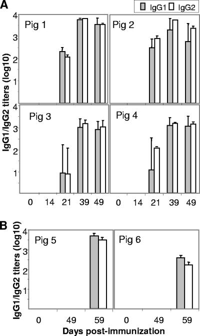
Likewise, total IgG antibodies to FMDV were detected for pigs 1 to 4 by ELISA at 14 days postimmunization with peptide B4T, reaching maximum values at day 39 and showing no increase after FMDV challenge (Table 3). Similar IgG1 and IgG2 titers were found on day 21, both isotypes being highly boosted after the second dose of B4T, with titers reaching at least 3 log units (Fig. 1A). Conversely, contact pigs 5 and 6 developed specific IgGs only after FMDV inoculation, with isotype profiles similar to those observed for the peptide-immunized animals (Table 3 and Fig. 1B).
FIG. 1.
Serum IgG1- and IgG2-specific responses in pigs following FMDV infection. (A) Peptide-immunized animals. (B) Contact pigs. Titers are expressed as reciprocals of the last dilution of sera (log10), calculated by interpolation to give an A492 of 1.0 OD unit. Each bar corresponds to geometric mean of at least two determinations ± standard error. At day 21, pigs 1 to 4 were immunized with a second dose of peptide. Day 49 corresponds to 10 days postchallenge (pigs 1 to 4) or 10 days postcontact (pigs 5 and 6). Day 59 corresponds to 10 days postinoculation for pigs 5 and 6.
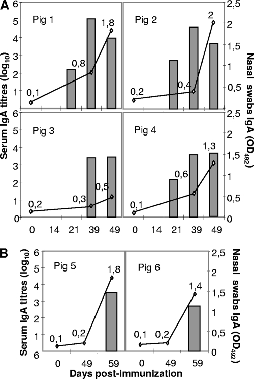
Inhalation of airborne FMDV, leading to virus replication in the respiratory tract, is considered as the most common route for natural transmission of this virus. Therefore, we also examined the effect of vaccination on systemic and local IgA responses. Remarkably, immunization with peptide B4T induced a strong systemic IgA-specific response (Fig. 2). Sera from three out of four immunized pigs showed FMDV-specific IgA from day 21, with titers above 2 log units. The titers increased after the second dose, reaching by day 39 (challenge) values higher than those seen for contact pigs 10 days after inoculation. Specific IgA titers were also found for nasal fluids of B4T-immunized pigs (Fig. 2A). This mucosal response was associated with significant titers of neutralizing activity in fluids from nasal samples (Table 3). On the other hand, whereas FMDV challenge caused no boost in systemic IgA levels of peptide-immunized animals, mucosal titers did experience a substantial increase (Fig. 2A). Conversely, for contact pigs 5 and 6, IgAs were detected only after inoculation, with mucosal titers reaching levels similar to those seen for immunized pigs (Fig. 2B).
FIG. 2.
IgA-specific responses to FMDV. Shown are sera (bars) and nasal fluids (open circles) collected at different days after peptide immunization (pigs 1 to 4) (A) or after contact with peptide-immunized pigs (animals 5 and 6) (B). Mean titers of IgA in sera are expressed as described in the legend to Fig. 1. Titers of IgA in nasal fluids are expressed as the OD at 492 nm (OD492) value (shown above each time point) obtained with a 1/10 serum dilution.
Peptide B4T induces FMDV-specific T-cell responses.
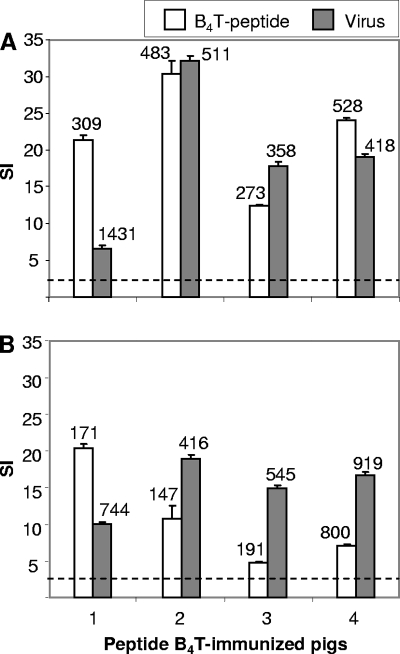
Induction of FMDV-specific T cells was detected in lymphoproliferation assays with PBMC of peptide-immunized pigs. High specific responses (SI, >6) against either FMDV or peptide B4T were found for lymphocytes from pigs 1 to 4 collected at day 39 (Fig. 3A). Proliferative responses were also detected after virus challenge; in this case, the SI was on average slightly lower than those determined before challenge (Fig. 3B). No stimulation was observed with PBMC either from pigs 1 to 4 or from contact pigs 5 and 6 prior to FMDV inoculation (data not shown).
FIG. 3.
Specific T-cell responses in peptide-immunized pigs. Samples were collected at days 39 (day of challenge) (A) and 49 (10 days postinfection) (B). Peak lymphoproliferative responses of pigs 1 to 4 to peptide B4T (20 μg/ml) and to FMDV C-S8c1 (105 TCID50/ml) are shown. Data are shown as SI (see Materials and Methods) and standard deviations are indicated. The cpm obtained in the corresponding control cultures are shown above each bar.
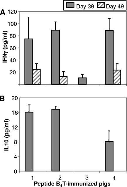
Production of IFN-γ, a Th1-cytokine, was detected in supernatants of PBMC from immunized pigs collected at day 39 in response to in vitro stimulation with peptide B4T but not in mock-stimulated cultures (Fig. 4A). IFN-γ levels correlated with the lymphoproliferative responses found. Thus, IFN-γ amounts higher than 70 pg/ml were detected for stimulated lymphocytes from pigs 1, 2, and 4, while pig 3 had a lower level (15 pg/ml). Conversely, amounts of IL-10 above the sensitivity of the assay were detected only for lymphocytes from pigs 1 and 2 (Fig. 4B). This pattern of cytokine production suggested that peptide B4T elicited FMDV-specific T cells that, when stimulated in vitro, developed a response dominated by Th1. After FMDV challenge (day 49), a clear decrease in IFN-γ and IL-10 release was detected (Fig. 4).
FIG. 4.
In vitro stimulation of cytokines. IFN-γ (A) and IL-10 (B) released by PBMC from peptide-immunized pigs stimulated in vitro with 20 μg/ml of peptide B4T. Peak values (in pg/ml) were detected by ELISA at 48 h and 72 h of in vitro stimulation for IL-10 and IFN-γ, respectively (see details in Materials and Methods). The detection levels for both cytokines in control cultures (medium alone) were below the sensitivity of the assays (7 pg/ml for IFN-γ and 15 pg/ml for IL-10).
Immunization with peptide B4T allows differentiation of infected and vaccinated pigs.
To assess whether peptide immunization induced an antibody response distinguishable from that of infected animals, serum samples from pigs 1 to 4 collected at days 39 and 49 (10 days postchallenge) were analyzed for antibodies to NS protein 3ABC. None of the sera tested positive. On the other hand, for pigs 5 and 6 no anti-3ABC response was detected during the contact period, whereas consistent titers were found after viral inoculation (Table 2) that were similar to those detected for pigs 7 and 8, controls for infection (data not shown).
DISCUSSION
Mimicking protective responses using synthetic peptides poses an attractive and multidisciplinary challenge. Despite the potential advantages of this approach—such as innocuousness, thermal stability, and easy scale-up—the complexity of the interactions between pathogens and host immune responses have limited the development of successful peptide vaccines (31, 34, 43).
Multimerization of peptides, such as that provided by dendrimeric constructs, has long been recognized as one of the most effective tools for enhancing peptide immunogenicity (11, 54). In the present work, we report solid protection of pigs against FMDV challenge conferred by a dendrimeric peptide containing one B- and one T-cell immunodominant epitope. Intramuscular immunization with peptide B4T prevented the emergence of clinical signs and contact transmission in the four pigs studied upon a severe FMDV challenge. The two control naive pigs that were housed with the immunized animals during the challenge period did not show clinical signs but developed them upon subsequent FMDV infection. The failure to detect FMDV RNA in either serum or nasal samples, as well as the absence of transient leucopenia associated with FMDV infection (18), supported by the lack of reduction of the proliferative response to the mitogen concanavalin A in the pigs immunized with peptide B4T (data not shown), confirmed that the levels of virus replication in these animals were below those required for virus spread and disease triggering.
This solid protection conferred by peptide B4T correlated with the detection of high levels of circulating neutralizing FMDV antibodies that were boosted after the second peptide dose, with titers around 2 log values, i.e., about 1 log unit lower than those developed by contact pigs after 10 days of infection. Differences in the IgG1/IgG2 ratio induced by FMDV vaccination or infection versus those found for peptide-immunized animals have been correlated with the lack of solid protection conferred by the latter (36, 53). The IgG1 and IgG2 titers induced by peptide B4T were similar to those detected for infected contact pigs, suggesting no major differences in the isotype modulation induced.
Interestingly, FMDV-specific IgA titers were detected for three of the pigs as soon as 21 days after the first immunization with peptide B4T and were boosted for all animals after the second B4T dose, reaching 4- to 5-log values for pigs 1 and 2 and somewhat lower levels for pigs 3 and 4 (3 to 4 log units, similar to what was seen for infected control pigs). Remarkably, specific IgA antibodies were also detected for nasal samples from three of the four B4T-vaccinated pigs before challenge. After virus challenge, the nasal IgA titers were boosted for the four immunized animals, reaching values similar to those observed for infected contact pigs. In contrast to other vaccines having to rely on mucosal delivery to access the natural inductive pathway (29, 35, 39), B4T can induce high mucosal immune responses by parenteral administration. Several different, nonexclusive mechanisms have been proposed to explain the production of secretory antibodies after parenteral administration of the antigen, including direct diffusion of soluble or phagocytosed antigens to mucosa-associated lymphoid tissue or activation of antigen-presenting cells at draining lymph nodes, which then migrate to mucosa-associated lymphoid tissue (12). In any event, parenterally administered antigens capable of inducing strong mucosal immunity, such as our B4T dendrimer or a few other immunogens so far described (28, 37, 56), appear to be good candidates for future mucosal vaccines. They can overcome some of the main problems associated with mucosal administration, such as local degradation or physical ejection of the vaccine, both of which hamper the control of the dose delivered.
The intradermal route for FMDV inoculation was chosen according to potency requirements for FMD vaccines (47). The correlation found between solid protection and IgA induction raises the interesting possibility of assessing the protection conferred by B4T peptide by use of respiratory/exposure challenge conditions, which are likely to be more similar to field transmission.
The functional role in protection of the T cells induced by FMDV and of the balance of cytokines they release remains to be well established (2). Immunization with peptide B4T elicited T cells that consistently proliferated when stimulated with the peptide as well as with FMDV. After FMDV challenge, while the lymphoproliferative response to peptide was reduced in three out of four animals, proliferation to the virus was instead maintained. This suggests that immunization with peptide B4T primes T cells that can recognize the viral epitopes presented in the context of a subsequent virus encounter. Upon in vitro peptide stimulation, the primed T cells released IFN-γ and to a lower extent IL-10. IFN-γ is a major activator of macrophages, enhancing their antimicrobial activity and their capacity for processing and presenting antigens to T lymphocytes. In pigs, the majority of IFN-γ-producing cells are αβ T lymphocytes, and within these the majority are of the double-positive CD4+ CD8+ subset, which contains memory T cells (42). It has been reported that IFN-γ stimulates MHC expression in antigen-presenting cells and efficiently inhibits FMDV replication (60). Altogether, our results suggest that priming of porcine T cells with peptide B4T induces a T-cell activation that efficiently contributes to FMDV protection.
Animal-to-animal variations have been reported for the protective responses to peptide vaccines, including those against FMDV (16, 53), and have been associated with the MHC-restricted recognition of the T-cell epitopes included in their compositions (25). Interestingly, despite the differences in the B- and T-cell responses elicited by the four animals in this study, all of them turned out to be protected after FMDV challenge, suggesting that peptide B4T can elicit B- and T-cell responses sufficient to cope with virus replication, thus minimizing the chances of selecting escape virus (53).
The mechanisms leading to the solid protection conferred by peptide B4T remain to be known in detail. Peptides where the present B- and T-cell epitopes were simply juxtaposed in a linear fashion elicited only partial protection in pigs and low levels of FMDV-specific systemic IgAs (C. Cubillos, I. Avalos, E. Borras, B. G. de la Torre, J. Bárcena, D. Andreu, F. Sobrino, and E. Blanco, unpublished results). This suggests that either the dendrimeric presentation of the B-cell site or the inclusion of a T-cell site separated from the dendrimeric B sites by a cathepsin D cleavage site or both could be relevant for the antibody response observed. Also, a role in protection of FMDV-specific T-cell epitopes has been found for animals immunized with vaccinia virus recombinants expressing FMDV three-dimensional protein in the absence of B-cell capsid antigenic sites (24).
Differentiation between vaccinated and infected animals is of great interest, both to monitor virus circulation and to avoid trade restrictions on animals or animal products from countries applying vaccination by those not applying it. This is a controversial issue for the control of animal diseases, particularly FMD (40). Interestingly, our B4T peptide induced a serological response compatible with its use as a vaccine facilitating the differentiation of infected from vaccinated animals, since it did not elicit antibodies to the NS protein 3ABC, which are diagnostic for FMDV replication (13).
The number of animals in this study is similar to those in many other introductory studies on vaccine candidates, although it is not enough for statistical demonstration. Even so, the results strongly support that immunization with peptide B4T can elicit in pigs an immune response similar to that induced in control animals after FMDV infection. This response associates not only with the prevention of clinical disease but also with the inhibition of the local replication and the airborne excretion of FMDV. These results point to this dendrimeric peptide approach as an interesting candidate for the improvement of peptide subunit vaccines. To address how useful the immunogenic potential of this approach can become, experiments are in progress to assess whether analogous peptide constructs including B-cell epitopes from other FMDV serotypes can confer solid protection against FMDV challenge in pigs and cattle.
Supplementary Material
Acknowledgments
We thank Javier Domínguez and Angel Ezquerra for fruitful discussions.
Work at CBMSO and INIA was supported by Spanish grants from CICYT (BIO2002-04091-C03-02/03 and BIO2005-07592-C02-01), MEC (CSD2006-0007), and Fundación Severo Ochoa and by an EU grant (SSP503603). Work at UPF was supported by the Spanish Ministry of Education and Science (grant BIO2002-04091-C03-01 and BIO2005-07592-CO2-02) and by Generalitat de Catalunya (SGR00494 and CIDEM-BAPP). C.C. was supported by a predoctoral fellowship from Ministerio de Educación y Ciencia (FPI).
Footnotes
Published ahead of print on 30 April 2008.
Supplemental material for this article may be found at http://jiv.asm.org/.
REFERENCES
- 1.Acharya, R., E. Fry, D. Stuart, G. Fox, D. Rowlands, and F. Brown. 1989. The three-dimensional structure of foot-and-mouth disease virus at 2.9 A resolution. Nature 337709-716. [DOI] [PubMed] [Google Scholar]
- 2.Barnard, A. L., A. Arriens, S. Cox, P. Barnett, B. Kristensen, A. Summerfield, and K. C. McCullough. 2005. Immune response characteristics following emergency vaccination of pigs against foot-and-mouth disease. Vaccine 231037-1047. [DOI] [PubMed] [Google Scholar]
- 3.Barnett, P. V., L. Pullen, L. Williams, and T. R. Doel. 1996. International bank for foot-and-mouth disease vaccine: assessment of Montanide ISA 25 and ISA 206, two commercially available oil adjuvants. Vaccine 141187-1198. [DOI] [PubMed] [Google Scholar]
- 4.Barteling, S. J. 2004. Modern inactivated foot-and-mouth disease (FMD) vaccines: historical background and key elements in production and use. Horizon Bioscience, Norfolk, United Kingdom.
- 5.Baxt, B., and E. Rieder. 2004. Molecular aspects of foot-and-mouth disease virus virulence and host range: role of host cell receptors and viral factors, p. 145-172. In F. Sobrino and E. Domingo (ed.), Foot-and-mouth disease. Current perspectives. Horizon Bioscience, Norfolk, United Kingdom.
- 6.Belsham, G. J. 2005. Translation and replication of FMDV RNA. Curr. Top. Microbiol. Immunol. 28843-70. [DOI] [PubMed] [Google Scholar]
- 7.Bittle, J. L., R. A. Houghten, H. Alexander, T. M. Shinnick, J. G. Sutcliffe, R. A. Lerner, D. J. Rowlands, and F. Brown. 1982. Protection against foot-and-mouth disease by immunization with a chemically synthesized peptide predicted from the viral nucleotide sequence. Nature 29830-33. [DOI] [PubMed] [Google Scholar]
- 8.Blanco, E., M. Garcia-Briones, A. Sanz-Parra, P. Gomes, E. De Oliveira, M. L. Valero, D. Andreu, V. Ley, and F. Sobrino. 2001. Identification of T-cell epitopes in nonstructural proteins of foot-and-mouth disease virus. J. Virol. 753164-3174. [DOI] [PMC free article] [PubMed] [Google Scholar]
- 9.Blanco, E., K. McCullough, A. Summerfield, J. Fiorini, D. Andreu, C. Chiva, E. Borras, P. Barnett, and F. Sobrino. 2000. Interspecies major histocompatibility complex-restricted Th cell epitope on foot-and-mouth disease virus capsid protein VP4. J. Virol. 744902-4907. [DOI] [PMC free article] [PubMed] [Google Scholar]
- 10.Blanco, E., L. J. Romero, M. El Harrach, and J. M. Sanchez-Vizcaino. 2002. Serological evidence of FMD subclinical infection in sheep population during the 1999 epidemic in Morocco. Vet. Microbiol. 8513-21. [DOI] [PubMed] [Google Scholar]
- 11.Borras-Cuesta, F., Y. Fedon, and A. Petit-Camurdan. 1988. Enhancement of peptide immunogenicity by linear polymerization. Eur. J. Immunol. 18199-202. [DOI] [PubMed] [Google Scholar]
- 12.Bouvet, J. P., N. Decroix, and P. Pamonsinlapatham. 2002. Stimulation of local antibody production: parenteral or mucosal vaccination? Trends Immunol. 23209-213. [DOI] [PubMed] [Google Scholar]
- 13.Brocchi, E., I. E. Bergmann, A. Dekker, D. J. Paton, D. J. Sammin, M. Greiner, S. Grazioli, F. De Simone, H. Yadin, B. Haas, N. Bulut, V. Malirat, E. Neitzert, N. Goris, S. Parida, K. Sorensen, and K. De Clercq. 2006. Comparative evaluation of six ELISAs for the detection of antibodies to the non-structural proteins of foot-and-mouth disease virus. Vaccine 246966-6979. [DOI] [PubMed] [Google Scholar]
- 14.Carrillo, C., E. R. Tulman, G. Delhon, Z. Lu, A. Carreno, A. Vagnozzi, G. F. Kutish, and D. L. Rock. 2005. Comparative genomics of foot-and-mouth disease virus. J. Virol. 796487-6504. [DOI] [PMC free article] [PubMed] [Google Scholar]
- 15.Collen, T. 1994. Foot-and-mouth disease (aphthovirus): viral T cell epitopes. In B. M. L. Goddeevis and I. Morrison (ed.), Cell mediated immunity in ruminants. CRC Press Inc., Boca Raton, FL.
- 16.Collen, T., R. Dimarchi, and T. R. Doel. 1991. A T cell epitope in VP1 of foot-and-mouth disease virus is immunodominant for vaccinated cattle. J. Immunol. 146749-755. [PubMed] [Google Scholar]
- 17.Collen, T., and T. R. Doel. 1990. Heterotypic recognition of foot-and-mouth disease virus by cattle lymphocytes. J. Gen. Virol. 71309-315. [DOI] [PubMed] [Google Scholar]
- 18.Diaz-San Segundo, F., F. J. Salguero, A. de Avila, M. M. de Marco, M. A. Sanchez-Martin, and N. Sevilla. 2006. Selective lymphocyte depletion during the early stage of the immune response to foot-and-mouth disease virus infection in swine. J. Virol. 802369-2379. [DOI] [PMC free article] [PubMed] [Google Scholar]
- 19.DiMarchi, R., G. Brooke, C. Gale, V. Cracknell, T. Doel, and N. Mowat. 1986. Protection of cattle against foot-and-mouth disease by a synthetic peptide. Science 232639-641. [DOI] [PubMed] [Google Scholar]
- 20.Doel, T. R. 2003. FMD vaccines. Virus Res. 9181-99. [DOI] [PubMed] [Google Scholar]
- 21.Domingo, E., M. G. Mateu, M. A. Martínez, J. Dopazo, A. Moya, and F. Sobrino. 1990. Genetic variability and antigenic diversity of foot-and-mouth disease virus, vol. 2. Academic Press Inc., London, United Kingdom.
- 22.Eble, P. L., A. Bouma, K. Weerdmeester, J. A. Stegeman, and A. Dekker. 2007. Serological and mucosal immune responses after vaccination and infection with FMDV in pigs. Vaccine 251043-1054. [DOI] [PubMed] [Google Scholar]
- 23.Francis, M. J., and L. Black. 1983. Antibody response in pig nasal fluid and serum following foot-and-mouth disease infection or vaccination. J. Hyg. 91329-334. [DOI] [PMC free article] [PubMed] [Google Scholar]
- 24.Garcia-Briones, M. M., E. Blanco, C. Chiva, D. Andreu, V. Ley, and F. Sobrino. 2004. Immunogenicity and T cell recognition in swine of foot-and-mouth disease virus polymerase 3D. Virology 322264-275. [DOI] [PubMed] [Google Scholar]
- 25.Garcia-Briones, M. M., G. C. Russell, R. A. Oliver, C. Tami, O. Taboga, E. Carrillo, E. L. Palma, F. Sobrino, and E. J. Glass. 2000. Association of bovine DRB3 alleles with immune response to FMDV peptides and protection against viral challenge. Vaccine 191167-1171. [DOI] [PubMed] [Google Scholar]
- 26.Gerner, W., B. V. Carr, K. H. Wiesmuller, E. Pfaff, A. Saalmuller, and B. Charleston. 2007. Identification of a novel foot-and-mouth disease virus specific T-cell epitope with immunodominant characteristics in cattle with MHC serotype A31. Vet. Res. 38565-572. [DOI] [PubMed] [Google Scholar]
- 27.Golding, S. M., R. S. Hedger, and P. Talbot. 1976. Radial immuno-diffusion and serum-neutralisation techniques for the assay of antibodies to swine vesicular disease. Res. Vet. Sci. 20142-147. [PubMed] [Google Scholar]
- 28.Guthrie, T., C. G. Hobbs, V. Davenport, R. E. Horton, R. S. Heyderman, and N. A. Williams. 2004. Parenteral influenza vaccination influences mucosal and systemic T cell-mediated immunity in healthy adults. J. Infect. Dis. 1901927-1935. [DOI] [PubMed] [Google Scholar]
- 29.Holmgren, J., and C. Czerkinsky. 2005. Mucosal immunity and vaccines. Nat. Med. 11S45-S53. [DOI] [PubMed] [Google Scholar]
- 30.Kitching, R. P., A. M. Hutber, and M. V. Thrusfield. 2005. A review of foot-and-mouth disease with special consideration for the clinical and epidemiological factors relevant to predictive modelling of the disease. Vet. J. 169197-209. [DOI] [PubMed] [Google Scholar]
- 31.Leclerc, C. 2003. New approaches in vaccine development. Comp. Immunol. Microbiol. Infect. Dis. 26329-341. [DOI] [PubMed] [Google Scholar]
- 32.Lee, C. C., J. A. MacKay, J. M. Frechet, and F. C. Szoka. 2005. Designing dendrimers for biological applications. Nat. Biotechnol. 231517-1526. [DOI] [PubMed] [Google Scholar]
- 33.Mateu, M. G. 1995. Antibody recognition of picornaviruses and escape from neutralization: a structural view. Virus Res. 381-24. [DOI] [PubMed] [Google Scholar]
- 34.Meloen, R. H., J. P. Langeveld, W. M. Schaaper, and J. W. Slootstra. 2001. Synthetic peptide vaccines: unexpected fulfillment of discarded hope? Biologicals 29233-236. [DOI] [PubMed] [Google Scholar]
- 35.Moyle, P. M., R. P. McGeary, J. T. Blanchfield, and I. Toth. 2004. Mucosal immunisation: adjuvants and delivery systems. Curr. Drug Deliv. 1385-396. [DOI] [PubMed] [Google Scholar]
- 36.Mulcahy, G., C. Gale, P. Robertson, S. Iyisan, R. D. DiMarchi, and T. R. Doel. 1990. Isotype responses of infected, virus-vaccinated and peptide-vaccinated cattle to foot-and-mouth disease virus. Vaccine 8249-256. [DOI] [PubMed] [Google Scholar]
- 37.Musey, L., Y. Ding, M. Elizaga, R. Ha, C. Celum, and M. J. McElrath. 2003. HIV-1 vaccination administered intramuscularly can induce both systemic and mucosal T cell immunity in HIV-1-uninfected individuals. J. Immunol. 1711094-1101. [DOI] [PubMed] [Google Scholar]
- 38.Núñez, J. I., N. Molina, E. Baranowski, E. Domingo, S. Clark, A. Burman, S. Berryman, T. Jackson, and F. Sobrino. 2007. Guinea pig-adapted foot-and-mouth disease virus with altered receptor recognition can productively infect a natural host. J. Virol. 818497-8506. [DOI] [PMC free article] [PubMed] [Google Scholar]
- 39.Ogra, P. L., H. Faden, and R. C. Welliver. 2001. Vaccination strategies for mucosal immune responses. Clin. Microbiol. Rev. 14430-445. [DOI] [PMC free article] [PubMed] [Google Scholar]
- 40.Pasick, J. 2004. Application of DIVA vaccines and their companion diagnostic tests to foreign animal disease eradication. Anim. Health Res. Rev. 5257-262. [DOI] [PubMed] [Google Scholar]
- 41.Pereira, H. G. 1981. Foot-and-mouth disease. Academic Press Inc., London, United Kingdom.
- 42.Rodriguez-Carreno, M. P., L. Lopez-Fuertes, C. Revilla, A. Ezquerra, F. Alonso, and J. Dominguez. 2002. Phenotypic characterization of porcine IFN-gamma-producing lymphocytes by flow cytometry. J. Immunol. Methods 259171-179. [DOI] [PubMed] [Google Scholar]
- 43.Rowlands, D. 2004. Foot-and-mouth disease virus peptide vaccines. In F. Sobrino and E. Domingo (ed.), Foot-and-mouth disease. Current perspectives. Horizon Bioscience, Norfolk, United Kingdom.
- 44.Saiz, J. C., A. Rodriguez, M. Gonzalez, F. Alonso, and F. Sobrino. 1992. Heterotypic lymphoproliferative response in pigs vaccinated with foot-and-mouth disease virus. Involvement of isolated capsid proteins. J. Gen. Virol. 732601-2607. [DOI] [PubMed] [Google Scholar]
- 45.Saiz, M., D. B. De La Morena, E. Blanco, J. I. Nunez, R. Fernandez, and J. M. Sanchez-Vizcaino. 2003. Detection of foot-and-mouth disease virus from culture and clinical samples by reverse transcription-PCR coupled to restriction enzyme and sequence analysis. Vet. Res. 34105-117. [DOI] [PubMed] [Google Scholar]
- 46.Salt, J. S. 1993. The carrier state in foot and mouth disease—an immunological review. Br. Vet. J. 149207-223. [DOI] [PubMed] [Google Scholar]
- 47.Salt, J. S., G. Mulcahy, and R. P. Kitching. 1996. Isotype-specific antibody responses to foot-and-mouth disease virus in sera and secretions of “carrier” and “non-carrier” cattle. Epidemiol. Infect. 117349-360. [DOI] [PMC free article] [PubMed] [Google Scholar]
- 48.Samuel, A. R., and N. J. Knowles. 2001. Foot-and-mouth disease virus: cause of the recent crisis for the UK livestock industry. Trends Genet. 17421-424. [DOI] [PubMed] [Google Scholar]
- 49.Sanz-Parra, A., M. A. Jimenez-Clavero, M. M. Garcia-Briones, E. Blanco, F. Sobrino, and V. Ley. 1999. Recombinant viruses expressing the foot-and-mouth disease virus capsid precursor polypeptide (P1) induce cellular but not humoral antiviral immunity and partial protection in pigs. Virology 259129-134. [DOI] [PubMed] [Google Scholar]
- 50.Sobrino, F., M. Davila, J. Ortin, and E. Domingo. 1983. Multiple genetic variants arise in the course of replication of foot-and-mouth disease virus in cell culture. Virology 128310-318. [DOI] [PubMed] [Google Scholar]
- 51.Sobrino, F., and E. Domingo. 2001. Foot-and-mouth disease in Europe. EMBO Rep. 2459-461. [DOI] [PMC free article] [PubMed] [Google Scholar]
- 52.Sobrino, F., M. Saiz, M. A. Jimenez-Clavero, J. I. Nunez, M. F. Rosas, E. Baranowski, and V. Ley. 2001. Foot-and-mouth disease virus: a long known virus, but a current threat. Vet. Res. 321-30. [DOI] [PubMed] [Google Scholar]
- 53.Taboga, O., C. Tami, E. Carrillo, J. I. Nunez, A. Rodriguez, J. C. Saiz, E. Blanco, M. L. Valero, X. Roig, J. A. Camarero, D. Andreu, M. G. Mateu, E. Giralt, E. Domingo, F. Sobrino, and E. L. Palma. 1997. A large-scale evaluation of peptide vaccines against foot-and-mouth disease: lack of solid protection in cattle and isolation of escape mutants. J. Virol. 712606-2614. [DOI] [PMC free article] [PubMed] [Google Scholar]
- 54.Tam, J. P. 1996. Recent advances in multiple antigen peptides. J. Immunol. Methods 19617-32. [DOI] [PubMed] [Google Scholar]
- 55.Tam, J. P., Y. A. Lu, and J. L. Yang. 2002. Antimicrobial dendrimeric peptides. Eur. J. Biochem. 269923-932. [DOI] [PubMed] [Google Scholar]
- 56.Thompson, J. M., A. C. Whitmore, J. L. Konopka, M. L. Collier, E. M. Richmond, N. L. Davis, H. F. Staats, and R. E. Johnston. 2006. Mucosal and systemic adjuvant activity of alphavirus replicon particles. Proc. Natl. Acad. Sci. USA 1033722-3727. [DOI] [PMC free article] [PubMed] [Google Scholar]
- 57.Toja, M., C. Escarmis, and E. Domingo. 1999. Genomic nucleotide sequence of a foot-and-mouth disease virus clone and its persistent derivatives. Implications for the evolution of viral quasispecies during a persistent infection. Virus Res. 64161-171. [DOI] [PubMed] [Google Scholar]
- 58.van Lierop, M. J., K. van Maanen, R. H. Meloen, V. P. Rutten, M. A. de Jong, and E. J. Hensen. 1992. Proliferative lymphocyte responses to foot-and-mouth disease virus and three FMDV peptides after vaccination or immunization with these peptides in cattle. Immunology 75406-413. [PMC free article] [PubMed] [Google Scholar]
- 59.Van Noort, J. M., J. Boon, A. C. Van der Drift, J. P. Wagenaar, A. M. Boots, and C. J. Boog. 1991. Antigen processing by endosomal proteases determines which sites of sperm-whale myoglobin are eventually recognized by T cells. Eur. J. Immunol. 211989-1996. [DOI] [PubMed] [Google Scholar]
- 60.Zhang, Z. D., G. Hutching, P. Kitching, and S. Alexandersen. 2002. The effects of gamma interferon on replication of foot-and-mouth disease virus in persistently infected bovine cells. Arch. Virol. 1472157-2167. [DOI] [PubMed] [Google Scholar]
Associated Data
This section collects any data citations, data availability statements, or supplementary materials included in this article.


