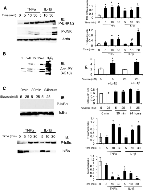
FIG. 7.
Cytokines, but not high glucose conditions, induce MAPK phosphorylation, tyrosine phosphorylation, and IκBα phosphorylation and degradation in HRECs. A: Since high glucose conditions did not activate or stimulate HRECs, the direct effects of TNF-α and IL-1β on HRECs were tested. HRECs were serum starved overnight and stimulated with 5 ng/ml TNF-α or 1 ng/ml IL-1β for different time periods as indicated. The activation of ERK and JNK signaling pathways was assessed by immunoblot analyses using anti–phospho-ERK1/2 and anti–phospho JNK antibodies as indicated. Equal amounts of protein were added to each lane as confirmed by actin levels. Both cytokines induced marked ERK1/2 and JNK phosphorylation. Representative results from at least three independent experiments are shown on the left and quantified and presented as means ± SD on the right. *P < 0.01 compared with control. B: Tyrosine phosphorylation in HRECs was assessed by immunoblot using anti-phospho-tyrosine antibody after treatment with high glucose for 0.5–78 h with or without IL-1β (1 ng/ml). Stimulation for 10 min with either 0.5 mmol/l H2O2 (positive control) or 1 ng/ml IL-1β led to a marked increase in tyrosine-phosphorylated proteins. Twenty-four–hour exposure data are shown; similar results were obtained for 0.5- to 78-h time points. C: The effects of high glucose (top) conditions, TNF-α, and IL-1β (bottom image) on IκBα phosphorylation and degradation, the first step in NF-κB pathway activation, were examined in HRECs. High glucose had no effect, whereas 5 ng/ml TNF-α or 1 ng/ml IL-1β for 0–30 min led to a dramatic increase in IκBα phosphorylation and degradation. Representative results from at least three independent experiments are shown on the left and quantified and presented as means ± SD on the right. *P < 0.01 compared with control.

