

Abstract
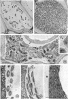
The rod-shaped rickettsia-like bacteria of Pierce's disease measure about 0.25 to 0.50 μm in diameter and 1.0 to 4.0 μm long. The bacteria have a cell wall consisting of a trilaminar outer membrane and two intermediate low-density layers separated by a dense intermediate layer. A trilaminar cytoplasmic membrane is also present, resulting in a total wall complex thickness of 25 to 40 nm. A periodic infolding of the outer membrane and intermediate layers of the wall give the wall surface a ridged apperance. The ridges appear to go around the long axis of the cell, possibly in the form of spirals. Ribosomes and nuclear regions with easily visible deoxyribonucleic acid strands and clumps are distributed throughout the cytoplasm. Binary fission, during which the cell wall and cytoplasmic membrane folded inward to partition the cell, was observed. In the xylem of infected grapes, the bacteria are either distributed evenly throughout the lumen of the xylem vessel or appressed along the inner surface of the vessel walls in an electron-lucent matrix.
Full text
PDF


Images in this article


Selected References
These references are in PubMed. This may not be the complete list of references from this article.
- ANACKER R. L., FUKUSHI K., PICKENS E. G., LACKMAN D. B. ELECTRON MICROSCOPIC OBSERVATIONS OF THE DEVELOPMENT OF COXIELLA BURNETII IN THE CHICK YOLK SAC. J Bacteriol. 1964 Oct;88:1130–1138. doi: 10.1128/jb.88.4.1130-1138.1964. [DOI] [PMC free article] [PubMed] [Google Scholar]
- Anacker R. L., Pickens E. G., Lackman D. B. Details of the ultrastructure of Rickettsia prowazekii grown in the chick yolk sac. J Bacteriol. 1967 Jul;94(1):260–262. doi: 10.1128/jb.94.1.260-262.1967. [DOI] [PMC free article] [PubMed] [Google Scholar]
- Anderson D. R., Hopps H. E., Barile M. F., Bernheim B. C. Comparison of the ultrastructure of several rickettsiae, ornithosis virus, and Mycoplasma in tissue culture. J Bacteriol. 1965 Nov;90(5):1387–1404. doi: 10.1128/jb.90.5.1387-1404.1965. [DOI] [PMC free article] [PubMed] [Google Scholar]
- Bird R. G., Kordová N., Rehácek J. Fine structure of Rickettsia prowazeki in the haemocytes of ticks Hyalomma dromedarii. Acta Virol. 1967 Jan;11(1):60–62. [PubMed] [Google Scholar]
- Brezina R. Advances in rickettsial research. Curr Top Microbiol Immunol. 1969;47:20–39. doi: 10.1007/978-3-642-46160-6_2. [DOI] [PubMed] [Google Scholar]
- Brinton L. P., Burgdorfer W. Fine structure of Rickettsia canada in tissues of Dermacentor andersoni Stiles. J Bacteriol. 1971 Mar;105(3):1149–1159. doi: 10.1128/jb.105.3.1149-1159.1971. [DOI] [PMC free article] [PubMed] [Google Scholar]
- Burgdorfer W., Anacker R. L., Bird R. G., Bertram D. S. Intranuclear growth of Rickettsia rickettsii. J Bacteriol. 1968 Oct;96(4):1415–1418. doi: 10.1128/jb.96.4.1415-1418.1968. [DOI] [PMC free article] [PubMed] [Google Scholar]
- Burton P. R., Kordová N., Paretsky D. Electron microscopic studies of the rickettsia Coxiella burneti: entry, lysosomal response, and fate of rickettsial DNA in L-cells. Can J Microbiol. 1971 Feb;17(2):143–150. doi: 10.1139/m71-025. [DOI] [PubMed] [Google Scholar]
- CARO L. G., JACKSON E. B., SMADEL J. E., WISSIG S. L. Electron microscopic observations on intracellular rickettsiae. Am J Pathol. 1956 Nov-Dec;32(6):1117–1133. [PMC free article] [PubMed] [Google Scholar]
- Costerton J. W. The structure and function of the cell envelope of gram-negative bacteria. Rev Can Biol. 1970 Sep;29(3):299–316. [PubMed] [Google Scholar]
- Glauert A. M., Thornley M. J. The topography of the bacterial cell wall. Annu Rev Microbiol. 1969;23:159–198. doi: 10.1146/annurev.mi.23.100169.001111. [DOI] [PubMed] [Google Scholar]
- Hopkins D. L., Mollenhauer H. H. Rickettsia-like Bacterium Associated with Pierce's Disease of Grapes. Science. 1973 Jan 19;179(4070):298–300. doi: 10.1126/science.179.4070.298. [DOI] [PubMed] [Google Scholar]
- ITO S., VINSON J. W. FINE STRUCTURE OF RICKETTSIA QUINTANA CULTIVATED IN VITRO AND IN THE LOUSE. J Bacteriol. 1965 Feb;89:481–495. doi: 10.1128/jb.89.2.481-495.1965. [DOI] [PMC free article] [PubMed] [Google Scholar]
- Maillet P. L. Présence de particules de type rickettsien dans la salive d'un homoptére vecteur de la phyllodie du trèfle, Euscelis lineolatus Brullé (Homoptera jasidae. Rev Can Biol. 1970 Dec;29(4):391–393. [PubMed] [Google Scholar]
- Shkolnik L. Y., Zatulovsky B. G., Shestopalova N. M. Ultrastructure of Rickettsia prow azeki. An electron microscope study of ultrathin sections from infected louse guts and chick embryo yolk sacs. Acta Virol. 1966 May;10(3):260–265. [PubMed] [Google Scholar]
- Silva M. T., Sousa J. C. Ultrastructure of the cell wall and cytoplasmic membrane of gram-negative bacteria with different fixation techniques. J Bacteriol. 1973 Feb;113(2):953–962. doi: 10.1128/jb.113.2.953-962.1973. [DOI] [PMC free article] [PubMed] [Google Scholar]
- Spurr A. R. A low-viscosity epoxy resin embedding medium for electron microscopy. J Ultrastruct Res. 1969 Jan;26(1):31–43. doi: 10.1016/s0022-5320(69)90033-1. [DOI] [PubMed] [Google Scholar]