

Abstract
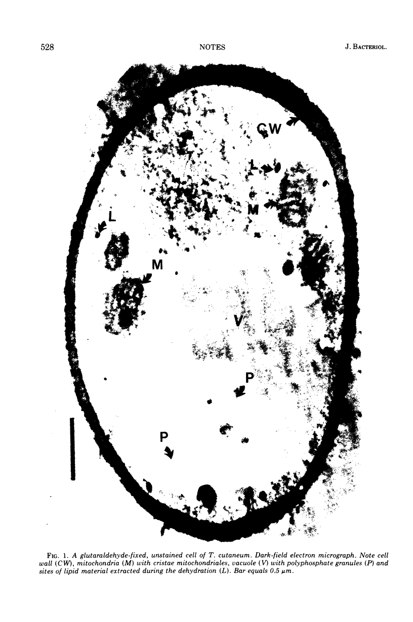
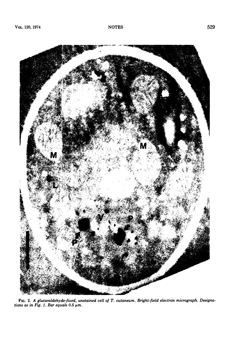
Dark-field electron micrographs of aldehyde-fixed, unstained yeast-phase cells of Trichosporon cutaneum exhibit remarkable contrast and good resolution when the sections studied are less than 25 nm thick and conical illumination is used.
Full text
PDF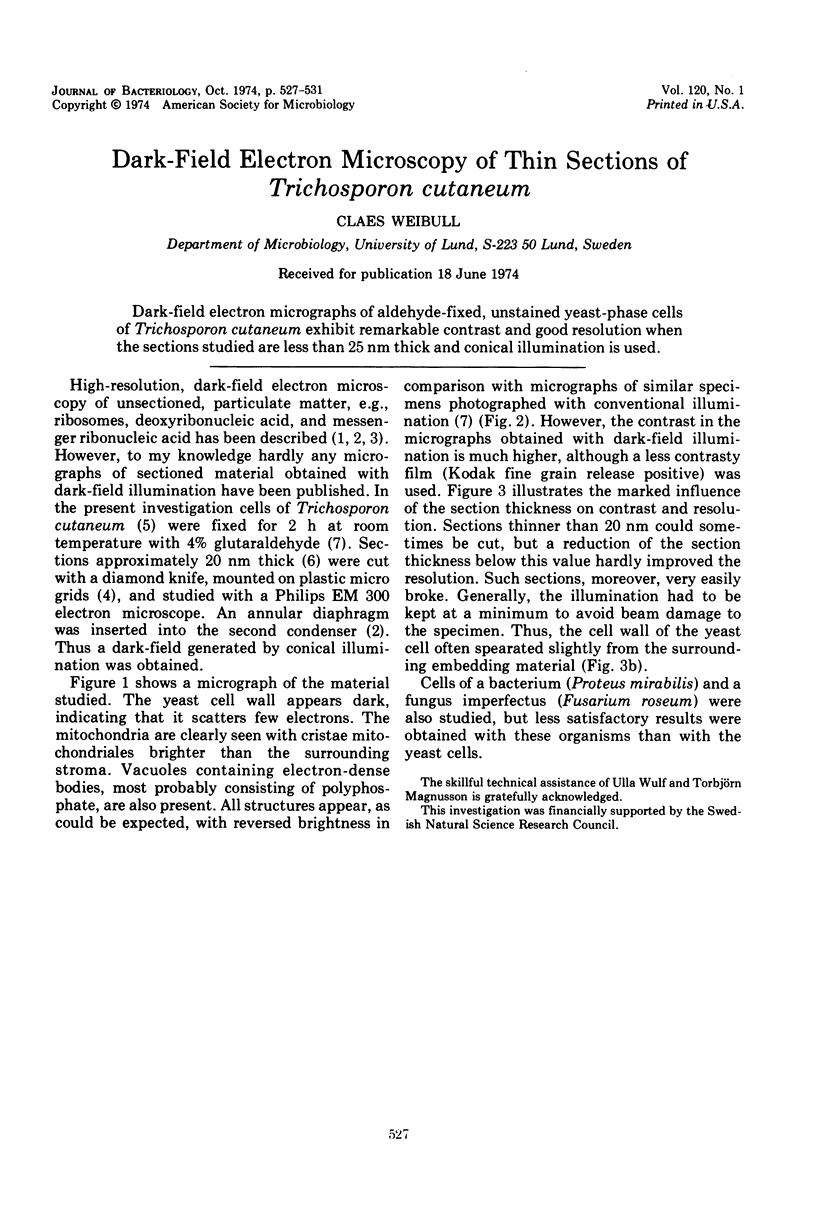
Images in this article
Selected References
These references are in PubMed. This may not be the complete list of references from this article.
- Brakenhoff G. J., Nanninga N., Pieters J. Relative mass determination from darkfield electron micrographs with an application to ribosomes. J Ultrastruct Res. 1972 Nov;41(3):238–257. doi: 10.1016/s0022-5320(72)90067-6. [DOI] [PubMed] [Google Scholar]
- Dubochet J., Ducommun M., Zollinger M., Kellenberger E. A new preparation method for dark-field electron microscopy of biomacromolecules. J Ultrastruct Res. 1971 Apr;35(1):147–167. doi: 10.1016/s0022-5320(71)80148-x. [DOI] [PubMed] [Google Scholar]
- Fukami A., Adachi K. A new method of preparation of a self-perforated micro plastic grid and its application. J Electron Microsc (Tokyo) 1965;14(2):112–118. [PubMed] [Google Scholar]
- Neujahr H. Y., Varga J. M. Degradation of phenols by intact cells and cell-free preparations of Trichosporon cutaneum. Eur J Biochem. 1970 Mar 1;13(1):37–44. doi: 10.1111/j.1432-1033.1970.tb00896.x. [DOI] [PubMed] [Google Scholar]
- Weibull C. Electron microscope studies on aldehyde-fixed, unstained microbial cells. J Ultrastruct Res. 1973 Apr;43(1):150–159. doi: 10.1016/s0022-5320(73)90075-0. [DOI] [PubMed] [Google Scholar]
- Weibull C. Estimation of the thickness of films used in electron microscopy. Z Allg Mikrobiol. 1972;12(6):487–490. doi: 10.1002/jobm.3630120607. [DOI] [PubMed] [Google Scholar]