Abstract
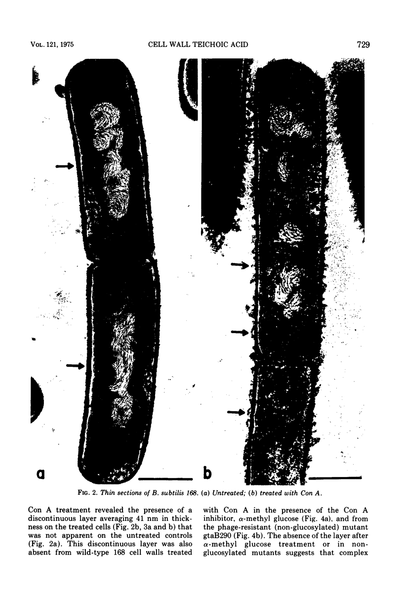
The phytohemagglutinin, concanavalin A (Con A), interacts specifically and reversibly with the polyglucosyl glycerol phosphate teichoic acid of Bacillus subtilis 168 cell walls. Advantage has been taken of this interaction to examine the organization of the surface teichoic acid at the ultrastructural level. Con A-treated whole cells and cell walls contain an irregular, fluffy layer 25 to 60 nm thick which is absent in untreated or alpha-methyl glucoside-treated preparations. This discontinuous layer is present only on the outer profile of Con-A-treated cell walls. The surface teichoic acid is proposed to be oriented perpendicular to the long axis of the cell. Fixation and embedment for electron microscopy result in condensation of this layer which then contributes to the stainable portion of the wall. Con A treatment binds adjacent teichoic acid molecules in their native configuration producing the irregular, fluffy layer visualized.
Full text
PDF


Images in this article
Selected References
These references are in PubMed. This may not be the complete list of references from this article.
- Agrawal B. B., Goldstein I. J. Protein-carbohydrate interaction. VI. Isolation of concanavalin A by specific adsorption on cross-linked dextran gels. Biochim Biophys Acta. 1967 Oct 23;147(2):262–271. [PubMed] [Google Scholar]
- Archibald A. R., Baddiley J., Heckels J. E. Molecular arrangement of teichoic acid in the cell wall of Staphylococcus lactis. Nat New Biol. 1973 Jan 3;241(105):29–31. doi: 10.1038/newbio241029a0. [DOI] [PubMed] [Google Scholar]
- Burger M. M. Teichoic acids: antigenic determinants, chain separation, and their location in the cell wall. Proc Natl Acad Sci U S A. 1966 Sep;56(3):910–917. doi: 10.1073/pnas.56.3.910. [DOI] [PMC free article] [PubMed] [Google Scholar]
- Chatterjee A. N. Use of bacteriophage-resistant mutants to study the nature of the bacteriophage receptor site of Staphylococcus aureus. J Bacteriol. 1969 May;98(2):519–527. doi: 10.1128/jb.98.2.519-527.1969. [DOI] [PMC free article] [PubMed] [Google Scholar]
- Coyette J., Ghuysen J. M. Structure of the cell wall of Staphylococcus aureus, strain Copenhagen. IX. Teichoic acid and phage adsorption. Biochemistry. 1968 Jun;7(6):2385–2389. doi: 10.1021/bi00846a048. [DOI] [PubMed] [Google Scholar]
- Doyle R. J., Birdsell D. C. Interaction of concanavalin A with the cell wall of Bacillus subtilis. J Bacteriol. 1972 Feb;109(2):652–658. doi: 10.1128/jb.109.2.652-658.1972. [DOI] [PMC free article] [PubMed] [Google Scholar]
- Doyle R. J. Modification of bacteriophage phi 25 adsorption to Bacillus subtilis by concanavalin A. J Bacteriol. 1973 Jan;113(1):198–202. doi: 10.1128/jb.113.1.198-202.1973. [DOI] [PMC free article] [PubMed] [Google Scholar]
- Fan D. P., Beckman B. E., Beckman M. M. Cell wall turnover at the hemispherical caps of Bacillus subtilis. J Bacteriol. 1974 Mar;117(3):1330–1334. doi: 10.1128/jb.117.3.1330-1334.1974. [DOI] [PMC free article] [PubMed] [Google Scholar]
- GOLDSTEIN I. J., HOLLERMAN C. E., SMITH E. E. PROTEIN-CARBOHYDRATE INTERACTION. II. INHIBITION STUDIES ON THE INTERACTION OF CONCANAVALIN A WITH POLYSACCHARIDES. Biochemistry. 1965 May;4:876–883. doi: 10.1021/bi00881a013. [DOI] [PubMed] [Google Scholar]
- Glaser L., Ionesco H., Schaeffer P. Teichoic acids as components of a specific phage receptor in Bacillus subtilis. Biochim Biophys Acta. 1966 Aug 24;124(2):415–417. doi: 10.1016/0304-4165(66)90211-x. [DOI] [PubMed] [Google Scholar]
- Higgins M. L., Shockman G. D. Procaryotic cell division with respect to wall and membranes. CRC Crit Rev Microbiol. 1971 May;1(1):29–72. doi: 10.3109/10408417109104477. [DOI] [PubMed] [Google Scholar]
- Mauck J., Glaser L. On the mode of in vivo assembly of the cell wall of Bacillus subtilis. J Biol Chem. 1972 Feb 25;247(4):1180–1187. [PubMed] [Google Scholar]
- Parks W. P., Melnick J. L., Rongey R., Mayor H. D. Physical assay and growth cycle studies of a defective adeno-satellite virus. J Virol. 1967 Feb;1(1):171–180. doi: 10.1128/jvi.1.1.171-180.1967. [DOI] [PMC free article] [PubMed] [Google Scholar]
- REYNOLDS E. S. The use of lead citrate at high pH as an electron-opaque stain in electron microscopy. J Cell Biol. 1963 Apr;17:208–212. doi: 10.1083/jcb.17.1.208. [DOI] [PMC free article] [PubMed] [Google Scholar]
- RYTER A., KELLENBERGER E., BIRCHANDERSEN A., MAALOE O. Etude au microscope électronique de plasmas contenant de l'acide désoxyribonucliéique. I. Les nucléoides des bactéries en croissance active. Z Naturforsch B. 1958 Sep;13B(9):597–605. [PubMed] [Google Scholar]
- Spizizen J. TRANSFORMATION OF BIOCHEMICALLY DEFICIENT STRAINS OF BACILLUS SUBTILIS BY DEOXYRIBONUCLEATE. Proc Natl Acad Sci U S A. 1958 Oct 15;44(10):1072–1078. doi: 10.1073/pnas.44.10.1072. [DOI] [PMC free article] [PubMed] [Google Scholar]
- Spurr A. R. A low-viscosity epoxy resin embedding medium for electron microscopy. J Ultrastruct Res. 1969 Jan;26(1):31–43. doi: 10.1016/s0022-5320(69)90033-1. [DOI] [PubMed] [Google Scholar]
- Young F. E. Requirement of glucosylated teichoic acid for adsorption of phage in Bacillus subtilis 168. Proc Natl Acad Sci U S A. 1967 Dec;58(6):2377–2384. doi: 10.1073/pnas.58.6.2377. [DOI] [PMC free article] [PubMed] [Google Scholar]


