Abstract
The C-C chemokine receptor 5 (CCR5) plays a crucial role in facilitating the entry of macrophage-tropic strains of the HIV-1 into cells, but the mechanism of this phenomenon is completely unknown. To explore the role of CCR5-derived signal transduction in viral entry, we introduced mutations into two cytoplasmic domains of CCR5 involved in receptor-mediated function. Truncation of the terminal carboxyl-tail to eight amino acids or mutation of the highly conserved aspartate-arginine-tyrosine, or DRY, sequence in the second cytoplasmic loop of CCR5 effectively blocked chemokine-dependent activation of classic second messengers, intracellular calcium fluxes, and the cellular response of chemotaxis. In contrast, none of the mutations altered the ability of CCR5 to act as an HIV-1 coreceptor. We conclude that the initiation of signal transduction, the prototypic function of G protein coupled receptors, is not required for CCR5 to act as a coreceptor for HIV-1 entry into cells.
Keywords: viral entry
The recent demonstration that select chemokine receptors (1), including C-C chemokine receptor 5 (CCR5; refs. 2–6), act as coreceptors in concert with CD4 to mediate internalization of HIV-1 represents a significant advance in our understanding of the pathogenesis of acquired immunodeficiency syndrome. The relative contributions of CD4 and such coreceptors to the HIV entry process have not been elucidated. Previous analyses of the CD4 molecule indicated that the intracellular domain was not essential for this activity (7, 8). Likewise, normal pathways of CD4 internalization are dispensable for HIV-1 entry (7, 8). Furthermore, CD4 itself is fully dispensable for cell entry by some strains of HIV-2 (9). Thus, recent studies have focused on the critical interactions between the HIV envelope glycoprotein and the coreceptor (10–12), and the findings are consistent with a possible primary role for these chemokine receptors in the fusion event.
Chemokine receptors are members of the seven-transmembrane domain superfamily of receptors, which activate cells by coupling to heterotrimeric G proteins (reviewed in refs. 13–15). A great deal is known about the mechanisms of chemokine-induced receptor activation, including the common modes of interaction with G proteins (16, 17). In contrast, the mechanism by which CCR5 facilitates internalization of HIV-1 is entirely unknown. We recently demonstrated that chimeras of CCR5 that failed to signal in response to chemokines remained fully functional as coreceptors for HIV-1 (18). These observations raised the possibility that CCR5 facilitated HIV-1 entry by a mechanism that was not dependent upon activation of intracellular signaling pathways. The present study addresses this hypothesis directly and provides evidence that neither agonist-dependent signaling nor the ability to induce chemotaxis is necessary for CCR5 to act as an effective coreceptor for HIV-1.
MATERIALS AND METHODS
Reagents.
Recombinant human chemokines were obtained from R & D Systems. Indo-1 AM was purchased from Molecular Probes. Lipofectamine, G418 sulfate, and minimal essential medium with Earle’s balanced salt solution were obtained from Life Technologies (Grand Island, NY). Bolton Hunter reagent was from DuPont/NEN. Fetal calf serum was obtained from HyClone. [2,8-3H]adenine and myo-[2-3H(N)]inositol were obtained from DuPont/NEN.
CCR5 Mutations.
CCR5 (19) was subcloned into the mammalian cell expression vector pCMV-1 (20). To facilitate determination of surface expression, the prolactin signal sequence, followed by the FLAG epitope (DYKDDDDK), was added at the amino terminus of the wild-type and mutated forms of CCR5, as described (19). CCR5 was truncated at the carboxyl end of the receptor by using PCR to introduce stop codons after Phe-320 (CCR5Δ1) or Leu-308 (CCR5Δ2). Mutations of Asp-125 to glycine (D125G), Arg-126 to glycine (R126G), Tyr-127 to alanine (Y127A), and Phe-135 to alanine (F135A) were introduced by overlapping PCR as described (21). All DNA sequence changes were confirmed by dideoxy sequencing.
Cells.
Human embryonic kidney (HEK)-293T cells stably expressing large T antigen (22) were a generous gift of David Baltimore (Massachusetts Institute of Technology, Cambridge, MA) and were grown in DMEM supplemented with penicillin and streptomycin. 300-19 Pre-B cells (23) were a generous gift of G. La Rosa (Leuko-Site, Cambridge, MA) and were grown in RPMI medium 1640 supplemented with antibiotics, glutamine, and 2-mercaptoethanol (55 μM). Transient transfections into HEK-293T cells were performed using Lipofectamine according to the manufacturer’s instructions. Receptor expression at the cell surface was quantitated by ELISA (17). Truncated and mutated forms of CCR5 were expressed at ≈50% of the level of wild-type CCR5 in these cells. Stable transfection of 300–19 cells was performed by electroporation and subsequent selection in medium supplemented with 800 μg/ml G418. Electroporation was done with 30 μg of DNA and 1.6 × 10⁶ cells in a total volume of 400 μl of media (250 mV; capacitance, 960 μF). Surface expression of wild-type CCR5 and CCR5-GGAA in 300–19 cells was analyzed by flow cytometry and found to be comparable.
Assays.
Chemokine binding to CCR5-transfected HEK-293T cells was determined as described (19). Calcium fluorimetry and determination of cyclic AMP levels were performed as described for CCR2 (24). For the inositol phosphate assay, the cells were cotransfected with CCR5 and Gqi5, as described (19). Chemotaxis assays were performed using a modification of the method of Campbell et al. (25). Briefly, 300–19 cells stably transfected with mutated or wild-type CCR5 were resuspended in RPMI medium 1640 plus BSA (1 mg/ml). After adjusting the cell density to 5 × 10⁶ cells/ml, 500,000 cells in 100 μl were added to the top chamber of a 24-well transwell chamber (6.5-mm diameter, 5-μm pore size, Costar 3421, Corning Costar) and incubated for 3 h at 37°C in an atmosphere containing 5% CO2. Cells that passed through the membrane were collected from the lower well and counted in a FACScan (Becton Dickinson). Cell numbers were also determined using a Coulter counter and found to be in excellent agreement with the results of the fluorescence-activated cell sorter analysis. HIV entry analysis was performed using a transient transfection-infection system largely as described previously (18). In the present studies, HEK-293T cells were used, and plasmids encoding CD4 and the indicated chemokine receptor variants were introduced by the calcium phosphate transfection method.
RESULTS
Cytoplasmic Tail Truncations.
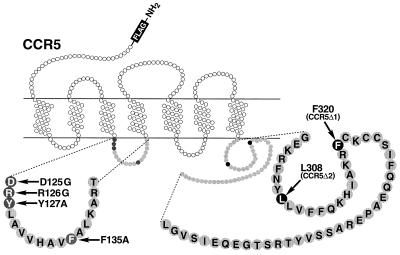
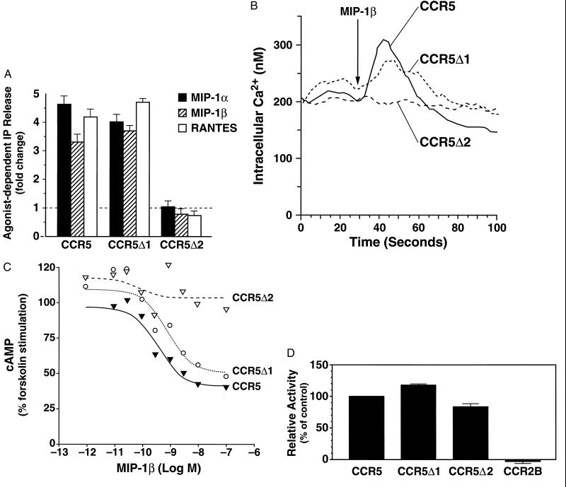
To investigate the importance of signal transduction in the HIV-1 coreceptor activity of chemokine receptors, we created mutations in CCR5 designed to greatly impair or eliminate its ability to couple to heterotrimeric G proteins. The first two mutations truncated the carboxyl-terminal tail of CCR5 from 52 amino acids in the wild-type receptor (19) to either 20 (CCR5Δ1) or eight amino acids (CCR5Δ2) (Fig. 1). Agonist-dependent signaling by the two constructs was assessed in transiently transfected HEK-293T cells. As shown in Fig. 2A, chemokine-dependent phosphatidyl inositol turnover by CCR5Δ1 was similar to that of wild-type CCR5, whereas CCR5Δ2 failed to induce detectable inositol phosphate hydrolysis. Consistent with these data, agonist-dependent increases in intracellular calcium were observed in cells transfected with CCR5 and CCR5Δ1 but not those transfected with CCR5Δ2 (Fig. 2B). As a third measure of signaling, the ability of each truncation mutant to inhibit adenylyl cyclase was examined. As shown in Fig. 2C, CCR5Δ1, but not CCR5Δ2, mediated potent, dose-dependent cyclase inhibition. Thus, truncation of the CCR5 carboxyl-terminal tail to <20 amino acids severely impaired the receptor’s ability to initiate signaling.
Figure 1.
CCR5 truncations and mutations. Wild-type CCR5 was truncated by introducing stop codons after Phe-320 (F320, CCR5Δ1) or Leu-308 (L308, CCR5Δ2). Mutations of the indicated amino acids in the second intracellular loop were introduced by PCR to functionally uncouple the receptor.
Figure 2.
Signal transduction and HIV-1 coreceptor activity mediated by CCR5 and CCR5 truncation mutants. HEK-293T cells were transiently transfected with CCR5, CCR5Δ1, and CCR5Δ2. (A) Inositol phosphate (IP) release. A fold increase of 1.0 corresponds to no activation. Shown is one of three similar experiments. (B) Calcium mobilization in response to MIP-1β (100 nM). Transiently transfected cells were loaded with indo-1AM, and intracellular calcium levels were measured as described (24). (C) Inhibition of adenylyl cyclase induced by MIP-1β. Transiently transfected cells were incubated with forskolin in the presence of the indicated concentrations of MIP-1β, and cAMP levels were determined as described (24). (D) The HIV-1 coreceptor activity of each truncation mutant was calculated as a percentage of the activity of each wild-type CCR5. The activity of CCR5 was defined as 100%. HIV-1 coreceptor activity was determined as described (18) using Ba-L. HEK-293T cells were cotransfected with human CD4 and the indicated chemokine receptor. CCR2 was included as a negative control, and the coreceptor activity of wild-type CCR5 was set at 100%. (Bars = SEM; n = 4.)
However, for entry of the CCR-dependent HIV strain Ba-L into cells, CCR5Δ2 was fully as active as wild-type CCR5 (Fig. 2D). These data suggested that neither an intact carboxyl-terminal tail nor the ability to activate classic second messengers was required for CCR5 to facilitate internalization of HIV-1.
Mutations in the Second Cytoplasmic Loop.
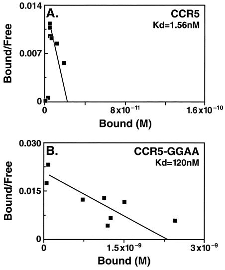
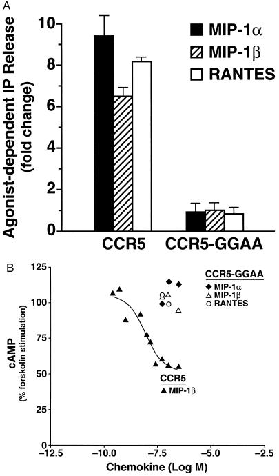
A second line of evidence that signaling by CCR5 was not required for HIV-1 coreceptor activity was sought by mutating CCR5 within the highly conserved aspartate-arginine-tyrosine (or DRY) sequence, which represents a motif found at the end of the third transmembrane domain of many seven-transmembrane domain receptors, and is thought to be critical for G protein coupling. In an attempt to uncouple CCR5 from both Gαi and Gαq, we mutated Asp-125 to glycine (D125G), Arg-126 to glycine (R126G), Tyr-127 to alanine (Y127A), and Phe-135 to alanine (F135A). The resulting receptor, designated CCR5-GGAA, bound macrophage inflammatory protein 1β (MIP-1β) with markedly lower affinity than wild-type CCR5 (Fig. 3). Similar results were obtained with MIP-1α (data not shown). Addition of CCR5 ligands to CCR5-GGAA-transfected cells failed to induce detectable phosphatidyl inositol turnover or inhibit adenylyl cyclase, even when the chemokines were present at concentrations approximating or exceeding the dissociation constant for binding (Fig. 4 A and B). Agonist-dependent increases in intracellular calcium were also not detected in response to the appropriate chemokines (data not shown). These results are consistent with a failure of CCR5-GGAA to couple to G proteins.
Figure 3.
Binding of MIP-1β to cells transfected with CCR5 and CCR5-GGAA. HEK-293T cells were transiently transfected with CCR5 (A) or CCR5-GGAA (B) and incubated with 125I-labeled MIP-1β (5 nM, CCR5; 50 nM, CCR5-GGAA), as described (19). Shown is the Scatchard analysis of competition binding experiments with unlabeled MIP-1β. Similar results were obtained with radiolabeled MIP-1α. One of three similar experiments is presented.
Figure 4.
Signal transduction mediated by CCR5 and CCR5-GGAA in transiently transfected HEK-293T cells. (A) Inositol phosphate (IP) release from HEK-293T cells transfected with CCR5 or CCR5-GGAA in response to the indicated chemokines (100 nM). A fold increase of 1.0 corresponds to no activation. Shown is one of three similar experiments. Similar results were seen with up to 600 nM MIP-1α, MIP-1β, and RANTES (regulated on activation, normal T cell expressed and secreted). (B) Inhibition of adenylyl cyclase in transiently transfected cells was determined as described in Fig. 1. The dose-response curve was obtained with wild-type CCR5 and MIP-1β. No response was seen to the indicated concentrations of MIP-1α, MIP-1β, or RANTES in cells transfected with CCR5-GGAA. (Bars = SEM.)
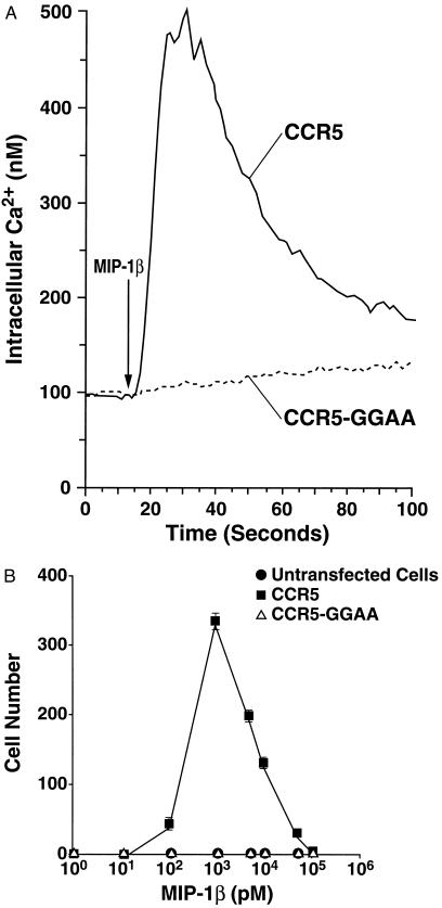
Further evidence that the mutated receptor was functionally uncoupled was obtained using 300-19 cells, a murine pre-B cell line (23). Stably transfected cells had similar surface expression of CCR5 and CCR5-GGAA, but the mutated receptor failed to flux calcium in response to MIP-1β (Fig. 5A) or other chemokines (data not shown). Chemotaxis is the prototypic function of the chemokines, and thus represents a biologically relevant functional assay of receptor activation. CCR5-transfected 300-19 cells exhibited a classic, biphasic chemotactic response upon incubation with increasing concentrations of MIP-1β (Fig. 5B). In contrast, CCR5-GGAA-transfected cells failed to migrate in response to MIP-1β or other chemokines.
Figure 5.
CCR5-GGAA failed to mobilize intracellular calcium or induce chemotaxis in 300–19 pre-B cells stably transfected with CCR5 or CCR5-GGAA as described. (A) Calcium mobilization in response to MIP-1β (100 nM) was measured as described in Fig. 2. (B) Chemotaxis was determined as described.
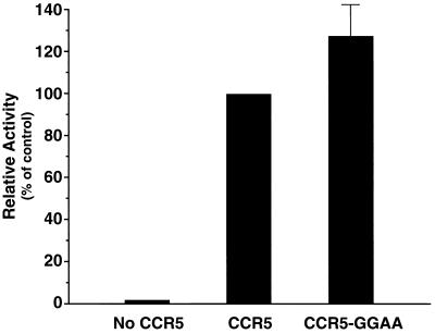
Despite the failure to induce chemotaxis or generate second messengers, CCR5-GGAA remained an excellent coreceptor for HIV-1 internalization (Fig. 6), consistent with the hypothesis that signal transduction is not a component of this mechanism.
Figure 6.
HIV-1 coreceptor activity of CCR5 and CCR5-GGAA in HEK-293T cells transfected with CD4 and either CCR5 or CCR5-GGAA, as described in Fig. 2. HIV-1 coreceptor activity was determined for Ba-L. CCR5 activity was set at 100%. (Bars = SEM, n = 3.)
DISCUSSION
In this study, we mutated two intracellular regions of CCR5 to determine if signal transduction is a necessary component of the mechanism by which this receptor acts in concert with CD4 to facilitate internalization of macrophage-tropic strains of HIV-1. Several lines of evidence support the conclusion that the coreceptor activity of CCR5 is not dependent upon its ability to initiate signaling through known G protein-dependent pathways. First, truncation of the carboxyl-terminal tail yielded a receptor that failed to induce hydrolysis of inositol phosphate, inhibit adenylyl cyclase, or mobilize intracellular calcium, but had no adverse effect on HIV-1 coreceptor activity. Second, mutation of the highly conserved aspartate-arginine-tyrosine (or DRY) sequence at the end of the third transmembrane domain prevented coupling to Gαi, but not coreceptor activity; this mutation also failed to support agonist-dependent intracellular calcium mobilization and chemotaxis when stably expressed in a lymphocyte-like cell line, confirming that the receptor was functionally uncoupled. Finally, we (data not shown) and others (26) have found that pretreatment with pertussis toxin, which completely blocks chemotaxis, has no effect on CCR5 coreceptor activity. We conclude from these data that agonist-dependent signaling, as represented by prototypic activation of G proteins and downstream pathways, is not required for CCR5 to act as an HIV-1 coreceptor.
We recently showed that a chimeric receptor in which the amino-terminal extracellular domain of CCR5 was substituted for the corresponding portion of CCR2 functioned well as an HIV-1 coreceptor, but failed to signal in response to known CCR2 or CCR5 ligands (18). These data raised the possibility that G protein coupling and signaling by CCR5 were not critical to its function as an HIV-1 coreceptor and were consistent with reports that pretreatment of cells with pertussis toxin did not block the ability of CCR5 and CXCR4 to facilitate HIV-1 internalization (26). Although pertussis toxin selectively blocks the ability of G protein-coupled receptors to activate Gαi, we and others have shown that chemokine receptors couple to members of the Gαq family in addition to Gαi (16, 17). It was thus possible that pertussis toxin–insensitive G proteins were used for coreceptor activity. We therefore sought to disrupt CCR5 coupling to G proteins by mutating critical cytoplasmic regions of the receptor. Truncation of the carboxyl-terminal tail of the angiotensin II receptor 1 impairs coupling to Gαi, but leaves Gαq-mediated responses completely intact (27). Uncoupling from both Gαi and Gαq has been achieved in the angiotensin II receptor 1, β- and α-adrenergic, and muscarinic m1 receptors by mutation of the highly conserved DRYXXV(I)XXPL sequence at the end of the third transmembrane domain (27–30). In the case of CCR5, signal transduction was effectively abolished either by truncating the carboxyl tail to eight amino acids or by mutating the aspartate-arginine-tyrosine (or DRY), sequence to GGALAVVHAVA (CCR5-GGAA). The reduced affinity of chemokine binding to CCR5-GGAA was also consistent with uncoupling of the receptor from G proteins (31). That CCR5-GGAA remained fully active as an HIV-1 coreceptor provided strong evidence that the ability to couple to G proteins and activate classic second messengers is noncontributory to HIV entry competence.
Agonist-dependent phosphorylation of carboxyl-terminal tail serine and threonine residues induces desensitization and internalization of G protein-coupled receptors through the binding of arrestins (reviewed in ref. 32). Neither serines nor threonines were retained in the two truncation mutants of CCR5 (CCR5Δ1 and CCR5Δ2) in the present study. Nevertheless, these mutants exhibited robust coreceptor activity for HIV-1. These observations demonstrate that phosphorylation of carboxyl-terminal tail serine and threonine residues is dispensable for viral entry and suggest that the mechanism of entry of the virus is distinct from the normal agonist-dependent pathways of receptor internalization. This conclusion is particularly intriguing because the same conclusion was reached with regard to the CD4 molecule (7, 8). The molecular basis of entry thus remains unknown.
The most thoroughly characterized function of chemokines is to initiate directed migration, or chemotaxis, of leukocytes. Since the signaling pathways leading to chemotaxis are not yet well understood, it was important to directly measure the ability of CCR5-GGAA to mediate this downstream, functional end point of receptor activation. The failure of cells expressing CCR5-GGAA to migrate provided further evidence that the mutated receptor was functionally uncoupled and is also the most direct evidence to date that G protein coupling is essential for chemotaxis. These data provide strong evidence that CCR5 acts through a novel mechanism, unrelated to G protein activation, in promoting the internalization of HIV-1.
Recent reports by Wu et al. (12) and Trkola et al. (11) have demonstrated that in the context of CD4, gp120 binds to CCR5 with nanomolar affinity. Considered together with studies from our laboratories (18) and others (11, 12), these data suggest a model in which multiple extracellular domains of CCR5 interact with gp120 to capture the virus. The molecular details of how the virus is subsequently internalized are completely unknown, but the results presented here indicate that activation of CCR5 is not a necessary component of this mechanism. Elucidation of the mechanism of viral entry may reveal important new functional paradigms for seven-transmembrane domain receptors.
Acknowledgments
We thank Dr. Robert E. Pitas for reading the manuscript, Angela Chen for manuscript preparation, John Carroll, Brian Clark, and Amy Corder for preparation of the figures, and Gary Howard and Stephen Ordway for editorial assistance. This work was funded in part by National Institutes of Health HL52773 (I.F.C.) and by Pfizer, Inc. (M.A.G.).
ABBREVIATIONS
- CCR2 and CCR5
C-C chemokine receptors 2 and 5
- HEK
human embryonic kidney
- MIP-1α and MIP-1β
macrophage inflammatory proteins 1α and 1β
References
- 1.Feng Y, Broder C C, Kennedy P E, Berger E A. Science. 1996;272:872–877. [Google Scholar]
- 2.Alkhatib G, Combadiere C, Broder C C, Feng Y, Kennedy P E, Murphy P M, Berger E A. Science. 1996;272:1955–1958. doi: 10.1126/science.272.5270.1955. [DOI] [PubMed] [Google Scholar]
- 3.Choe H, Farzan M, Sun Y, Sullivan N, Rollins B, Ponath P D, Wu L, Mackay C R, LaRosa G, Newman W, Gerard N, Gerard C, Sodroski J. Cell. 1996;85:1135–1148. doi: 10.1016/s0092-8674(00)81313-6. [DOI] [PubMed] [Google Scholar]
- 4.Deng H, Liu R, Ellmeier W, Choe S, Unutmaz D, Burkhart M, Di Marzio P, Marmon S, Sutton R E, Hill C M, Davis C B, Peiper S C, Schall T J, Littman D R, Landau N R. Nature (London) 1996;381:661–666. doi: 10.1038/381661a0. [DOI] [PubMed] [Google Scholar]
- 5.Dragic T, Litwin V, Allaway G P, Martin S R, Huang Y, Nagashima K A, Cayanan C, Maddon P J, Koup R A, Moore J P, Paxton W A. Nature (London) 1996;381:667–673. doi: 10.1038/381667a0. [DOI] [PubMed] [Google Scholar]
- 6.Doranz B J, Rucker J, Yi Y, Smyth R J, Samson M, Peiper S C, Parmentier M, Collman R G, Doms R W. Cell. 1996;85:1149–1158. doi: 10.1016/s0092-8674(00)81314-8. [DOI] [PubMed] [Google Scholar]
- 7.Maddon P J, McDougal J S, Clapham P R, Dalgleish A G, Jamal S, Weiss R A, Axel R. Cell. 1988;54:865–874. doi: 10.1016/s0092-8674(88)91241-x. [DOI] [PubMed] [Google Scholar]
- 8.Bedinger P, Moriarty A, von Borstel R C, II, Donovan N J, Steimer K S, Littman D R. Nature (London) 1988;334:162–165. doi: 10.1038/334162a0. [DOI] [PubMed] [Google Scholar]
- 9.Endres M J, Clapham P R, Marsh M, Ahuja M, Turner J D, McKnight A, Thomas J F, Stoebenau-Haggarty B, Choe S, Vance P J, Wells T N C, Power C A, Sutterwala S S, Doms R W, Landau N R, Hoxie J A. Cell. 1996;87:745–756. doi: 10.1016/s0092-8674(00)81393-8. [DOI] [PubMed] [Google Scholar]
- 10.Lapham C K, Ouyang J, Chandrasekhar B, Nguyen N Y, Dimitrov D S, Golding H. Science. 1996;274:602–605. doi: 10.1126/science.274.5287.602. [DOI] [PubMed] [Google Scholar]
- 11.Trkola A, Dragic T, Arthos J, Binley J M, Olson W C, Allaway G P, Cheng-Mayer C, Robinson J, Maddon P J, Moore J P. Nature (London) 1996;384:184–187. doi: 10.1038/384184a0. [DOI] [PubMed] [Google Scholar]
- 12.Wu L, Gerard N P, Wyatt R, Choe H, Parolin C, Ruffing N, Borsetti A, Cardoso A A, Desjardin E, Newman W, Gerard C, Sodroski J. Nature (London) 1996;384:179–183. doi: 10.1038/384179a0. [DOI] [PubMed] [Google Scholar]
- 13.Murphy P M. Annu Rev Immunol. 1994;12:593–633. doi: 10.1146/annurev.iy.12.040194.003113. [DOI] [PubMed] [Google Scholar]
- 14.Schall T J. In: The Cytokine Handbook. Thomson A W, editor. London: Academic; 1994. pp. 419–460. [Google Scholar]
- 15.Gerard C, Gerard N P. Annu Rev Immunol. 1994;12:775–808. doi: 10.1146/annurev.iy.12.040194.004015. [DOI] [PubMed] [Google Scholar]
- 16.Kuang Y, Wu Y, Jiang H, Wu D. J Biol Chem. 1996;271:3975–3978. doi: 10.1074/jbc.271.8.3975. [DOI] [PubMed] [Google Scholar]
- 17.Arai H, Charo I F. J Biol Chem. 1996;271:21814–21819. doi: 10.1074/jbc.271.36.21814. [DOI] [PubMed] [Google Scholar]
- 18.Atchison R E, Gosling J, Monteclaro F S, Franci C, Digilio L, Charo I F, Goldsmith M A. Science. 1996;274:1924–1926. doi: 10.1126/science.274.5294.1924. [DOI] [PubMed] [Google Scholar]
- 19.Raport C J, Gosling J, Schweickart V L, Gray P W, Charo I F. J Biol Chem. 1996;271:17161–17166. doi: 10.1074/jbc.271.29.17161. [DOI] [PubMed] [Google Scholar]
- 20.Andersson S, Davis D L, Dahlbäck H, Jörnvall H, Russell D W. J Biol Chem. 1989;264:8222–8229. [PubMed] [Google Scholar]
- 21.Picard V, Ersdal-Badju E, Lu A, Bock S C. Nucleic Acids Res. 1994;22:2587–2591. doi: 10.1093/nar/22.13.2587. [DOI] [PMC free article] [PubMed] [Google Scholar]
- 22.Pear W S, Nolan G P, Scott M L, Baltimore D. Proc Natl Acad Sci USA. 1993;90:8392–8396. doi: 10.1073/pnas.90.18.8392. [DOI] [PMC free article] [PubMed] [Google Scholar]
- 23.Reth M G, Ammirati P, Jackson S, Alt F W. Nature (London) 1985;317:353–355. doi: 10.1038/317353a0. [DOI] [PubMed] [Google Scholar]
- 24.Myers S J, Wong L M, Charo I F. J Biol Chem. 1995;270:5786–5792. doi: 10.1074/jbc.270.11.5786. [DOI] [PubMed] [Google Scholar]
- 25.Campbell J J, Qin S, Bacon K B, Mackay C R, Butcher E C. J Cell Biol. 1996;134:255–266. doi: 10.1083/jcb.134.1.255. [DOI] [PMC free article] [PubMed] [Google Scholar]
- 26.Cocchi F, DeVico A L, Garzino-Demo A, Cara A, Gallo R C, Lusso P. Nat Med. 1996;2:1244–1247. doi: 10.1038/nm1196-1244. [DOI] [PubMed] [Google Scholar]
- 27.Shibata T, Suzuki C, Ohnishi J, Murakami K, Miyazaki H. Biochem Biophys Res Commun. 1996;218:383–389. doi: 10.1006/bbrc.1996.0067. [DOI] [PubMed] [Google Scholar]
- 28.Dixon R A F, Sigal I S, Strader C D. Cold Spring Harbor Symp Quant Biol. 1988;53:487–497. doi: 10.1101/sqb.1988.053.01.056. [DOI] [PubMed] [Google Scholar]
- 29.Fraser C M, Wang C-D, Robinson D A, Gocayne J D, Venter J C. Mol Pharmacol. 1989;36:840–847. [PubMed] [Google Scholar]
- 30.Fraser C M, Chung F-Z, Wang C-D, Venter J C. Proc Natl Acad Sci USA. 1988;85:5478–5482. doi: 10.1073/pnas.85.15.5478. [DOI] [PMC free article] [PubMed] [Google Scholar]
- 31.Didsbury J R, Uhing R J, Tomhave E, Gerard C, Gerard N, Snyderman R. FEBS Lett. 1992;297:275–279. doi: 10.1016/0014-5793(92)80555-u. [DOI] [PubMed] [Google Scholar]
- 32.Hausdorff W P, Caron M G, Lefkowitz R J. FASEB J. 1990;4:2881–2889. [PubMed] [Google Scholar]


