Abstract
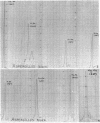
In Aspergillus fumigatus, X-ray microanalysis applied in wavelength dispersive spectroscopy enabled us to detect the presence of calcium, potassium, sulfur, phosphorus, and magnesium; and in A. niger we detected the presence of calcium, potassium, chloride, sulfur, phosphorus, sodium, and magnesium. These various elements were identified by their K α lines.
Full text
PDF


Images in this article
Selected References
These references are in PubMed. This may not be the complete list of references from this article.
- BOYDE A., SWITSUR V. R., FEARNHEAD R. W. Application of the scanning electron-probe x-ray microanalyser to dental tissues. J Ultrastruct Res. 1961 Jun;5:201–207. doi: 10.1016/s0022-5320(61)90015-6. [DOI] [PubMed] [Google Scholar]
- Berry J. P. Néphrocalcinose expérimentale par injection de parathrmone. Etude au microanalyseur à sonde électronique. Nephron. 1970;7(2):97–116. doi: 10.1159/000179813. [DOI] [PubMed] [Google Scholar]
- Galle P., Berry J. P. Cytochimie élémentaire ultrastructurale sur coupes ultrafines de poumon. Poumon Coeur. 1969;25(3):307–317. [PubMed] [Google Scholar]


