

Abstract
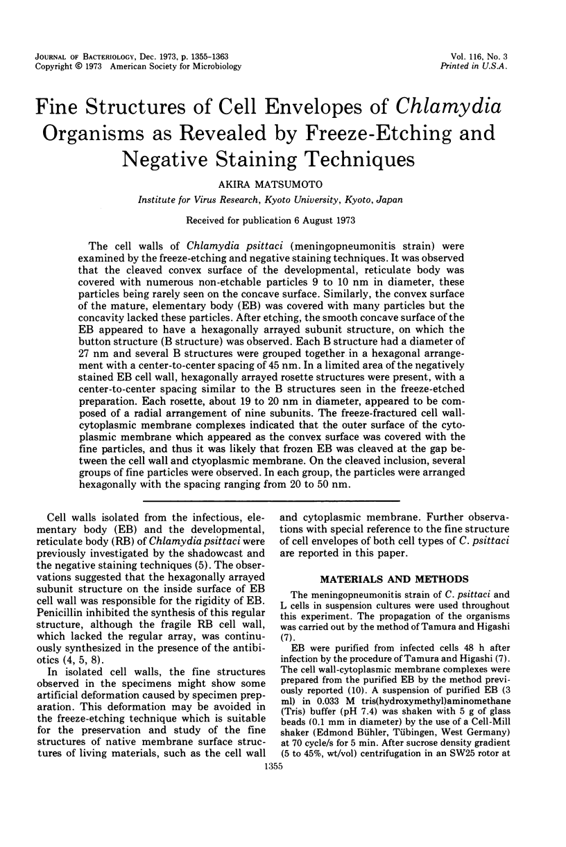
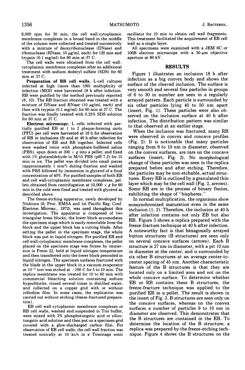
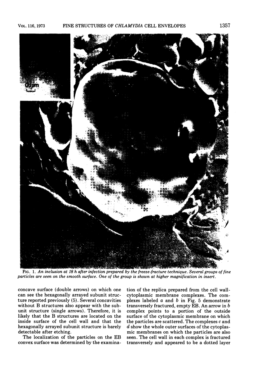
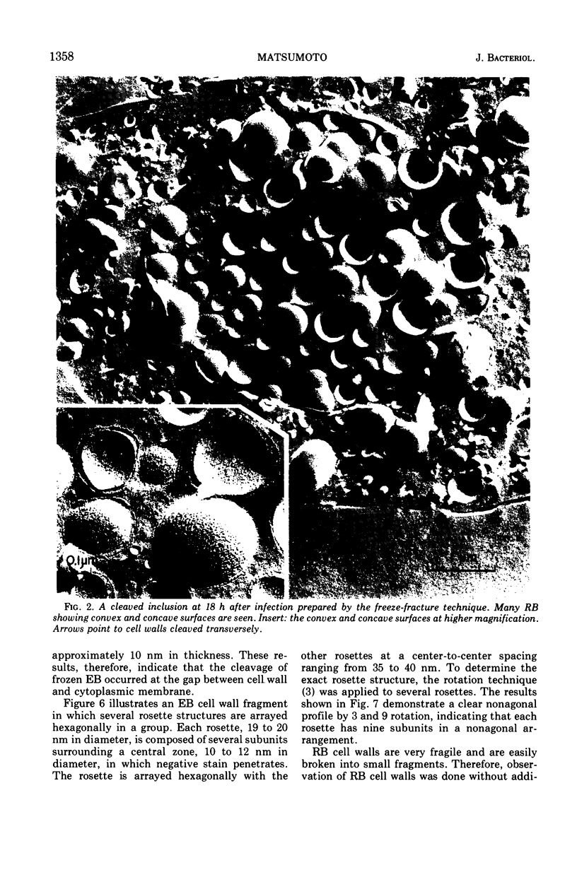
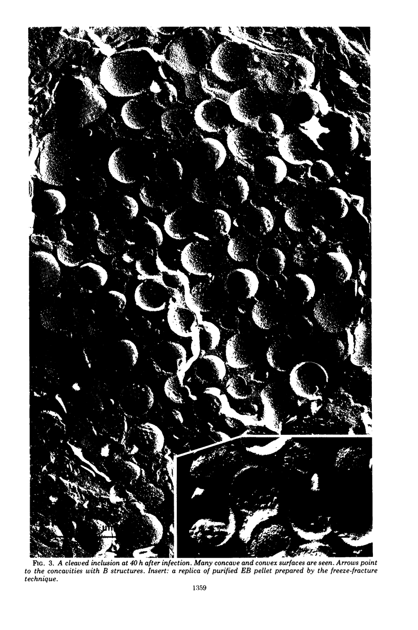
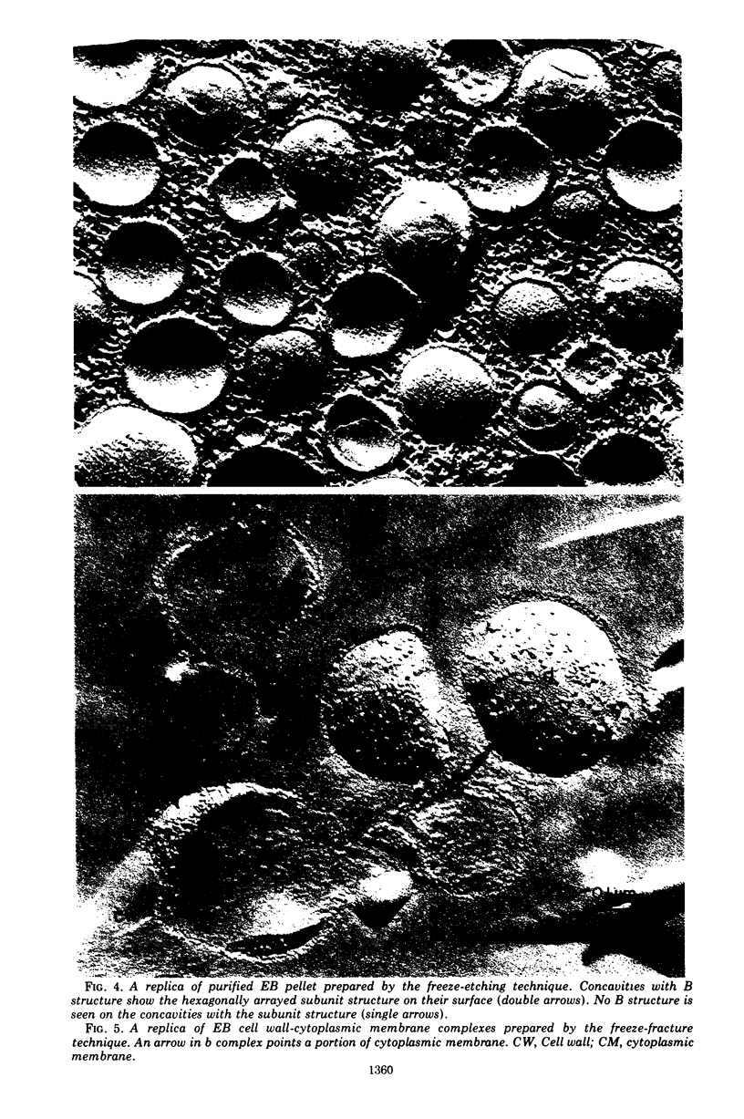
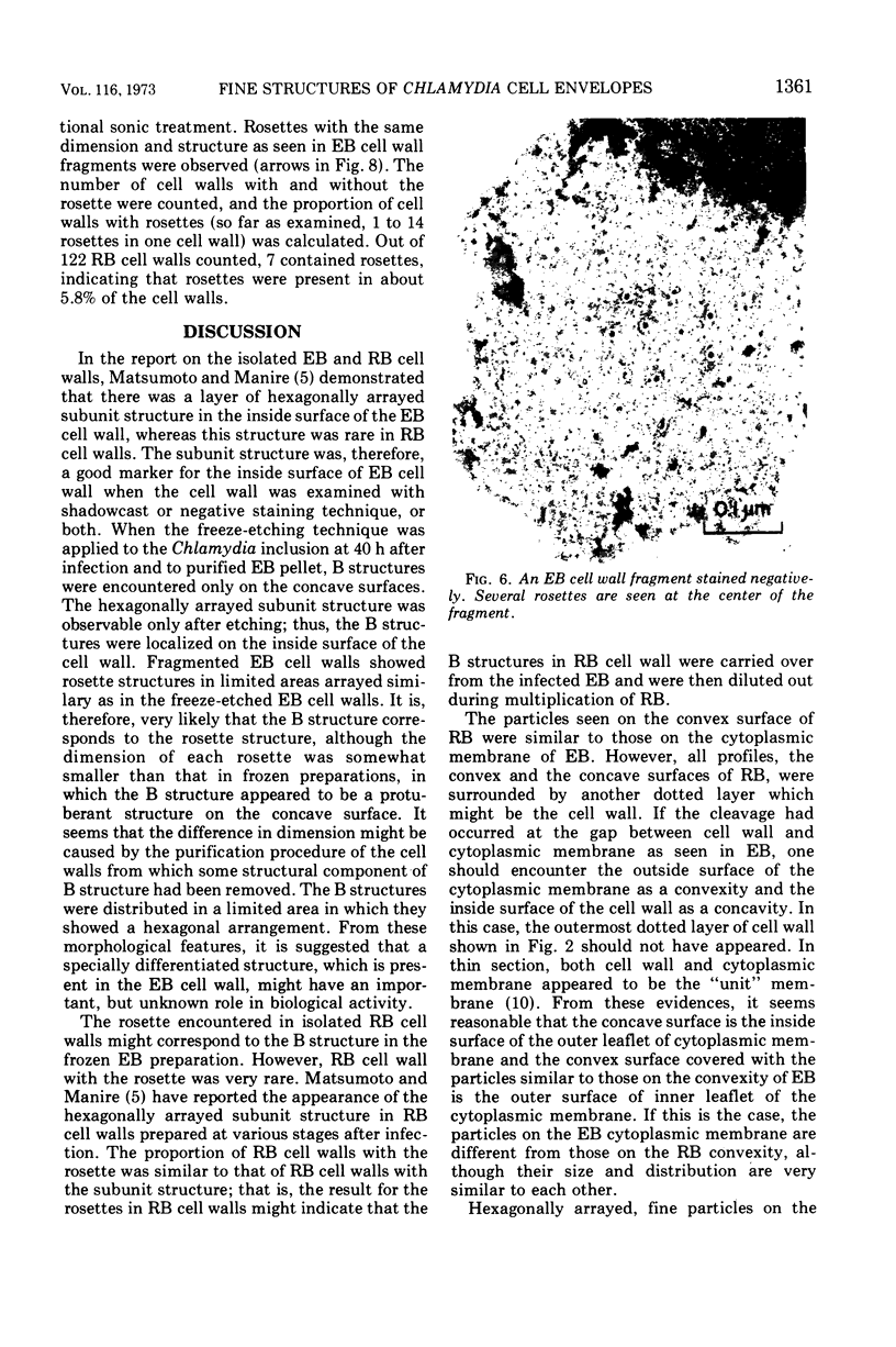
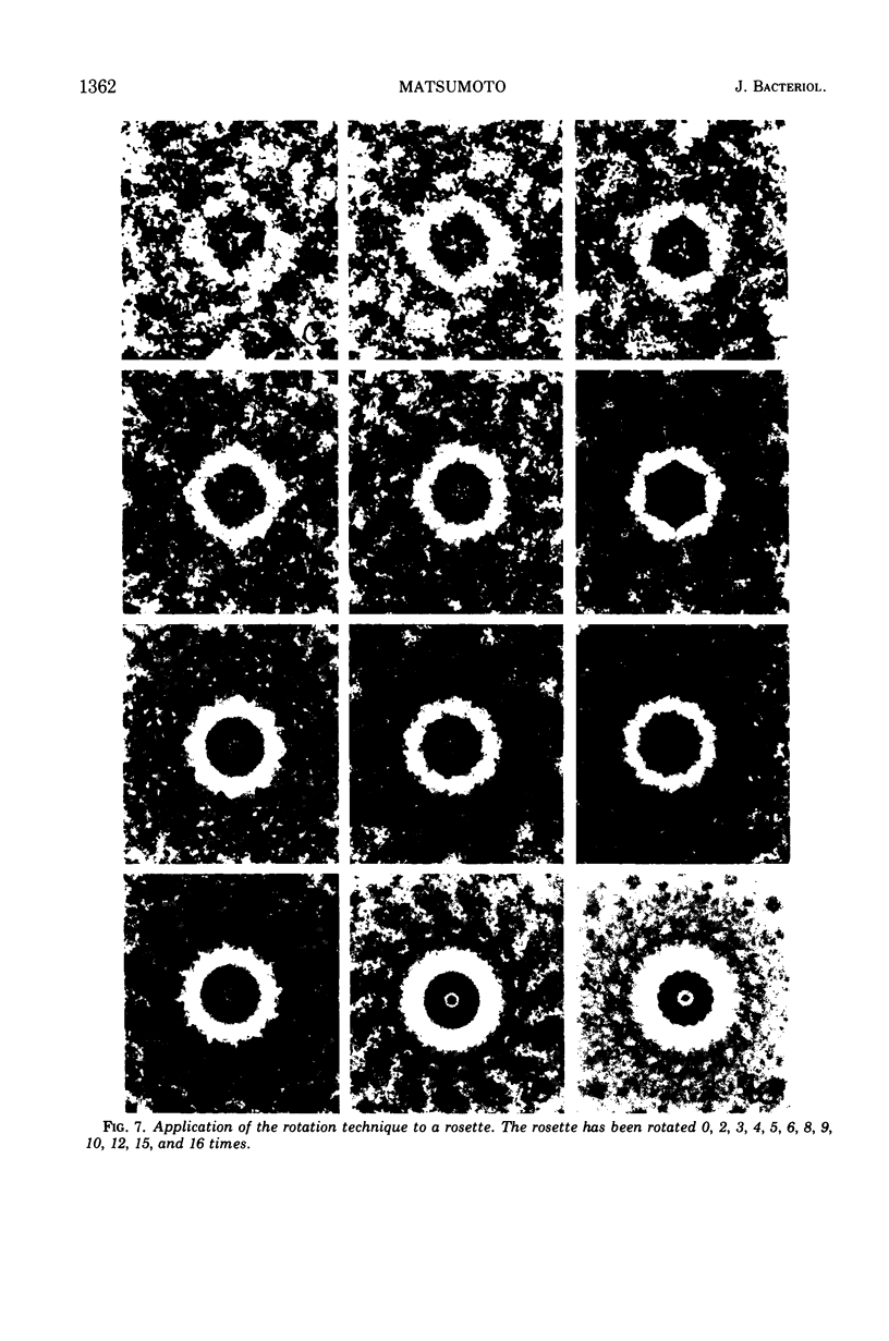
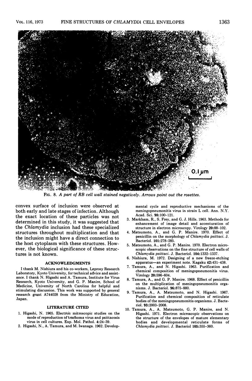
The cell walls of Chlamydia psittaci (meningopneumonitis strain) were examined by the freeze-etching and negative staining techniques. It was observed that the cleaved convex surface of the developmental, reticulate body was covered with numerous non-etchable particles 9 to 10 nm in diameter, these particles being rarely seen on the concave surface. Similarly, the convex surface of the mature, elementary body (EB) was covered with many particles but the concavity lacked these particles. After etching, the smooth concave surface of the EB appeared to have a hexagonally arrayed subunit structure, on which the button structure (B structure) was observed. Each B structure had a diameter of 27 nm and several B structures were grouped together in a hexagonal arrangement with a center-to-center spacing of 45 nm. In a limited area of the negatively stained EB cell wall, hexagonally arrayed rosette structures were present, with a center-to-center spacing similar to the B structures seen in the freeze-etched preparation. Each rosette, about 19 to 20 nm in diameter, appeared to be composed of a radial arrangement of nine subunits. The freeze-fractured cell wall-cytoplasmic membrane complexes indicated that the outer surface of the cytoplasmic membrane which appeared as the convex surface was covered with the fine particles, and thus it was likely that frozen EB was cleaved at the gap between the cell wall and ctyoplasmic membrane. On the cleaved inclusion, several groups of fine particles were observed. In each group, the particles were arranged hexagonally with the spacing ranging from 20 to 50 nm.
Full text
PDF
Images in this article
Selected References
These references are in PubMed. This may not be the complete list of references from this article.
- HIGASHI N. ELECTRON MICROSCOPIC STUDIES ON THE MODE OF REPRODUCTION OF TRACHOMA VIRUS AND PSITTACOSIS VIRUS IN CELL CULTURES. Exp Mol Pathol. 1965 Feb;76:24–39. doi: 10.1016/0014-4800(65)90021-3. [DOI] [PubMed] [Google Scholar]
- HIGASHI N., TAMURA A., IWANAGA M. Developmental cycle and reproductive mechanism of the meningopneumonitis virus in strain L cells. Ann N Y Acad Sci. 1962 Mar 5;98:100–121. doi: 10.1111/j.1749-6632.1962.tb30536.x. [DOI] [PubMed] [Google Scholar]
- Matsumoto A., Manire G. P. Electron Microscopic Observations on the Fine Structure of Cell Walls of Chlamydia psittaci. J Bacteriol. 1970 Dec;104(3):1332–1337. doi: 10.1128/jb.104.3.1332-1337.1970. [DOI] [PMC free article] [PubMed] [Google Scholar]
- Matsumoto A., Manire G. P. Electron microscopic observations on the effects of penicillin on the morphology of Chlamydia psittaci. J Bacteriol. 1970 Jan;101(1):278–285. doi: 10.1128/jb.101.1.278-285.1970. [DOI] [PMC free article] [PubMed] [Google Scholar]
- TAMURA A., HIGASHI N. PURIFICATION AND CHEMICAL COMPOSITION OF MENINGOPNEUMONITIS VIRUS. Virology. 1963 Aug;20:596–604. doi: 10.1016/0042-6822(63)90284-8. [DOI] [PubMed] [Google Scholar]
- Tamura A., Manire G. P. Effect of penicillin on the multiplication of meningopneumonitis organisms (Chlamydia psittaci). J Bacteriol. 1968 Oct;96(4):875–880. doi: 10.1128/jb.96.4.875-880.1968. [DOI] [PMC free article] [PubMed] [Google Scholar]
- Tamura A., Matsumoto A., Higashi N. Purification and chemical composition of reticulate bodies of the meningopneumonitis organisms. J Bacteriol. 1967 Jun;93(6):2003–2008. doi: 10.1128/jb.93.6.2003-2008.1967. [DOI] [PMC free article] [PubMed] [Google Scholar]
- Tamura A., Matsumoto A., Manire G. P., Higashi N. Electron microscopic observations on the structure of the envelopes of mature elementary bodies and developmental reticulate forms of Chlamydia psittaci. J Bacteriol. 1971 Jan;105(1):355–360. doi: 10.1128/jb.105.1.355-360.1971. [DOI] [PMC free article] [PubMed] [Google Scholar]