

Abstract
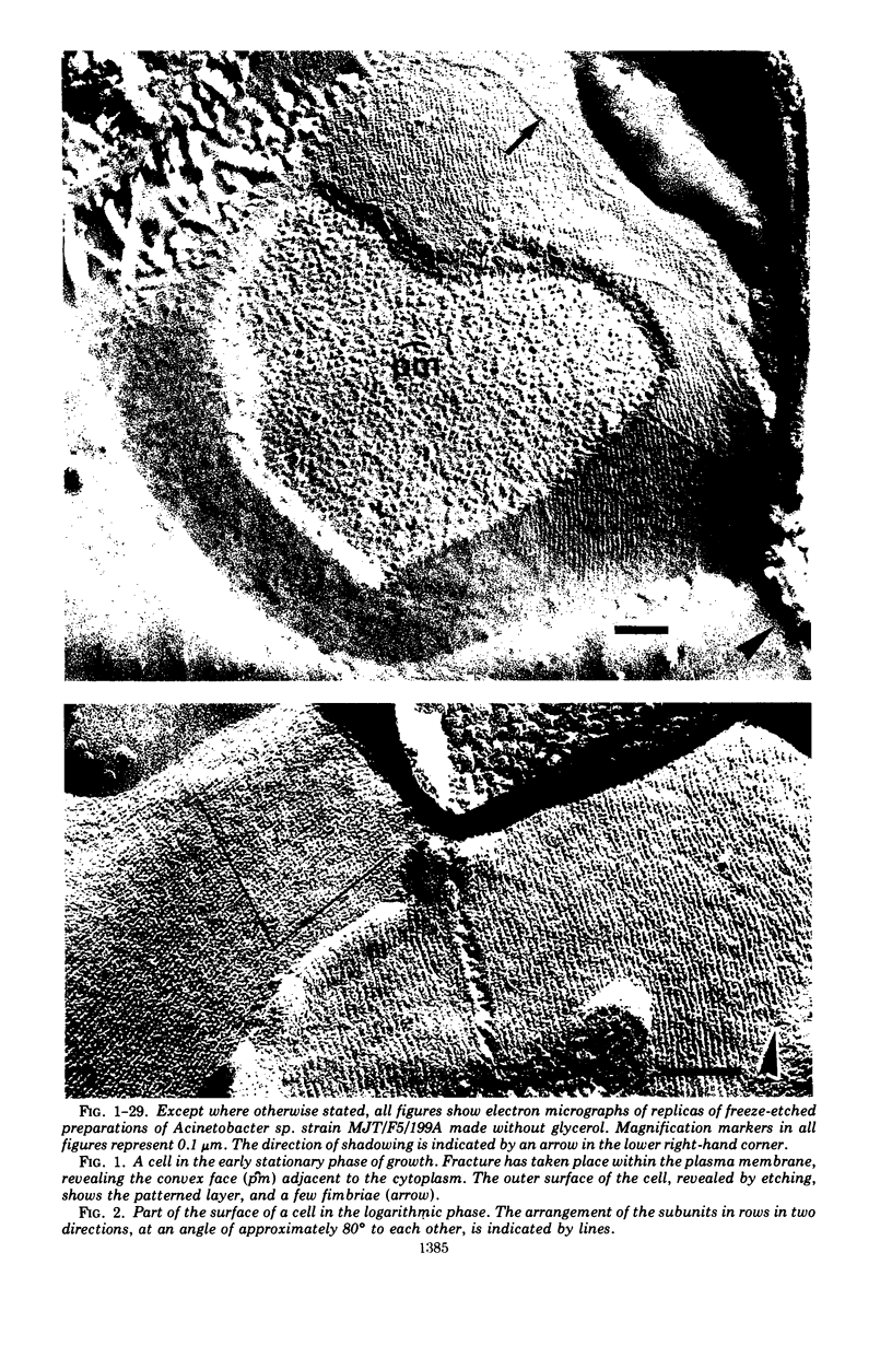
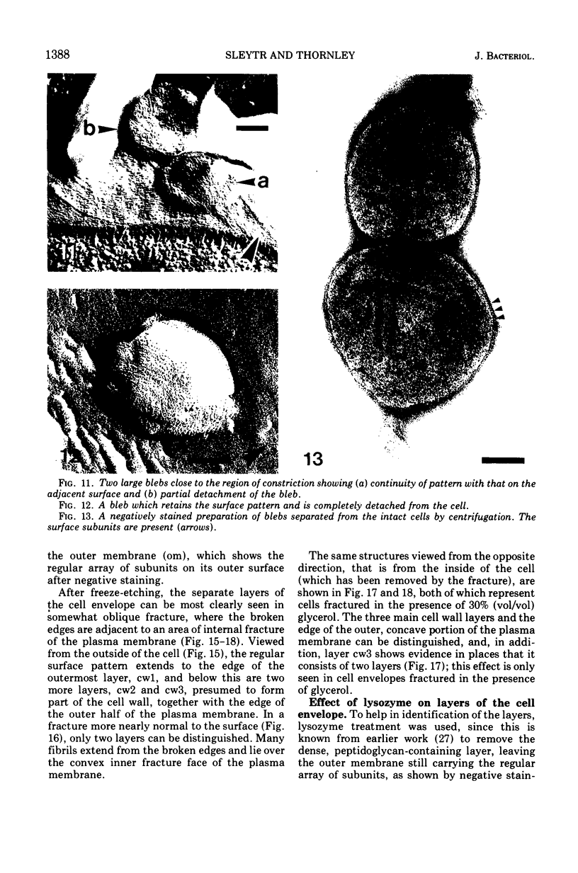
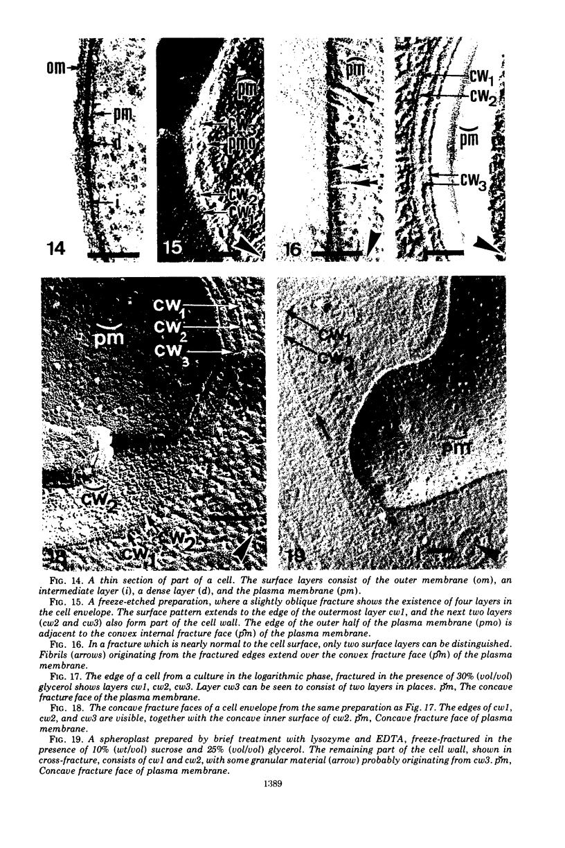
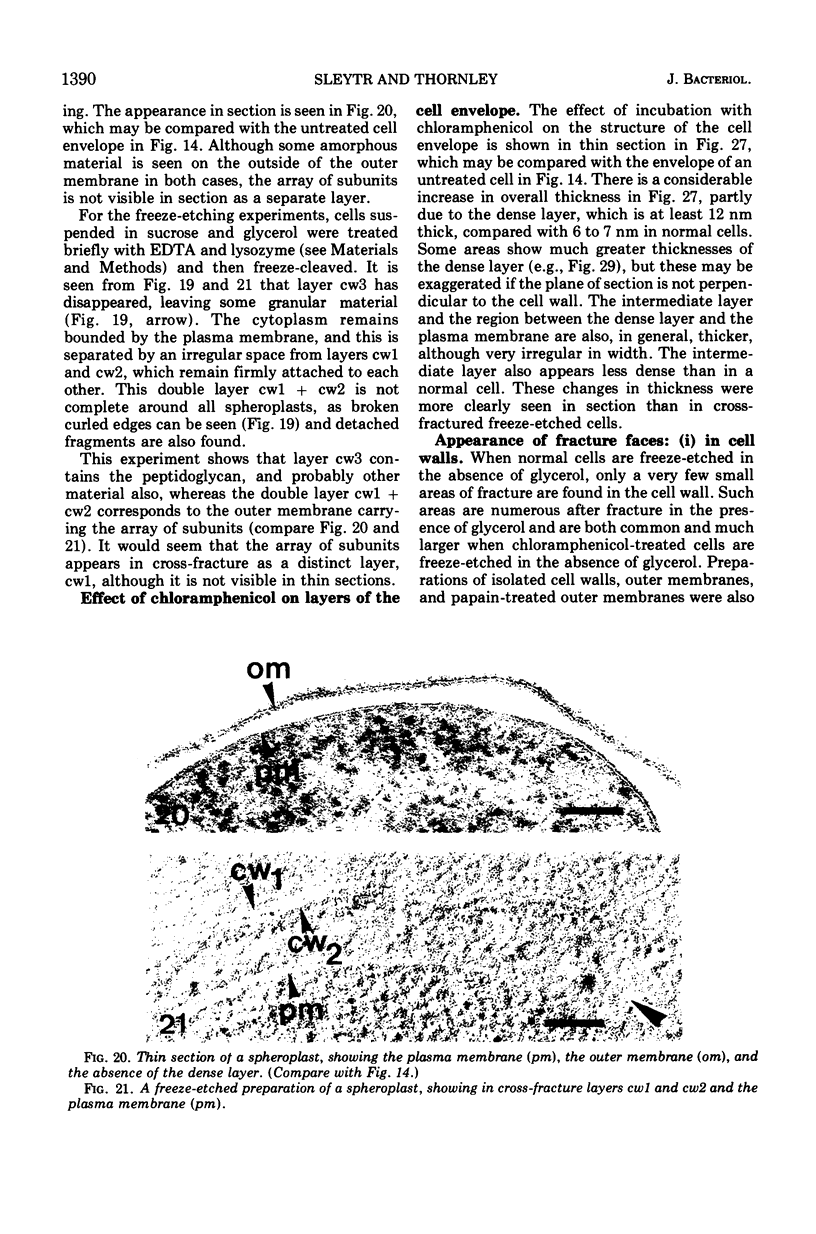
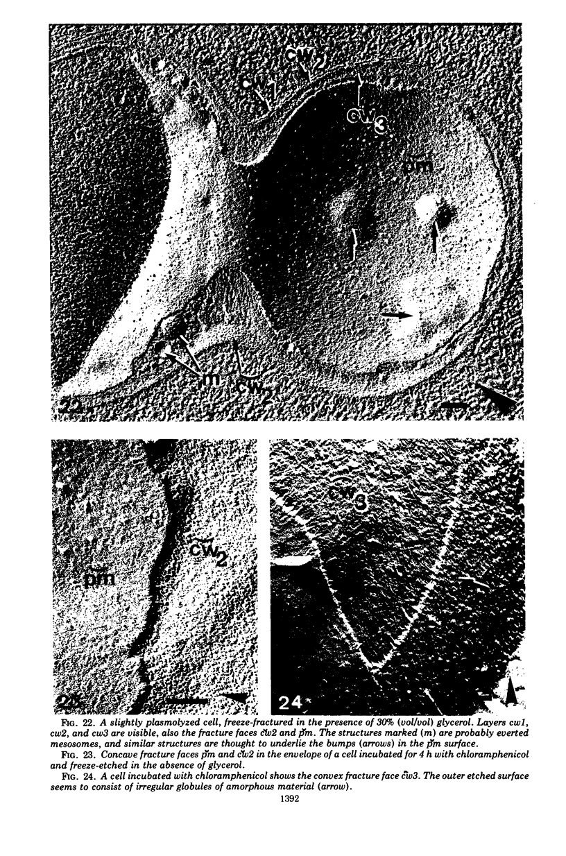
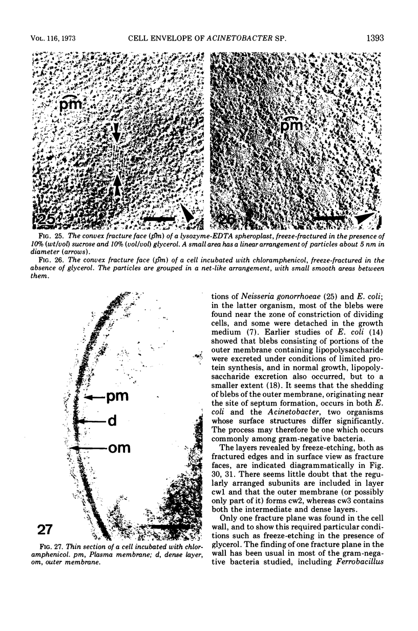
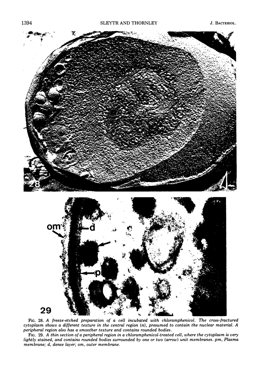
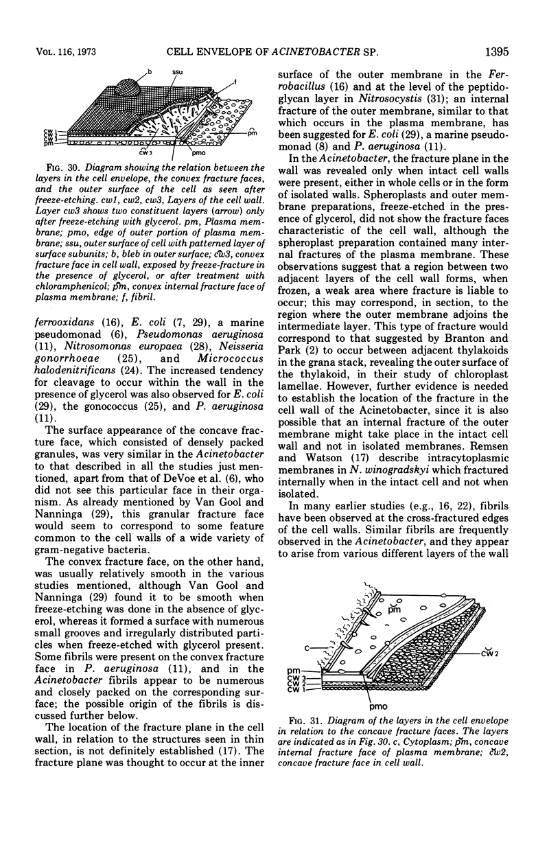
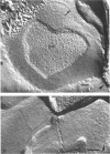
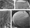
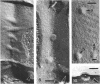
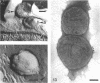
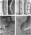
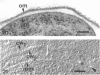
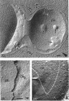
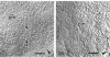
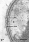
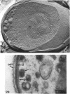
Freeze-etching was applied to preparations, with and without glycerol, of Acinetobacter sp. strain MJT/F5/199A, consisting of intact cells after normal growth or after incubation with chloramphenicol, spheroplasts, and isolated cell walls and outer membranes. Etched preparations show that a regular array of subunits forms the surface of normal cells. Near the zones of constriction in dividing cells, blebs and irregularities are seen, and some blebs, consisting of both surface subunits and outer membrane, are released from the cells. The cross-fractured cell envelope shows four layers which are related to the structures seen in section as follows: cw1, which is not visible in section, contains the surface subunits; cw2 consists of all or part of the outer membrane; cw3 includes the intermediate and dense, peptidoglycan-containing layers; within these cell wall layers is the plasma membrane. Internal fracture of the plasma membrane occurs under all conditions tested, but the fracture plane in the cell wall is only revealed in chloramphenicol-treated cells or normal cells freeze-fractured with glycerol present; the characteristic fracture faces are not seen in spheroplasts or isolated outer membranes. The concave fracture face c̆w2 consists of densely packed granules, while the convex face c̆w3 is fibrillar. The probable location of this fracture plane is discussed. After incubation with chloramphenicol, the outer surface of the cells is obscured by extracellular material, the dense peptidoglycan-containing layer is increased in thickness, and the cytoplasm contains rounded bodies bounded by one or more unit membranes.
Full text
PDF

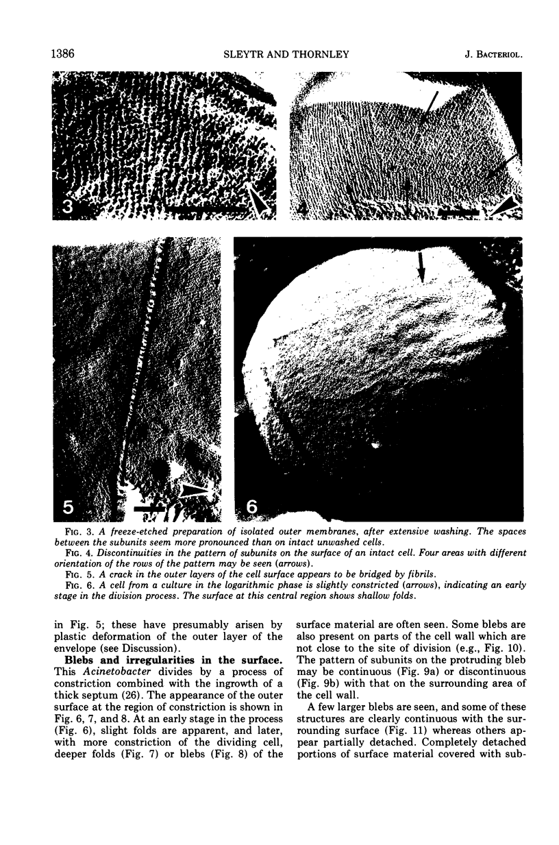
Images in this article
Selected References
These references are in PubMed. This may not be the complete list of references from this article.
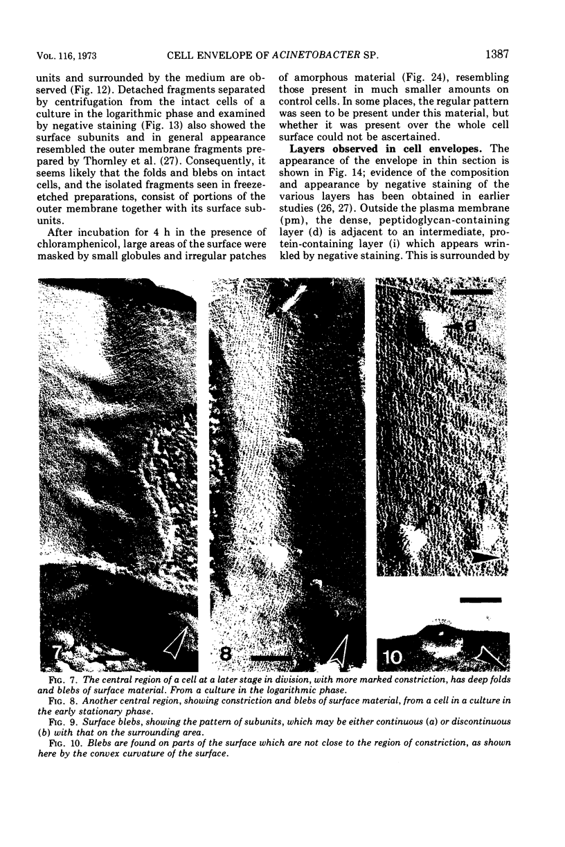
- Bayer M. E., Remsen C. C. Structure of Escherichia coli after freeze-etching. J Bacteriol. 1970 Jan;101(1):304–313. doi: 10.1128/jb.101.1.304-313.1970. [DOI] [PMC free article] [PubMed] [Google Scholar]
- Branton D., Park R. B. Subunits in chloroplast lamellae. J Ultrastruct Res. 1967 Aug;19(3):283–303. doi: 10.1016/s0022-5320(67)80222-3. [DOI] [PubMed] [Google Scholar]
- Bullivant S., Rayns D. G., Bertaud W. S., Chalcroft J. P., Grayston G. F. Freeze-fractured myosin filaments. J Cell Biol. 1972 Nov;55(2):520–524. doi: 10.1083/jcb.55.2.520. [DOI] [PMC free article] [PubMed] [Google Scholar]
- Clark A. W., Branton D. Fracture faces in frozen outer segments from the guinea pig retina. Z Zellforsch Mikrosk Anat. 1968;91(4):586–603. doi: 10.1007/BF00455276. [DOI] [PubMed] [Google Scholar]
- Cole R. M. Symposium on the fine structure and replication of bacteria and their parts. 3. Bacterial cell-wall replication followed by immunofluorescence. Bacteriol Rev. 1965 Sep;29(3):326–344. doi: 10.1128/br.29.3.326-344.1965. [DOI] [PMC free article] [PubMed] [Google Scholar]
- DeVoe I. W., Costerton J. W., MacLeod R. A. Demonstration by freeze-etching of a single cleavage plane in the cell wall of a gram-negative bacterium. J Bacteriol. 1971 May;106(2):659–671. doi: 10.1128/jb.106.2.659-671.1971. [DOI] [PMC free article] [PubMed] [Google Scholar]
- Fiil A., Branton D. Changes in the plasma membrane of Escherichia coli during magnesium starvation. J Bacteriol. 1969 Jun;98(3):1320–1327. doi: 10.1128/jb.98.3.1320-1327.1969. [DOI] [PMC free article] [PubMed] [Google Scholar]
- Forge A., Costerton J. W., Kerr K. A. Freeze-etching and x-ray diffraction of the isolated double-track layer from the cell wall of a gram-negative marine pseudomonad. J Bacteriol. 1973 Jan;113(1):445–451. doi: 10.1128/jb.113.1.445-451.1973. [DOI] [PMC free article] [PubMed] [Google Scholar]
- Frehel C., Beaufils A. M., Ryter A. Etude au microscope électronique de la croissance de la paroi chez B. subtilis et B. megaterium. Ann Inst Pasteur (Paris) 1971 Aug;121(2):139–148. [PubMed] [Google Scholar]
- Giesbrecht P., Ruska H. Uber Veränderungen der Feinstrukturen von Bakterien unter der Einwirkung von Chloramphenicol. Klin Wochenschr. 1968 Jun 1;46(11):575–582. doi: 10.1007/BF01747836. [DOI] [PubMed] [Google Scholar]
- Gilleland H. E., Jr, Stinnett J. D., Roth I. L., Eagon R. G. Freeze-etch study of Pseudomonas aeruginosa: localization within the cell wall of an ethylenediaminetetraacetate-extractable. J Bacteriol. 1973 Jan;113(1):417–432. doi: 10.1128/jb.113.1.417-432.1973. [DOI] [PMC free article] [PubMed] [Google Scholar]
- Knox K. W., Vesk M., Work E. Relation between excreted lipopolysaccharide complexes and surface structures of a lysine-limited culture of Escherichia coli. J Bacteriol. 1966 Oct;92(4):1206–1217. doi: 10.1128/jb.92.4.1206-1217.1966. [DOI] [PMC free article] [PubMed] [Google Scholar]
- Remsen C. C., Watson S. W. Freeze-etching of bacteria. Int Rev Cytol. 1972;33:253–296. doi: 10.1016/s0074-7696(08)61452-7. [DOI] [PubMed] [Google Scholar]
- Remsen C., Lundgren D. G. Electron microscopy of the cell envelope of Ferrobacillus ferrooxidans prepared by freeze-etching and chemical fixation techniques. J Bacteriol. 1966 Dec;92(6):1765–1771. doi: 10.1128/jb.92.6.1765-1771.1966. [DOI] [PMC free article] [PubMed] [Google Scholar]
- Rothfield L., Pearlman-Kothencz M. Synthesis and assembly of bacterial membrane components. A lipopolysaccharide-phospholipid-protein complex excreted by living bacteria. J Mol Biol. 1969 Sep 28;44(3):477–492. doi: 10.1016/0022-2836(69)90374-x. [DOI] [PubMed] [Google Scholar]
- Schwarz U., Asmus A., Frank H. Autolytic enzymes and cell division of Escherichia coli. J Mol Biol. 1969 May 14;41(3):419–429. doi: 10.1016/0022-2836(69)90285-x. [DOI] [PubMed] [Google Scholar]
- Shockman G. D. Symposium on the fine structure and replication of bacteria and their parts. IV. Unbalanced cell-wall synthesis: autolysis and cell-wall thickening. Bacteriol Rev. 1965 Sep;29(3):345–358. doi: 10.1128/br.29.3.345-358.1965. [DOI] [PMC free article] [PubMed] [Google Scholar]
- Sleytr U. B. Fracture faces in intact cells and protoplasts of Bacillus stearothermophilus. A study by conventional freeze-etching and freeze-etching of corresponding fracture moieties. Protoplasma. 1970;71(3):295–312. doi: 10.1007/BF01279638. [DOI] [PubMed] [Google Scholar]
- Sleytr U., Adam H., Klaushofer H. Die Feinstruktur der Zellwand und Cytoplasmamembran von Clostridium nigrificans, dargestellt mit Hilfe der Gefrierätz- und Ultradünnschnittechnik. Arch Mikrobiol. 1969;66(1):40–58. [PubMed] [Google Scholar]
- Sleytr U. Gefrierätzung verschiedener Stämme von Bacillus sphaericus. Arch Mikrobiol. 1970;72(3):238–251. [PubMed] [Google Scholar]
- Sleytr U., Kocur M. Structure of Micrococcus denitrificans and M. halodenitrificans revealed by freeze-etching. J Appl Bacteriol. 1973 Mar;36(1):19–22. doi: 10.1111/j.1365-2672.1973.tb04068.x. [DOI] [PubMed] [Google Scholar]
- Swanson J. Studies on gonococcus infection. II. Freeze-fracture, freeze-etch studies on gonocci. J Exp Med. 1972 Nov 1;136(5):1258–1271. doi: 10.1084/jem.136.5.1258. [DOI] [PMC free article] [PubMed] [Google Scholar]
- Thornley M. J., Glauert A. M. Fine structure and radiation resistance in Acinetobacter: studies on a resistant strain. J Cell Sci. 1968 Jun;3(2):273–294. doi: 10.1242/jcs.3.2.273. [DOI] [PubMed] [Google Scholar]
- Thornley M. J., Glauert A. M., Sleytr U. B. Isolation of outer membranes with an ordered array of surface subunits from Acinetobacter. J Bacteriol. 1973 Jun;114(3):1294–1308. doi: 10.1128/jb.114.3.1294-1308.1973. [DOI] [PMC free article] [PubMed] [Google Scholar]
- Van Gool A. P. Ultrastructure of Nitrosomonas europaea cells as revealed by freeze-etching. Arch Mikrobiol. 1972;82(2):120–127. doi: 10.1007/BF01890403. [DOI] [PubMed] [Google Scholar]
- Watson S. W., Remsen C. C. Cell envelope of Nitrosocystis oceanus. J Ultrastruct Res. 1970 Oct;33(1):148–160. doi: 10.1016/s0022-5320(70)90122-x. [DOI] [PubMed] [Google Scholar]
- van Gool A. P., Nanninga N. Fracture faces in the cell envelope of Escherichia coli. J Bacteriol. 1971 Oct;108(1):474–481. doi: 10.1128/jb.108.1.474-481.1971. [DOI] [PMC free article] [PubMed] [Google Scholar]