Abstract
Objective
Excessive frontal plane knee movement during forward lunge movements might be associated with the occurrence of knee injuries in tennis. Here, we attempt to determine whether hip muscle strength is related to the frontal plane motion of the knee during a functional lunge movement.
Design
A correlational study.
Participants
A total of 84 healthy subjects (76 men, 8 women), with no history of knee or lower leg complaints.
Interventions
Muscle strength of six hip muscle groups was measured using a handheld dynamometer. Subjects were videotaped during a forward lunge and peak knee valgus or varus angles were determined using a digital video analysis software program.
Main outcome measurements
A correlation was examined between hip muscle strength and the amount of frontal plane movement of the knee during a forward lunge.
Results
There were no significant differences in hip muscle strength between the valgus group and the varus group during the forward lunge movement. No significant correlation was found between the strength of the assessed hip muscles and the amount of movement into valgus/varus. In the varus group a moderate positive correlation was found between the External Rotation/Internal Rotation force ratio and the amount of knee varus during the forward lunge movement (r = 0.31, p = 0.03).
Conclusions
The findings suggest that in healthy subjects hip muscle strength is not correlated to the amount of valgus/varus movement of the knee during a forward lunge. This suggests that other factors (eg, proprioception, core hip stability) might be more important in controlling knee movement during this tennis‐specific movement.
Tennis is one of the most popular sports worldwide. Due to the rising number of participants practicing tennis, the increasing pressure to practice, higher expectations of performance and, hence, increased demands on the human body, the injuries associated with tennis are becoming a matter of increasing concern in the world of sports medicine.1 Modern tennis involves powerful movements that place a heavy load on the musculoskeletal system and thus exposes tennis players to a high risk of overuse injuries.2 Lower extremity injury occurs consistently more frequently than other injuries in tennis, and in tennis more than other racquet sports.3,4,5 The knee accounts for the majority of lower limb injuries with tendon injuries, patellofemoral problems and intra‐articular knee injuries predominating in tennis.1,3,6,7 A study by Hutchison et al,8 which followed 1440 tennis players over a 6 year period, showed that the lower extremities provided the majority of sprain type injuries with 87,5% of ligament sprains coming from the knee and ankle.
To our knowledge, in the current literature there are no studies regarding the movement patterns of the lower extremities during tennis. However, when one observes the lower limbs of players during tennis, a forward lunge movement is one of the most frequently exerted movements during the game. Because of the load these movements place on the knee joint, problems in the knee are related to the constant pounding that occurs during play.9,10 Therefore, as forward lunges are performed very frequently during tennis, it is of great importance that the player exerts this movement in a correct physiological way with respect to the knee joint to prevent injury. This implies that during the forward lunge the movement of the knee in the frontal plane is to be kept within “its physiological limits”, ie, a quadriceps (Q) angle not exceeding the purported pathological limit of 15–20°.12
The Q angle is a clinical measure of the alignment of the quadriceps femoris musculature relative to the underlying skeletal structures of the pelvis, femur and tibia.11 This angle is formed by the imaginary line from the anterior superior iliac spine to the centre of the patella and from the centre of the patella to the middle of the anterior tibial tuberosity.
It has been stated that when the Q angle exceeds 15–20°, it is commonly thought to contribute to knee extensor dysfunction and hence knee injuries such as patellofemoral pain.12
In the literature it is an accepted fact that proximal core hip strength is needed for control of distal segments.13 Therefore, the force of the muscles surrounding the hip might play an important role in controlling the movement of the knee in the frontal and transversal plane. Ireland et al14 postulated that uncontrolled femoral adduction and internal rotation secondary to hip weakness results in an increase in the dynamic Q angle at the knee. Repetitive activities with this malalignment could eventually lead to knee injury. Hence, hip muscle weakness might be associated with impaired biomechanics and postures of the leg that contribute to lower extremity injuries.15 It is therefore currently targeted as one of the possible predisposing factors for knee overuse injuries, such as anterior knee pain and iliotibial band friction syndrome, and lower leg injuries such as medial tibial stress syndrome.13,14,16,17
Although there is reason to assume that hip muscle weakness could effect the frontal plane movement of the knee during lunge movements in tennis, to our knowledge no studies have been published that have investigated the relationship between hip muscle strength and the movement pattern of the knee in the frontal plane during this manoeuvre. Therefore, the purpose of this study was to test the hypothesis that the strength of the muscles around the hip are related to the frontal plane motion of the knee during a functional lunge movement.
Materials and methods
Subjects
A total of 84 officer cadets (76 men, 8 women) of the Belgian Royal Military Academy, who were without a history of any hip, knee or lower leg complaints, were recruited for this study. The average age of the subjects was 19.2 years (range 18–30 years). The cadets had an average height of 177.7 cm (range 160.0–192.0 cm) and an average weight of 70.2 kg (range 42–91 kg). The aim of the study was explained to each subject, and all subjects signed an informed consent. The study was approved by the Ethical Commission of Belgian Defence. Before testing, all cadets visited the same sports physician for a comprehensive injury history and a clinical examination of the knee joint. Subjects who had a history of a surgical procedure involving the hip, knee, lower leg, ankle or foot or a history of an injury to the hip, knee, lower leg, ankle or foot within 6 months of the start of the study were excluded.
Evaluation
The muscle strength of the six major muscle groups of the subject's hips was measured using a handheld dynamometer. The frontal plane movement of the subject's knees during a forward lunge was recorded using a Sony HC20E camera (Sony, Corp., Tokyo, Japan) that was placed in front of the subject perpendicular to the frontal plane.
Muscle strength testing
Strength testing of the hip muscles was performed using a Microfet handheld dynamometer (Hoggan Health Industries, West Jordan, Utah, USA). The test–retest reliability of muscle testing in the lower extremity using a handheld dynamometer has shown intraclass correlation coefficient values of 0.95 to 0.99,18 0.68 to 0.7919 and 0.74 to 0.80.20 The six major muscle groups of the subject's hips were tested in a randomly determined order. The tested muscles were: hip flexors, extensors, abductors, adductors, internal and external rotators. During the test procedure the subject applied a maximum isometric muscle contraction to the examiner's hand, holding the dynamometer in a fixed position (make method). After a practice trial, three trials were performed. The muscle contraction was held for 5 s with 15 s of rest between trials.
Muscle testing was performed in consistency with the methods of muscle testing described by Reese.21 During the test the subjects were instructed to hold their arms crossed over their chest to prevent them from self stabilising by holding their hands on the table. Hip flexion was tested in a seated position. Resistance was applied with the dynamometer placed on the anterior aspect of the distal thigh at 2 cm proximal to the knee. Hip extension was tested in a prone position with resistance applied on the posterior aspect of the distal thigh at 2 cm proximal to the popliteal crease. Hip abduction was tested supine with the hip of the limb to be tested abducted and in neutral position with the knee extended. Resistance was applied on the lateral aspect of the distal thigh at 2 cm proximal to the lateral epicondyle of the knee. Hip adduction was also tested supine with the untested limb in full abduction, the test limb in adduction and the knees extended. Resistance was applied at 2 cm proximal to the medial epicondyle of the knee. Internal and external hip rotation were tested in a seated position with resistance applied 2 cm proximal to respectively the lateral and medial malleolus.
The peak force from the three trials was used for data analysis. In addition, flexion/extension (Flex/Ext), abduction/adduction (Abd/Add) and external rotation/internal rotation (ER/IR) force ratios were calculated and used for analysis. Prior to data analysis, strength measurements, recorded in Newton, were normalised to body weight for each subject.
Evaluation of knee frontal plane movement
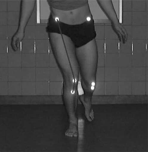
Subjects were videotaped as they performed a series of forward lunges (fig 1). A Sony HC20E camera was positioned on a 60 cm high stand in front of the subject, perpendicular to the frontal plane of the knee at a distance of 2.5 meters from the subject. In this way, 2D video data of the frontal plane movement of the knee were collected. For both legs the subjects were asked to perform a series of three forward lunges starting from a standing position with feet at shoulder width. The subjects performed the lunge movement barefoot. The knee flexion angle of the weight‐bearing extremity during the lunge movement was limited to 45° by varying the distance over which the lunge had to be performed.
Figure 1 Frontal plane posture of a subject's knee during a forward lunge. Informed consent was obtained for publication of this figure.
To determine the knee valgus/varus angles for each subject reflective markers were fixed to the skin over anatomical landmarks. Markers were placed on the right and left anterior superior iliac spines, the centre of the left and right patella and the middle of the left and right anterior tibial tuberosity. A digital video analysis software program, Darttrainer 2.5 (Dartfish video software solutions, Fribourg, Switzerland), was used to determine the peak knee valgus or varus angles for each subject during the three forward lunge movements with each leg. The average valgus or varus angle of the left and right leg was calculated and used for statistical analysis.
Statistical analysis
Based on the video analysis of the frontal plane movement of the knees during the recorded forward lunge movements, subjects could be divided into two groups: a group that moved their knee into valgus (valgus group) and a group that moved their knee into varus (varus group) during forward lunge.
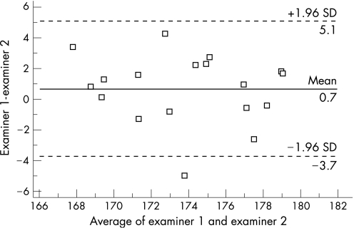
Statistical analysis was performed with SPSS for Windows V.12.0 (SPSS Inc, Chicago, Illinois). To assess the repeatability of the digital video analysis of the peak knee valgus and varus angles during the lunge movement the Bland–Altman plot was used to show the range of agreement between the first and the second examiner.22 In the graph the difference between the two examiners' scores was plotted against the average of the measurements. The Kolmogorov–Smirnov test was used in order to indicate a normal distribution of the data. For the tested six major muscle groups, Independent samples t tests were used to compare hip muscle strength differences between the valgus group and the varus group.
In both groups the Pearson product–moment correlation coefficient was used to examine the relationship between the force and agonist/antagonist force ratios of the hip muscles and the amount of knee movement in the frontal plane during a forward lunge. Statistical significance was accepted at the level of α⩽0.05.
Results
The Bland–Altman plot (fig 2) shows that the differences within mean (SD) 1.96 (0.69 (4.39)) are not clinically relevant, confirming a good repeatability of the score of the digital video analysis of the peak knee valgus and varus angles as 95% of the differences were less than two SD.
Figure 2 Bland–Altman plot difference between two examiner's scores against average of the digital video analysis scores of the peak knee valgus and varus angles during the lunge movement.
There were no significant differences in muscle strength for none of the six tested muscle groups between the valgus group and the varus group (table 1).
Table 1 Mean, SD and p values of the t test of the strength (in Newton, normalised to body weight) of the six tested hip muscle groups of the valgus and varus group (α = 0.05).
| Hip muscle strength (N) | Mean valgus group | SD valgus group | Mean varus group | SD varus group | Significance (t test) |
|---|---|---|---|---|---|
| Flexor | 389.08 | 68.16 | 375.48 | 57.25 | 0.41 |
| Extensor | 559.11 | 152.86 | 571.52 | 116.50 | 0.73 |
| Abductor | 371.26 | 57.41 | 391.79 | 38.98 | 0.12 |
| Adductor | 377.38 | 79.83 | 381.62 | 61.63 | 0.82 |
| Internal rotator | 238.23 | 42.87 | 241.04 | 38.40 | 0.79 |
| External rotator | 244.88 | 48.69 | 254.47 | 33.39 | 0.39 |
Neither in the valgus group nor in the varus group, a significant correlation could be revealed between the strength of any of the assessed muscle groups and the amount of movement into valgus/varus (table 2).
Table 2 Correlation coefficients (r) and p values of the correlations between the strength of the hip muscle groups (in Newton) and the amount of movement into valgus/varus (α = 0.05).
| Hip muscle strength (N) | Valgus group | Varus group | ||
|---|---|---|---|---|
| Correlation coefficient (r) | p Value | Correlation coefficient (r) | p Value | |
| Flexor | 0.19 | 0.23 | 0.03 | 0.89 |
| Extensor | 0.11 | 0.49 | −0.20 | 0.35 |
| Abductor | −0.002 | 0.99 | −0.41 | 0.06 |
| Adductor | 0.03 | 0.84 | −0.32 | 0.12 |
| Internal rotator | −0.22 | 0.15 | −0.39 | 0.06 |
| External rotator | −0.05 | 0.75 | 0.21 | 0.33 |
In the valgus group no significant correlation was found between the force ratios of the hip muscles, Flex/Ext (r = −0.08, p = 0.40), Abd/Add (r = −0.15, p = 0.12) and ER/IR (r = 0.14, p = 0.15), and the amount of valgus movement at the knee. However, in the varus group the ER/IR force ratio was positively related to the amount of knee varus during the forward lunge movement (r = 0.31, p = 0.03).
No statistical significant differences could be detected for the results of the assessed strength of the six tested hip muscle groups, the agonist/antagonist force ratios or the amount of knee movement in the frontal plane during the forward lunge between the male and female subjects.
Discussion
It has been demonstrated that muscle weakness proximal to a symptomatic area is often present in lower‐extremity injury conditions.14,16,17,23,24,25 The closed kinetic chain theory suggests that sufficient proximal hip strength is needed for control of distal segments to prevent injury. If a joint of the lower extremity is not functioning properly, injuries can be manifested in other joints, particularly those that are distal to the affected joint. It is therefore speculated that hip muscle weakness could play a role in knee overuse injuries.13
In this current study no significant strength differences of the six major muscle groups around the hip were found between the group of subjects whose knees moved into valgus and the group whose knees moved into varus during a forward lunge movement. In addition, a significant relationship was not found between the strength of the assessed hip muscles and the amount of knee valgus/varus movement during the lunge in either group. Therefore, the idea that the strength of the muscles around the hip is related to the amount of knee movement into valgus or varus during movements such as a forward lunge might not be valid in healthy individuals.
As previously stated by Ireland et al,14 to date, studies that have investigated the relationship between lower‐extremity frontal plane stability and the prevention of knee injuries are scarce. A study by Hewett et al26 demonstrated that following a 6 week training program including lower‐extremity plyometric drills and general strength and flexibility exercises, there was a 50% reduction in the adduction/abduction moments at the knee during the landing phase of a vertical jump. Although the training program did not focus on the hip muscles specifically, the decrease in frontal plane knee moment was the only significant predictor of the athlete's risk for knee ligamentous injury. A subsequent prospective study by the same authors showed that female athletes who participated in the same training program had a significantly lower incidence of severe knee ligament injury than those who did not.27
In this current study, except for the ER/IR force ratio in the varus group, no relationship could be established between hip muscle strength and frontal plane movement of the knee during a tennis lunge. However in distance runners who presented with iliotibial band syndrome, Fredericson et al28 postulated that strengthening of the gluteus medius fostered increased control of thigh adduction and internal rotation tendencies during running, thereby minimising the valgus vector at the knee. Also, Ireland et al14 indicated that strengthening of the abductors and external rotators of the hip might benefit individuals suffering from anterior knee pain by improving stability of the lower extremity in the frontal and transverse planes of motion during sport‐specific activities.
In this current study, however, we did find that in the group of subjects whose knees moved into varus the ER/IR force ratio at the hip was moderately, yet significantly positively related to the amount of knee varus during a forward lunge movement. This indicates that when the external rotator muscle strength exceeds the muscle strength of the internal rotators of the hip, the knee is moved more into varus during this particular movement. The positive correlation between the ER/IR force ratio and the degree of varus movement during the lunge movement, found in this study, might concur with the findings of Fredericson et al in a way that a stronger force of the external rotators of the hip relative to the internal rotator force might cause the knee to be moved more laterally during activities such as a forward lunge.
One possible explanation for the fact that in our study further no correlation between hip muscle strength and the amount of varus/valgus movement at the knee during the forward lunge could be established could be that the tested population consisted of all healthy subjects without any hip, knee or lower leg complaints at the moment of the study. It is plausible that a relationship between the investigated parameters could manifest itself in a population suffering from knee or other lower extremity injuries and presenting hip muscle weakness.
In addition, fatigue of the hip muscles could be a factor that might have an effect on the relationship between the strength of the hip muscles and the amount of knee varus/valgus during forward lunge movements in tennis. It might be possible that this relationship only reveals itself when the muscles are getting fatigued during prolonged tennis playing. As the duration of tennis matches is usually 90–120 min on grass and fast surfaces and 120–180 min on clay, an important subject is the time course of changes in muscle strength during this prolonged intermittent exercise.29 Girard et al29 found in well trained male tennis players that a progressive reduction in maximal voluntary strength of the knee extensors was highly correlated with increases in perceived exertion throughout a 3 h tennis match. If this is also the case for the hip musculature, this could possibly have an effect on the movement patterns of the knee in the frontal and transversal plane. This condition was not assessed in this study.
However, in the investigated population of this study the strength of the hip muscles did not seem to be related to the degree of knee valgus and varus during the forward lunge. As postulated earlier, this could also mean that, in an asymptomatic population, hip muscle strength might not be such an important factor in the control of the frontal plane knee posture during this kind of movement as has been hypothesised in the beginning of this study. This might suppose that other factors, such as good proprioception and sufficient core hip stability, instead of the absolute strength of the hip muscles could be more important in the control of knee movement during a forward lunge movement. Proprioception is granted to be of great importance in the coordination of skill‐demanding movements that are performed in tennis and therefore also contributes to lower limb control during tennis playing.30 As it has been predicted that tennis players' proprioception would be directly related to the amount of practice,30 based upon the results of this study, it could be assumed that proprioceptive training of the lower extremities might be more appropriate than purely strength training of the hip muscles to ensure the quality of a tennis‐specific movement such as a forward lunge.
No significant differences could be found between the male and female subjects, neither for the assessed strength of the six tested hip muscle groups and the agonist/antagonist force ratios, nor for the amount of knee movement in the frontal plane during the forward lunge. However, we believe that in this study the number of female subjects (n = 8) might be too small to detect possible differences between males and females in this regard.
What is already known on this topic
Proximal hip strength is needed for control of the static and dynamic alignment of distal segments to prevent injuries in intensive sports such as tennis.
What this study adds
This study investigated the relationship between hip muscle strength and the movement pattern of the knee in the frontal plane during a tennis‐specific forward lunge movement.
A limitation of this study is that only healthy subjects without self reported lower extremity problems were assessed, whereas possible relationships between hip muscle strength and frontal plane movement of the knee during sport‐specific activities might more likely to be found in a patient population suffering from lower limb injuries. However, it was the primary goal of this study to search for a possible relationship between hip muscle strength and the frontal plane posture of the knee during a forward lunge in an asymptomatic population. Future research should assess whether this relationship can be established in a patient population suffering from lower extremity injuries in which hip muscle weakness might be present. In addition, the question if hip muscle fatigue, which could be caused by prolonged tennis playing, has an effect on knee frontal plane movement during tennis‐specific lower limb movements should be addressed in future studies.
In this study, the isometric strength of the hip muscles was tested using a handheld dynamometer. Handheld dynamometry for measuring hip muscle strength was chosen as this method has been proven to be a reliable method.18,19,20
However, during a forward lunge there are not solely isometric contractions. Consequently, it might be more functional to examine the relationship between the frontal plane posture of the knee during a forward lunge and the isokinetic (concentric and eccentric) strength of the hip muscles. This should be addressed in future research.
Conclusions
In this study no significant differences in hip strength could be detected between the subjects whose knee moved into valgus and those whose knee moved into varus during a forward lunge movement. However, in the subjects whose knee moved into varus, ER/IR force ratio was moderately related to the amount of knee varus during the lunge movement. The results of this study suggest that in healthy individuals hip muscle strength is not correlated to the amount of valgus/varus movement of the knee during a forward lunge. This does suspect that instead of the absolute strength of the hip muscles other factors such as sufficient proprioception and core hip stability might be more important for controlling knee movement during this tennis‐specific movement.
Abbreviations
Q angle - quadriceps angle
Footnotes
Competing interests: None declared.
Informed consent was obtained for publication of figure 1.
References
- 1.Mohtadi N, Poole A. Racquet sports. In: Caine DJ, Caine CG, Lindner KJ, eds. Epidemiology of sports injuries. Champaign, Illinois: Human Kinetics, 1996301–311.
- 2.Maquirriain J, Ghisi J P. The incidence and distribution of stress fractures in elite tennis players. Br J Sports Med 200640454–459. [DOI] [PMC free article] [PubMed] [Google Scholar]
- 3.Pluim B M, Staal J B, Windler G E.et al Tennis injuries: occurrence, aetiology, and prevention. Br J Sports Med 200640415–423. [DOI] [PMC free article] [PubMed] [Google Scholar]
- 4.Berson B L, Rolnick A M, Ramos C G.et al An epidemiologic study of squash injuries. Am J Sports Med 19819103–106. [DOI] [PubMed] [Google Scholar]
- 5.Soderstrom C A, Doxanas M T. Racquetball: a game with preventable injuries. Am J Sports Med 198210180–183. [DOI] [PubMed] [Google Scholar]
- 6.Chard M D, Lachman S M. Racquet sport patterns of injury presenting to a sports injury clinic. Brit J Sports Med 198727150–153. [DOI] [PMC free article] [PubMed] [Google Scholar]
- 7.Barber F A, Sutker A N. Iliotibial band syndrome. Sports Med 199214144–148. [DOI] [PubMed] [Google Scholar]
- 8.Hutchinson M R, Laprade R F, Burnett Q M.et al Injury surveillance at the USTA boys tennis championships – A 6‐year study. Med Sci Sports Exerc 199527826–830. [PubMed] [Google Scholar]
- 9.Gecha S R, Torg E. Knee injuries in tennis. Clin Sports Med 19887435–452. [PubMed] [Google Scholar]
- 10.Leach R E. Leg and foot injuries in racquet sports. Clin Sports Med 19887359–370. [PubMed] [Google Scholar]
- 11.Livingston L A, Spaulding S J. OPTOTRAK maesurement of the quadriceps angle using standardized foot positions. J Athletic Train 200237252–255. [PMC free article] [PubMed] [Google Scholar]
- 12.Livingston L A. The quadriceps angle: a review of the literature. J Orthop Sports Phys Ther 199828105–109. [DOI] [PubMed] [Google Scholar]
- 13.Niemuth P E, Johnson R J, Myers M J.et al Hip muscle weakness and overuse injuries in recreational runners. Clin J Sport Med 20051514–21. [DOI] [PubMed] [Google Scholar]
- 14.Ireland M L, Willson J D, Ballantyne B T.et al Hip strength in females with and without patellofemoral pain. J Orthop Sports Phys Ther 200333671–676. [DOI] [PubMed] [Google Scholar]
- 15.Hollman J H, Kolbeck K E, Hitchcock J L.et al Correlations between hip strength and static foot and knee posture. J Sports Rehab 20061512–23. [Google Scholar]
- 16.Tyler T F, Nicholas S J, Mullaney M J.et al The role of hip muscle function in the treatment of patellofemoral pain syndrome. Am J of Sports Med 2006341–7. [DOI] [PubMed] [Google Scholar]
- 17.Mascal C L, Landel R, Powers C. Management of patellofemoral pain targeting hip, pelvis and trunk muscle function: two case reports. J Orthop Sports Phys Ther 200333647–660. [DOI] [PubMed] [Google Scholar]
- 18.Wang C Y, Olson S L, Protas E J. Test–retest strength reliability: hand‐held dynamometry in community‐dwelling elderly fallers. Arch Phys Med Rehab 200283811–815. [DOI] [PubMed] [Google Scholar]
- 19.Kimura I F, Jefferson L M, Gulick D T.et al Intra‐ and intertester reliability of Chatillon and Microfet hand‐ held dynamometers in measuring force production. J Sport Rehab 19965197–205. [Google Scholar]
- 20.Agre J C, Magness J L, Hull S Z.et al Strength testing with a portable dynamometer: reliability for the upper and lower extremities. Arch Phys Med Rehab 198768454–458. [PubMed] [Google Scholar]
- 21.Soderberg G L. Handheld dynamometry for muscle testing. In: Reese NB, ed. Muscle and sensory testing. Philadelphia, Pennsylvania, USA: WB Saunders Company, 1999405–410.
- 22.Bland J M, Altman D G. Statistical methods for assessing agreement between two methods of clinical measurement. Lancet 19861307–310. [PubMed] [Google Scholar]
- 23.Nicholas J A, Strizak A M, Veras G. A study of thigh muscle weakness in different pathological states of the lower extremity. Am J Sports Med 19764241–248. [DOI] [PubMed] [Google Scholar]
- 24.Jaramillo J, Worrell T W, Ingersoll C D. Hip isometric strength following knee surgery. J Orthop Sports Phys Ther 199420160–165. [DOI] [PubMed] [Google Scholar]
- 25.Janda V. Muscle strength in relation to muscle length, pain and muscle imbalance. In: Harms‐Ringdahl K, ed. Muscle strength (international perspectives in physical therapy). Edinburgh, UK: Churchill Livingstone, 199383–91.
- 26.Hewett T E, Stroupe A L, Nance T A.et al Plyometric training in female athletes. Decreased impact forces and increased hamstring torques. Am J Sports Med 199624765–773. [DOI] [PubMed] [Google Scholar]
- 27.Hewett T E, Lindenfeld T N, Riccobene J V.et al The effect of neuromuscular training on the incidence of knee injury in female athletes. A prospective study. Am J Sports Med 199927699–706. [DOI] [PubMed] [Google Scholar]
- 28.Fredericson M, Cookingham C L, Chaudhari A M.et al Hip abductor weakness in distance runners with iliotibial band syndrome. Clin J Sport Med 200010169–175. [DOI] [PubMed] [Google Scholar]
- 29.Girard O, Lattier G, Micallef J P.et al Changes in exercise characteristics, maximal voluntary contraction, and explosive strength during prolonged tennis playing. Br J Sports Med 200640521–526. [DOI] [PMC free article] [PubMed] [Google Scholar]
- 30.Lin C H, Lien Y H, Wang S F.et al Hip and knee proprioception in elite, amateur, and novice tennis players. Am J Phys Med Rehabil 200685216–221. [DOI] [PubMed] [Google Scholar]


