Abstract
Objectives
The aim of this study was to evaluate whether removing the calcifications in the rotator cuff tendons during surgical subacromial decompression improves outcome in patients with calcific tendonitis.
Methods
Two groups of 20 patients with a subacromial impingement syndrome and cuff calcifications were operated on. In group A, patients had an anterolateral acromioplasty according to Neer with excision of calcifications. In group B, the same procedure was performed without additional excision of calcifications. After a minimum follow‐up of 3 years the patients were assessed with the disabilities of arm, shoulder and hand score (DASH), the visual analogue scale (VAS) for pain, measurements of range of motion (ROM) in all planes, and satisfaction with treatment.
Results
The results for the DASH score, ROM, VAS and satisfaction with treatement showed no significant difference between the two groups.
Conclusion
The results of our study suggest that removal of calcific deposits with anterolateral acromioplasty does not influence patient outcome. Further prospective studies are needed to determine the optimal surgical treatment for calcific tendonitis.
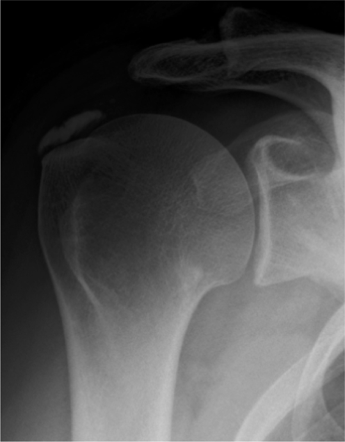
Calcific tendonitis of the shoulder is a common and painful disorder and is characterised by calcifications in the tendons of the rotator cuff (fig 1). The incidence in the healthy population is 2.7%, rising to 6.8% in patients with shoulder pain.1,2 The predominant age is 30–60 years and women are affected slightly more often than men. The calcifications are most often seen in the tendon of the supraspinatus muscle.1 Risk factors for shoulder pain due to problems of the rotator cuff include overhead activities and sports.3,4 The treatment of choice is primarily conservative. This includes rest, physiotherapy, non‐steroidal anti‐inflammatory drugs and at a later stage a subacromial infiltration with corticosteroids. When conservative treatment fails, surgery can be recommended. In most studies on surgical treatment of calcific tendonitis, removal of the calcifications in combination with a subacromial decompression is only recommended when there are signs of subacromial irritation.5,6,7,8,9,10,11 However, it has also been advocated that a subacromial decompression alone might be sufficient, stating that the calcifications will dissolve as a matter of natural course.12 The aim of this study was to evaluate whether it is beneficial for patient outcome to remove the calcifications of the tendons of the rotator cuff when performing a subacromial decompression.
Figure 1 Example of a calcification in the rotator cuff on an x ray of the shoulder.
Materials and methods
Patients
A total of 40 patients (27 women and 13 men) with calcific tendonitis were selected for this retrospective cohort study performed at the St. Elisabeth Hospital in Tilburg, the Netherlands. Two treatment groups (A and B) were defined and treated accordingly. In group A, an open acromioplasty was performed with removal of the calcifications of the rotator cuff. In group B, an open acromioplasty was performed without removal of the calcifications. Four orthopaedic surgeons in our hospital performed the surgical procedure. The decision as to whether to remove the calcifications depended upon the personal preference of the operating orthopaedic surgeon; two orthopaedic surgeons always removed the deposits, whereas the other two only performed subacromial decompressions. Patients were operated on at random as this hospital uses a general operating list. This created a pseudorandomisation of the patients in both groups. The inclusion criteria were presence of shoulder pain resistant to conservative treatment for at least 6 months and positive impingement signs: a positive Hawkins test and a positive effect of a subacromial infiltration with local anaesthetic. Further inclusion criteria were calcifications in the cuff on a conventional x ray of the shoulder, a sonographically intact rotator cuff and a follow‐up time of at least 3 years after the operation. Exclusion criteria were a rupture of the rotator cuff, previous surgery to the extremity of the painful shoulder, additional surgery at the time of the operation, such as resection of the lateral clavicle, or other pathology of the cervical spine, elbow or wrist.
A questionnaire was sent to all patients with a minimum follow‐up of 3 years after surgery. The questionnaire included the disabilities of the arm, shoulder and hand (DASH) score, which is a validated score for disabilities of arm, shoulder and hand,13,14 visual analogue scale for pain, and satisfaction with treatment. The range of motion was measured in all planes.
Surgical intervention
The patient was placed in the beach chair position. An anterolateral incision was made beginning at the anterolateral part of the acromion. The deltoid muscle was divided and the origin of the deltoid muscle was detached from the acromion medially and laterally over 1 cm. The coraco‐acromial ligament was left untouched. The bursal tissue was removed and the anterolateral corner of the acromion was removed with an osteotome. The rotator cuff was inspected for tears. In group A, the operating orthopaedic surgeon made a longitudinal incision over the tendon where he felt or saw the calcification. The calcifications were removed as much as possible. The longitudinal incision was closed with a vicryl suture. In both groups the deltoid muscle was reattached to the acromion using vicryl sutures. Then, the wound was closed over a drain in layers.
The postoperative treatment was similar for both groups. All patients received an arm sling for 4 weeks and they were allowed to move the arm within the limits of pain. No standard physiotherapy was given.
Statistical analysis
Baseline characteristics are presented according to treatment group. Means are presented for continuous variables and percentages for categorical variables.
Mean DASH score, mean range of motion and mean VAS for pain are presented according to treatment group. The Student's t test was used to test any differences for statistical significance. All tests were two‐sided with a cut‐off for statistical significance of 0.05.
Results
A total of 40 patients with a subacromial impingement syndrome were operated on. Patients' characteristics are listed in table 1. Two patients in group A and one in group B did not return the questionnaire and were lost to follow‐up.
Table 1 Baseline characteristics of both groups.
| Patient characteristics | A | B | p Value |
|---|---|---|---|
| Age, years | 51.2 (42–60) | 53.4 (41–62) | NS |
| Female (%) | 13 (65) | 14 (70) | NS |
| Right shoulder (%) | 12 (60) | 11 (55) | NS |
| Duration of symptoms (months) | 15 (6–36) | 14 (6–30) | NS |
| Size of the calcifications (mm) | 15.1 (9–22) | 14.9 (10–18) | NS |
NS, not significant.
Table 2 shows the range of motion in all six planes. There was no significant difference in the range of motion in all planes between both groups.
Table 2 Range of motion.
| Range of motion (°) | A | B | p Value |
|---|---|---|---|
| Abduction | 173 | 169 | NS |
| Adduction | 42 | 39 | NS |
| Elevation | 171 | 172 | NS |
| Retroflexion | 36 | 39 | NS |
| External rotation | 81 | 79 | NS |
| Internal rotation | 69 | 74 | NS |
NS, not significant.
Table 3 shows the VAS for pain that consisted of two items: pain during activity and pain at rest. No significant difference was seen between both groups.
Table 3 VAS for pain.
| VAS for pain | A | B | p Value |
|---|---|---|---|
| During activity | 4.3 | 4.2 | NS |
| At rest | 2.9 | 3.0 | NS |
NS, not significant.
Table 4 shows the results for the DASH score, which is a disability score. The score is divided in two sub‐groups: activities and symptoms. No significant differences between group A and B were seen in these two sub‐groups.
Table 4 Disability of arm shoulder and hand score.
| DASH score | A | B | p Value |
|---|---|---|---|
| Activities | 1.25 | 1.30 | NS |
| Symptoms | 1.89 | 1.74 | NS |
NS, not significant.
In group A 16 patients were satisfied and two were not. In group B 15 patients were satisfied and 4 were not. This difference is not statistically significant.
Discussion
The aetiology of calcific tendonitis is largely unknown. The condition might be related to fibrosis and necrosis within the tendon with subsequent degeneration,15 although this has been questioned by Bosworth.2 The disorder has four histological stages.1,2 The first stage involves fibrocartilaginous metaplasia within the tendon. Usually this is located 1 cm medial to the insertion of the supraspinatus tendon. The second stage is known as the formative phase in which calcific deposits form. In the third stage, the resorptive phase, the deposits are resolved by extravasation into the subacromial bursa. The final stage involves healing and repair of the rotator cuff.1 There is a natural cycle in which the tendon has the capacity to repair itself. In chronic calcific tendonitis this cycle can be blocked at any stage.2
On radiographs the different stages are characterised by their appearance. The most widely used classification is the one according to the French Arthroscopic Society on anteroposterior view.16 They defined four types of deposits. Type A deposits are sharply delineated, dense and homogenous. Type B deposits are sharply delineated, have a dense appearance and consist of multiple fragments. Type C deposits are heterogeneous in appearance and a fluffy deposit. Type D deposits are dystrophic calcifications at the tendon insertion. The last two types are associated with the resorptive stage, whereas type A and B seem to become blocked in the resorptive stage and are associated with chronic calcific tendonitis.
If conservative treatment fails other modalities are used. As it is the general opinion that calcific deposits are the cause of the chronic pain, the different treatment options usually focus on removing these deposits. Alternative treatment techniques have been developed, such as extra‐corporal shock wave therapy, a promising non‐invasive treatment that has been reported to be comparable to surgery in the long run.17,18,19
In a study conducted by Rompe et al surgical removal was superior to shock wave therapy in the treatment of type A calcifications; no difference in treatment outcome was seen for type B calcific deposits. In their discussion, they indicate that the prospective randomised trial was difficult to perform because patients preferred non‐invasive treatment over a surgical intervention.7
Another less invasive treatment technique uses fine needling (barbotage), whereby the calcifications are removed by sucking the substance out of the tendon. This technique has good results in 60% of the patients.20,21,22,23
What is already known on this topic
Calcific tendonitis is a common and painful disorder of the rotator cuff.
Treatment is primarily conservative.
In case conservative treatment fails, surgical removal of the calcifications can be recommended.
When there are signs of subacromial irritation, subacromial decompression can also be performed.
What this study adds
This study shows that there is no difference in patient outcome between surgical subacromial decompression with or without removal of the calcifications.
This raises the question whether it is really necessary to remove the calcifications in patients with calcific tendonitis.
Many studies advocate the surgical removal of the deposits, either by an open procedure or an arthroscopic procedure. A subacromial decompression is only performed when there are signs of subacromial irritation.6,7,8,9,10,11 Porcellini et al conclude that a successful outcome appears to be strongly related to the absence of calcium deposits in the tendon cuff only.5 By contrast, Tillander et al saw no difference in treatment outcome and reported the interesting finding that the calcifications had dissolved spontaneously. They proposed that the calcifications are not the cause of the pain, but might be regarded as an insignificant observation on radiographic evaluation with regard to the indication for treatment.9 We agree with this statement and postulate that the calcifications are a transient process that should neither be a reason for surgery itself, nor a reason to perform an open procedure instead of an arthroscopic procedure. Most studies on this topic present good long‐term results for removal of the calcifications. However, as they are all uncontrolled, these results should be interpreted with caution. We challenge the general belief that removal of the deposits is an essential treatment step. The results of our study suggest that removal of calcific deposits with anterolateral acromioplasty do not influence patient outcome.
A limitation of the present study is the retrospective design. However, due to the use of a general operating list in our hospital, which determined the operating orthopaedic surgeon, pseudorandomisation of the patients occurred as two surgeons always removed the deposits whereas the other two did not. A second limitation of the study is the omission of calcification staging on pre‐and postoperative radiographs. As we have seen above, only type A and B deposits are the ones that need further treatment. We are also unable to see what happens to the deposits after a subacromial decompression, as the retrospective character of the study did not include radiographs at follow‐up. In our study, no difference was seen in patient outcome, which suggest that the essential step in the operative treatment of calcific tendonitis is not the removal of the calcifications. It might also be possible that the operation and the tampering of the tendon itself restarts the natural cycle and moves into the resorptive stage. Further prospective studies between the different treatment modalities are necessary to find out in which patients' baseline characteristics a surgical procedure, and what kind of procedure will give the best results.
Conclusion
The results of our study suggest that removal of calcific deposits with anterolateral acromioplasty does not influence patient outcome. Further prospective studies are needed to determine the optimal surgical treatment for calcific tendonitis.
Abbreviations
DASH - disabilities of arm, shoulder and hand
ROM - range of motion
VAS - visual analogue scale
Footnotes
Competing interests: None declared.
References
- 1.Editorial Calcific tendonitis of the shoulder. New Engl J Med 19993401582–1584. [DOI] [PubMed] [Google Scholar]
- 2.Bosworth B M. Calcium deposits in the shoulder and subacromial bursitis; a survey of 12122 shoulders. JAMA 19411162477–2482. [Google Scholar]
- 3.Lo Y P, Hsu Yc, Chan K M. Epidemiology of shoulder impingement in upper arm sports events. Br J Sports Med 199024163–167. [DOI] [PMC free article] [PubMed] [Google Scholar]
- 4.Brasseur J L, Lucidarme O, Tardieu M.et al Ultrasonographic rotator‐cuff changes in veteran tennis players: the effect of hand dominance and comparison with clinical findings. Eur Radiol 200414857–864. [DOI] [PubMed] [Google Scholar]
- 5.Porcellini G, Paladini P, Campi F.et al Arthroscopic treatment of calcifying tendonitis of the shoulder: clinical and ultrasonographic follow‐up findings at two to five years. J Shoulder Elbow Surg 200413503–508. [DOI] [PubMed] [Google Scholar]
- 6.Rochwerger A, Franceschi J P, Viton J M.et al Surgical management of calcific tendonitis of the shoulder: an analysis of 26 cases. Clin Rheumatol 199918313–316. [DOI] [PubMed] [Google Scholar]
- 7.Rompe J, Zoellner J, Nafe B. Shock wave therapy versus vonventional surgery in the treatment of calcifying tendonitis of the shoulder. Clin Orthop 200138772–82. [DOI] [PubMed] [Google Scholar]
- 8.Rubenthaler F, Ludwig J, Wiese M.et al Prospective randomised surgical treatments for calcifying tendinopathy. Clin Orthop 2003410278–284. [DOI] [PubMed] [Google Scholar]
- 9.Seil R, Litzenburger H, Kohn P.et al Arthroscopic treatment of chronically painful calcifying tendonitis of the supraspinatus tendon. Arthroscopy 200622521–527. [DOI] [PubMed] [Google Scholar]
- 10.Wittenberg R H, Rubenthaler F, Wölk T.et al Surgical or conservative treatment for chronic rotator cuff calcifying tendinitis – a matched‐pair analysis of 100 patients. Arch Orthop Trauma Surg 200112156–59. [DOI] [PubMed] [Google Scholar]
- 11.Jerosch J, Strauss M, Schmiel S. Atrhroscopic treatment of calcific tendonitis of the shoulder. J Shoulder Elbow Surg 1998730–37. [DOI] [PubMed] [Google Scholar]
- 12.Tillander B M, Norlin R O. Change of calcifications after arthroscopic subacromial decompression. J Shoulder Elbow Surg 19987213–217. [DOI] [PubMed] [Google Scholar]
- 13.Gummesson C, Atroshi I, Ekdahl C. The disabilities of the arm, shoulder and hand (DASH) outcome questionnaire: longitudinal construct validity and measuring self‐rated health change after surgery. BMC Musculoskeletal Disorders 2003411–16. [DOI] [PMC free article] [PubMed] [Google Scholar]
- 14.Veehof M M, Sleegers E J, Van Veldhoven N H.et al Psychometric qualities of the dutch language version of the disabilities of the arm, shoulder, and hand questionnaire (DASH‐DLV). J Hand Ther 200215347–354. [DOI] [PubMed] [Google Scholar]
- 15.McLaughlin H L. Lesions of the muscolotendinous cuff of the shoulder: III. Observations on the pathology, course and treatment of calcific deposits. Ann Surg 1946124354–362. [PubMed] [Google Scholar]
- 16.Molé D, Kempf J F, Gleyze P. Results of endoscopic treatment of non‐broken tendinopathies of the rotator cuff. 2. Calcifications of the rotator cuff. Rev Chir Orthop Reparatrice Appar Mot 199379532–541. [PubMed] [Google Scholar]
- 17.Ebenblicher G R, Erdogmus C B, Resch K L.et al Ultrasound therapy for calcific tendonitis of the shoulder. New Engl J Med 19993401533–1538. [DOI] [PubMed] [Google Scholar]
- 18.Loew M, Decke W, Kusnierczak D.et al Shock‐wave therapy is effective for chronic calcifying tendonitis of the shoulder. J Bone Joint Surg 199981B983–987. [DOI] [PubMed] [Google Scholar]
- 19.Moretti B, Garofalo R, Genco S.et al Medium‐energy shock wave therapy in the treatment of rotator cuff calcifying tendinitis. Knee Surg Sports Traumatol Arthrosc 200513405–410. [DOI] [PubMed] [Google Scholar]
- 20.Chiou H J, Chou Y H, Wu J J.et al The role of high‐resolution ultrasonography in management of calcific tendonitis of the rotator cuff. Ultrasound Med Biol 200127735–743. [DOI] [PubMed] [Google Scholar]
- 21.Cooper G, Lutz G E, Adler R S. Ultrasound‐guided aspiration of symptomatic rotator cuff calcific tendonitis. Am J Phys Med Rehab 20058481. [DOI] [PubMed] [Google Scholar]
- 22.Farin P U, Räsänen H, Jaroma H, Harju A. Rotator cuff calcifications: treatment with ultrasound‐guided percutaneous needle aspiration and lavage. Skeletal Radiol 199625551–554. [DOI] [PubMed] [Google Scholar]
- 23.Farin P U. Consistency of rotator cuff calcifications: observations on plain radiography, sonography, computed tomography and at needle treatment. Investigative Radiology199631300–304. [DOI] [PubMed] [Google Scholar]


