Abstract
Background
Sural nerve injuries are an evident risk especially of minimal‐invasive surgical Achilles tendon repair. However, detailed anatomical studies focusing on the relationship of the sural nerve with the Achilles tendon at various levels are scarce, even pending in two planes.
Aim
To determine the position and course of the sural nerve in relation to the Achilles tendon in two planes after trans‐section and computer‐assisted determination.
Methods
The exact course of the sural nerve was determined in 10 cadavers (55.3 years, 19–89 years), using a computer‐assisted method in two planes (transversal/sagittal).
Results
The sural nerve crossed the Achilles tendon at 11 (8.7–12.4) cm proximal to the tuber calcanei. The distance between the lateral crossing and the proximal musculotendineus junction was 35 (20–58) mm. Starting from the tuber calcanei, the distance was 2/2 mm (transversal/sagittal plane) at 11 cm proximal to the tuber calcanei, 4/4 mm at 10 cm proximal, 5/6 mm at 9 cm, 8/10 mm at 5 cm and 11/18 mm at the tuber calcanei.
Conclusion
In the lateral crossing region of the sural nerve and the lateral proximal Achilles tendon 9–12 cm proximal to the tuber calcanei, a close relationship of both anatomical structures can be visualised using computer‐assisted measurements; caution is suggested to prevent sural nerve entrapment in either open or percutaneous Achilles tendon repair.
A number of surgical studies are focusing on the value of open versus percutaneous surgical repair of the Achilles tendon, which was first described by Ma and Griffith1 among 18 patients. A recent comparative study enrolled 132 consecutive patients treated percutaneously versus 105 patients with conventional open repair at the same institute without randomisation.2 They reported significantly fewer major complications (4.5% vs 12.4%, p = 0.03) in the percutaneous group. However, a slightly higher rate of re‐ruptures (3.7% vs 2.8%, p = 0.680) and more sural nerve disturbances (4.5% vs 2.8%, p = 0.487) were noted. In a report by Sutherland and Maffulli,3 31 patients who had undergone repair of an acute rupture through a “modified” percutaneous technique had a total of five (16%) sural nerve injuries, three of which were resolved in 6–9 months. One patient underwent exploration, and the sural nerve was found to be transfixed by a suture. But even open surgical repair may be associated with sural nerve palsy, as described before by Cretnik et al2 and Winter et al.4
However, the exact anatomical course of the sural nerve in relation to the Achilles tendon is camouflaged. The aim of this study was to more exactly determine the position and course of the sural nerve in relation to the Achilles tendon in two planes after trans‐section and computer‐assisted determination.
Methods
A total of 10 cadavers (5 men, median (range) age 55.3 (19–89) years) were analysed through sections in their right leg. The exact relationship of the sural nerve with the Achilles tendon was determined after exact trans‐section in all 10 cases. After preparation of the Achilles tendon and the sural nerve, an exact calibration and digitalisation of the site was performed, creating digital images with a high‐resolution digital camera in two planes—the lateral (transversal) and the dorsoventral (sagittal). The digital two‐plane images were imported and analysed using the DICOM software efilm V.1.8 by an expert examiner. Exact calibration was performed after digitalisation with the efilm software. All distances were determined in either millimetre or centimetre using the calibrated measurement tool of the efilm software with 0.1 mm accuracy. All values are expressed as median (range).
Results
Distance of the tuber calcanei to the proximal sural nerve/Achilles tendon crossing
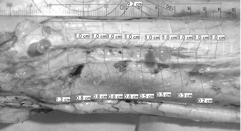
The proximal distance between the lateral crossing of the sural nerve and the lateral Achilles tendon at the proximal site and the tuber calcanei was 11 (8.7–12.4) cm (fig 1).
Figure 1 Trans‐section of the Achilles tendon and the sural nerve. Computer‐based quantification of the distances between both anatomical structures in two planes (displayed the transversal plane) at every centimetre starting from the tuber calcanei to the proximal calf.
Distance of the lateral sural nerve/Achilles tendon crossing to the gastrocnemius muscle
The distance between the lateral crossing of the sural nerve/Achilles tendon and the proximal musculotendineus junction of the Achilles tendon to the gastrocnemius muscle was only 35 (20–58) mm.
Relationship of the sural nerve with the Achilles tendon
Table 1 depicts the position of the sural nerve in relation to the Achilles tendon starting from the tuber calcanei, which is of much more practical value for the surgeon.
Table 1 Distance of the sural nerve to the Achilles tendon in the transversal (lateral) and the sagittal (dorsoventral) planes starting at the tuber calcanei.
| Distance from the tuber calcanei heading proximal to the calf, cm | Lateral distance of the sural nerve to the Achilles tendon (transversal plane), mm | Dorsoventral distance of the sural nerve to the Achilles tendon (sagittal plane), mm |
|---|---|---|
| 11 | 2 | 2 |
| 10 | 4 | 4 |
| 9 | 5 | 6 |
| 8 | 7 | 8 |
| 7 | 8 | 10 |
| 6 | 9 | 11 |
| 5 | 11 | 13 |
| 4 | 10 | 15 |
| 3 | 11 | 16 |
| 2 | 11 | 18 |
| 1 | 11 | 18 |
Discussion
The most important finding of this study is that the lateral crossing region of the sural nerve and the lateral proximal Achilles tendon is 9–12 cm proximal to the tuber calcanei, where a close relationship of both anatomical structures can be visualised. Therefore, caution is mandatory for the proximal suture in Achilles tendon repair to prevent sural nerve entrapment.
The sural nerve crosses the Achilles tendon from distal–lateral to proximal–medial. Even 3 cm distal to the sural nerve/Achilles tendon crossing, the transversal (lateral) distance of the sural nerve to the Achilles tendon is only 5 and 6 mm in the sagittal (ventral) plane. Proximal to the sural nerve/Achilles tendon crossing, we determined the musculotendineous junction to be within a median (range) of 35 (28–58) mm, where the sural nerve is encountered posterior to the dorsal paratendon of the Achilles tendon. Lawrence and Botte5 were the first to describe the sural nerve in an anatomical study that was limited only to one plane, the transversal plane. The same limitation holds true for the study by Webb et al,6 who determined the crossing of the sural nerve and the lateral border of the Achilles tendon 98 mm from the calcaneus, which was in line with our computer‐assisted calculation, and with the study by Mestdagh et al.7 However, to determine the exact position of an anatomical structure, two planes are mandatory, which was the purpose of this study.
This close sural nerve–Achilles tendon relationship has special implications for both the open and the percutaneous surgical repair of ruptured Achilles tendons. Modifications such as a limited open repair of Achilles tendon ruptures were suggested to reduce the incidence of sural nerve disturbances.8 In a case‐control study9 among 84 patients treated in two different hospitals using exactly the same surgical procedure, percutaneous repair, one hospital exposed the sural nerve in 38 patients, whereas the nerve was not exposed in the other hospital (46 patients). A total of 18% sural nerve‐related complications were noted, all in the non‐exposure group. Direct visualisation of the surval nerve seems to reduce sural nerve disturbances.
What is already known on this topic
Sural nerve injuries are evident in minimal‐invasive sutures of the Achilles tendon.
What this study adds
The lateral border of the Achilles tendon 9–12 cm proximal to the tuber calcanei is the “hot zone”, with a close relationship between the sural nerve and the Achilles tendon, where caution and direct visualisation is mandatory to reduce the risk of sural nerve injuries.
As a rule of thumb, the region 9–12 cm proximal to the tuber calcanei should be addressed as the “hot zone”, where the sural nerve enters the lateral border of the Achilles tendon. Within this area, no sutures should be applied without direct visualisation of the sural nerve in order to prevent sural nerve entrapment.
Limitations
We reported on a series of 10 adult cadavers, which is only a small number of subjects, and determined the position of the sural nerve in relation to the Achilles tendon in two planes, which have to be taken into account, given the anatomical variations.
Conclusion
In the lateral crossing region of the sural nerve and the lateral proximal Achilles tendon 9–12 cm proximal to the tuber calcanei, a close relationship of both anatomical structures can be visualised using computer‐assisted measurements, where caution is suggested to prevent sural nerve entrapment in either open or percutaneous Achilles tendon repair.
Footnotes
Competing interests: None declared.
References
- 1.Ma G W C, Griffith T G. Percutaneous repair of acute closed ruptured Achilles tendon: a new technique. Clin Orthop 1977128247–255. [PubMed] [Google Scholar]
- 2.Cretnik A, Kosanovic M, Smrkolj V. Percutaneous versus open repair of the ruptured Achilles tendon. Am J Sports Med 2005331369–1379. [DOI] [PubMed] [Google Scholar]
- 3.Sutherland A, Maffulli N. A modified technique of percutaneous repair of ruptured Achilles tendon. Oper Orthop Traumat 19997288–295. [Google Scholar]
- 4.Winter E, Weise K, Weller S.et al Surgical repair of Achilles tendon rupture. Comparison of surgical with conservative treatment. Arch Orthop Trauma Surg 1998117364–367. [DOI] [PubMed] [Google Scholar]
- 5.Lawrence S J, Botte M J. The sural nerve in the foot and ankle: an anatomic study with clinical and surgical implications. Foot Ankle Int 199415490–494. [DOI] [PubMed] [Google Scholar]
- 6.Webb J, Moorjani N, Radford M. Anatomy of the sural nerve and its relation to the Achilles tendon. Foot Ankle Int 200021475–477. [DOI] [PubMed] [Google Scholar]
- 7.Mestdagh H, Drizenko A, Maynou C.et al Origin and make up of the human sural nerve. Surg Radiol Anat 200123307–312. [DOI] [PubMed] [Google Scholar]
- 8.Assai M, Jung M, Stern R.et al Limited open repair of Achilles tendon ruptures. J Bone Joint Surg 200284A161–170. [PubMed] [Google Scholar]
- 9.Majewski M, Rohrbach M, Czaja S.et al Avoiding sural nerve injuries during percutaneous Achilles tendon repair. Am J Sports Med 200634793–798. [DOI] [PubMed] [Google Scholar]


