Abstract
Background
Tendinopathies of the rotator cuff muscles, biceps tendon and pectoralis major muscle are common causes of shoulder pain in athletes. Overuse insertional tendinopathy of pectoralis minor is a previously undescribed cause of shoulder pain in weightlifters/sportsmen.
Objectives
To describe the clinical features, diagnostic tests and results of an overuse insertional tendinopathy of the pectoralis minor muscle. To also present a new technique of ultrasonographic evaluation and injection of the pectoralis minor muscle/tendon based on use of standard anatomical landmarks (subscapularis, coracoid process and axillary artery) as stepwise reference points for ultrasonographic orientation.
Methods
Between 2005 and 2006, seven sportsmen presenting with this condition were diagnosed and treated at the Cape Shoulder Institute, Cape Town, South Africa.
Results
In five patients, the initiating and aggravating factor was performance of the bench‐press exercise (hence the term “bench‐presser's shoulder”). Medial juxta‐coracoid tenderness, a painful active‐contraction test and bench‐press manoeuvre, and decrease in pain after ultrasound‐guided injection of a local anaesthetic agent into the enthesis, in the absence of any other clinically/radiologically apparent pathology, were diagnostic of pectoralis minor insertional tendinopathy. All seven patients were successfully treated with a single ultrasound‐guided injection of a corticosteroid into the enthesis of pectoralis minor followed by a period of rest and stretching exercises.
Conclusions
This study describes the clinical features and management of pectoralis minor insertional tendinopathy, secondary to the bench‐press type of weightlifting. A new pain site‐based classification of shoulder pathology in weightlifters is suggested.
Tendinopathies are common in professional and recreational athletes; different forms have been described on the basis of the anatomical location of a lesion within the tendon (enthesis, main body of tendon and structures surrounding the tendon). Insertional tendinopathy (enthesiopathy) is one of the most common forms of tendinopathy and can affect any tendon in the body.1 Tendinopathy of the rotator cuff, long head of biceps and pectoralis major muscle are often implicated in the aetiology of shoulder pain in athletes.2,3,4
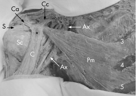
Pectoralis minor is a thin triangular muscle lying deep to the pectoralis major. It originates from the 3rd, 4th and 5th ribs near the costal cartilages. Its fibres ascend laterally and converge in a flat tendon that attaches to the medial border and upper surface of the coracoid process of the scapula. The axillary vessels and brachial plexus lie posterior to the muscle (fig 1). The function of the pectoralis minor is to tilt the scapula anteriorly—that is, to rotate the scapula about a coronal axis so that the coracoid process moves anteriorly and caudally, while the inferior angle moves posteriorly and medially.5 Sporting/training activities that involve this motion of the scapula (bench‐pressing, swimming and push‐up exercises) can theoretically result in overuse of this muscle, especially in the presence of training errors, poor technique or a rapid increase in training load, frequency and/or duration.1
Figure 1 Overview of anatomy and relationships of the pectoralis minor muscle. *, coracoid process tip; Ax, axillary artery; C, conjoint tendon; Ca, coracoacromial ligament; Cc, coracoclavicular ligaments; Pm, pectoralis minor muscle; S, supraspinatus muscle; Sc, subscapularis muscle; 3, 4, 5, origin of pectoralis minor muscle from the 3rd, 4th and 5th ribs.
This paper presents a previously undescribed condition, an overuse insertional tendinopathy of the pectoralis minor muscle, in seven sportsmen. The diagnostic tests and use of ultrasonography for evaluation of the pectoralis minor muscle/tendon are presented.
Materials and methods
Between 2005 and 2006, we diagnosed and successfully treated seven patients (five men, two women) presenting with an overuse tendinopathy of the pectoralis minor muscle. The mean age of the patients was 31.6 (range 17–62) years. Three patients were competitive sportsmen (rugby, swimming and body‐building) and four were non‐competitive ones (recreational body‐building/fitness exercise). The dominant upper extremity (right shoulder) was involved in three patients and non‐dominant one (left shoulder) in four patients. In five patients, the onset was subacute and the pain first occurred after a recent increase in the bench‐press exercise; the mean (range) duration of symptoms in this group was approximately 4.5 (1.5–12) weeks. In two patients, the onset was gradual (7–9 months) and was related to an increase in sporting activity in one of them.
All patients were evaluated by clinical, radiographic and ultrasonographic examinations. Clinical evaluation included a subjective evaluation of pain and functional status by interview, assessment of glenohumeral stability (apprehension and translation tests), assessment of rotator cuff integrity (Jobe's test, bear‐hug test and strength test using a portable dynamometer (ISOBEX 2.1, Cursor Ag, Bern, Switzerland)), assessment of long head of biceps tendon (Speed's test and Yergason's sign), and examination of the acromioclavicular joint (tenderness and cross‐body adduction).6,7,8 In addition, each shoulder was scored using the Constant and Murley9 method of functional assessment of the shoulder. Radiographic evaluation in three planes included true anteroposterior, axillary‐lateral and saggital‐Y views to exclude stress fractures or any other bony pathology of the coracoid process, acromioclavicular joint and glenohumeral joint. Ultrasonographic evaluation included assessment of the rotator cuff, biceps tendon and bicipital groove.
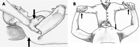
The pectoralis minor muscle was tested clinically by palpating for an area of tenderness medial to the coracoid along the inferior‐medial orientation of the muscle fibres. Active contraction of the muscle was tested as described in literature by Kendall and McCreary5: in the supine position, the patient thrusts the shoulder forward without exerting any downward pressure on the hand to force the shoulder forward. The examiner exerts pressure against the anterior aspect of the patient's shoulder, downward towards the table (fig 2A). A provocative test (bench‐press manoeuvre) was devised to assess the nature of symptoms experienced by the patients while performing bench‐press exercise. In the supine position, with both shoulders abducted and elbows flexed at 90° each, the patient exerts an upward force against resistance (fig 2B). Ultrasonographic evaluation of the pectoralis minor muscle consisted of assessment of muscle–tendon integrity (to exclude an isolated tear of pectoralis minor muscle) as described in the following (fig 3A,B). An ameliorative/therapeutic test, consisting of ultrasound‐guided injection of a local anaesthetic agent and a corticosteroid into the insertion of pectoralis minor tendon (medial juxta‐coracoid region), was performed to confirm and treat the insertional tendinopathy (fig 4).
Figure 2 (A) Active contraction test for the pectoralis minor muscle. The patient actively thrusts the shoulder forward (lower arrow) against resistance exerted by the examiner's hand (upper arrow). (B) Bench‐press manoeuvre for provocative testing of pectoralis minor insertional tendinopathy. The patient exerts an upward force (upper arrows) against resistance and mimicking the bench‐press exercise. Pain medial to the coracoid (lower arrow) suggests a positive test.
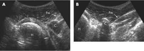
Figure 3 (A) Ultrasonographic visualisation of the anatomical relationship of the subscapularis muscle (Sc), coracoid process (C) and pectoralis minor tendon (Pm). (B) Ultrasonographic visualisation of the pectoralis minor muscle (Pm) and tendon inserting on the coracoid process (C). The axillary artery (A) is seen deep to the pectoralis minor muscle. H, humeral head.
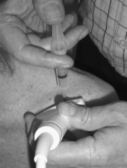
Figure 4 Position of the transducer and injection site for ultrasound‐guided injection of the pectoralis minor tendon.
All seven patients were followed up at 2, 4 and 6 weeks after the initial assessment. During this period, all sporting activities related to the upper limb were discontinued and stretching exercises of the pectoralis minor were prescribed.10 Thereafter, sporting activity was gradually resumed. A final functional assessment (clinical and ultrasonographic evaluation, Constant shoulder score) was performed at 12 weeks after the initial evaluation.
Statistical analysis of the data was performed to determine a significant difference between the preoperative and postoperative Constant scores. The data were analysed using a data analysis software system (STATISTICA, V.7.1, StatSoft, Inc., Oklahoma, USA). The preoperative and postoperative Constant scores were tested for equality of means with analysis of variance (significance level α = 0.05) and then with the non‐parametric Wilcoxon matched pairs test.
Technique of ultrasonographic identification, evaluation and injection of the pectoralis minor muscle tendon
Ultrasonographic identification of the pectoralis minor muscle is performed in a stepwise manner using the subscapularis muscle, coracoid process and axillary artery as standard and consistent reference landmarks. The examination proceeds in a lateral‐to‐medial direction. The first step is to identify the subscapularis and to follow it medially to the coracoid process. A curved array transducer (type C5‐2, Sonoline G50 Ultrasound Imaging System, Siemens Medical Solutions USA, Mountain View, California, USA) is placed on the proximal part of the humerus, perpendicular to the intertubercular/bicipital groove, and then moved medially. This manoeuvre identifies the insertion of the subscapularis tendon. The subscapularis is followed medially to the coracoid process, which is visualised as a strongly echogenic line with an anechoic shadow. This landmark can be confirmed by internal and external rotation of the shoulder, which shows a corresponding movement in the subscapularis under the static coracoid process (fig 3A). The next step is to identify the pectoralis minor muscle. This is achieved by moving the transducer further medially, in line with the orientation of pectoralis minor muscle fibres (inferior medial to superior lateral). When placed correctly, the axillary artery can be visualised as a pulsatile anechogenic structure. This can be further confirmed with the Doppler function of the system. The pectoralis minor muscle is identified as an echogenic layer, passing from medial to lateral, bounded inferiorly by the axillary artery and laterally by the coracoid process (fig 3B). Dynamic evaluation of the muscle can be performed by actively thrusting the shoulder forward and backward; the pectoralis minor is seen contracting and relaxing with this manoeuvre. The lesser echogenic layer above this is the pectoralis major muscle. Once the pectoralis minor has been identified, the probe is centred over the coracoid process. A 21‐guage needle is passed just medial to the coracoid process and can be seen entering the insertion of the pectoralis minor tendon at the coracoid process (fig 4). A solution of a mixture of a local anaesthetic agent (2 cm3 of lignocaine hydrochloride 1%) with a corticosteroid (1 cm3 of betamethasone acetate 3 mg) is then injected into the tendon and the needle is withdrawn.
Results
All seven patients subjectively described the pain to be moderate to severe in intensity, limiting their competitive/recreational sporting and daily activities. In five patients, the initiating and aggravating factor was the bench‐press exercise. In all seven patients, the glenohumeral joint was stable and tests for rotator cuff, biceps tendon and acromioclavicular joint pathology were negative. The mean (range) Constant and Murley score at initial assessment was 76% (71–79%). Three‐plane radiographic examination did not show any stress fractures or other bony/joint abnormality. Ultrasound evaluation showed normal rotator cuff and biceps tendon.
Medial (juxta‐coracoid) tenderness, pain in this region on performance of active contraction test and/or the bench‐press manoeuvre, and immediate reduction/disappearance of this tenderness/pain after injection of a local anaesthetic agent, were interpreted as positive tests for pectoralis minor insertional tendinopathy. Medial (juxta‐coracoid) pinpoint tenderness was positive in all seven patients. The active contraction test was positive in five patients, the provocative test (bench‐press manoeuvre) was positive in six patients, and the ameliorative test (ultrasound‐guided local anaesthetic injection) was positive in all seven patients.
At the final follow‐up (12 weeks), all seven patients had resumed pre‐injury level of sporting activity without any pain/discomfort. Clinical and ultrasonographic evaluation of the pectoralis minor muscle was normal and the mean (range) Constant and Murley score measured approximately 94% (91–96%). The Wilcoxon matched pairs test showed a significant increase (p<0.05) in the postoperative Constant scores (at final follow‐up) as compared with the preoperative scores.
Discussion
Weight training forms a standard fitness regimen in many sports. A high incidence of shoulder injuries has been reported in weightlifters and bodybuilders, and it is suggested that this may be a consequence of reduced professional supervision, especially at the amateur level.11 Within the shoulder, tendinopathies of the rotator cuff, long head of biceps and pectoralis major muscle and osteolysis of the distal clavicle have been described as complications of weight lifting.1,2,3,4,11 Insertional tendinopathy of pectoralis minor is a previously undescribed cause of shoulder pain in weightlifters/sportsmen. This study is an analysis of clinical and radiological findings in seven such cases. We describe this condition as “Bench‐Presser's Shoulder” as the bench‐press exercise was the initiating and aggravating factor in five of the seven cases.
Clinical evaluation of the pectoralis minor muscle was based on its anatomical location and action. Pectoralis minor tendon is the only significant structure that inserts on the medial surface of the coracoid process. Medial juxta‐coracoid tenderness would therefore suggest pathology of the pectoralis minor enthesis. Pain resulting from the active contraction test and the bench‐press manoeuvre, both of which result in contraction of pectoralis minor, also suggests a role of pectoralis minor muscle/enthesis. Confirmation of pectoralis minor insertional tendinopathy can be concurred by immediate reduction/disappearance of the pain (tenderness and test‐induced pain) after injection of a local anaesthetic agent into the insertion site, under ultrasound guidance.
Ultrasound evaluation of the pectoralis minor can be used to assess the integrity of the pectoralis minor tendon and to inject a local anaesthetic/corticosteroid into the insertion site. The technique described here is a simplified method, based on using standard anatomical landmarks for identification of the tendon and localisation of the insertion site. The subscapularis muscle, coracoid process and axillary artery are stepwise reference points that lead the examiner to the pectoralis minor muscle. Evaluation of the entire tendon is necessary to exclude the diagnosis of an isolated tear of the pectoralis minor, although this has been reported only once in literature.12 Corticosteroid‐induced tendon ruptures and resultant pain relief are known. Hence, follow‐up ultrasonographic evaluation of the tendon was performed to assess the status of the muscle/tendon of pectoralis minor.
We successfully treated this condition with a single ultrasound‐guided injection of a corticosteroid into the enthesis, a period of rest and stretching exercises for pectoralis minor muscle (6 weeks) and thereafter, gradual progression to pre‐injury level of sporting activity (6 weeks).10,12,13,14 On the basis of the results of this study and a review of literature, a simple symptom‐related algorithmic approach to shoulder pain in a weight‐training athlete can be suggested (table 1). 2,3,4,11,15
Table 1 A pain‐site‐based classification of pathology in a weight‐training athlete.
| Pain site | Probable pathology |
|---|---|
| Top of shoulder (acromioclavicular joint) | Osteolysis of distal clavicle, acromioclavicular joint arthritis |
| Lateral to coracoid | Bicipital tendinopathy, rotator cuff tendinopathy/rupture, pectoralis major tendinopathy/rupture |
| Medial juxta‐coracoid | Pectoralis minor insertional tendinopathy |
| Lateral juxta‐coracoid | Subcoracoid impingement |
In summary, we consider the following to be suggestive of an overuse tendinopathy of the pectoralis minor:
medial juxta‐coracoid pain/tenderness;
aggravated by active contraction of the pectoralis minor muscle;
reproduced by the bench‐press manoeuvre;
relieved by injection of a local anaesthetic into the tendon insertion;
ultrasonographically intact pectoralis minor muscle;
absence of any other clinically/radiologically apparent shoulder pathology.
What is already known on this topic
The shoulder is the most often injured joint in weightlifters. Within the shoulder, tendinopathies of the rotator cuff, long head of biceps and pectoralis major muscle, and osteolysis of the distal clavicle have been described in aetiology of shoulder pain in weight‐lifting athletes.
What this study adds
This study describes the clinical features and management of a previously undescribed condition of pectoralis minor insertional tendinopathy, secondary to bench‐press type of weightlifting.
A simplified technique of ultrasound‐guided injection of the pectoralis minor enthesis is described. A new pain‐site‐based classification of shoulder pathology in weightlifters is suggested.
Footnotes
Competing interests: None.
References
- 1.Maganaris C N, Narici M V, Almekinders L C.et al Biomechanics and pathophysiology of overuse tendon injuries: ideas on insertional tendinopathy. Sports Med 2004341005–1017. [DOI] [PubMed] [Google Scholar]
- 2.Teitz C C, Garrett W E, Jr, Miniaci A.et al Instructional Course Lectures, The American Academy of Orthopaedic Surgeons–tendon problems in athletic individuals. J Bone Joint Surg Am 199779138–152. [PubMed] [Google Scholar]
- 3.Chadwick C J. Tendinitis of the pectoralis major insertion with humeral lesions: A report of two cases. J Bone Joint Surg Br 198971816–818. [DOI] [PubMed] [Google Scholar]
- 4.Marone P J.Shoulder injuries in sports. London: Martin Dunitz, 199247–68.
- 5.Kendall F P, McCreary E K.Muscles: testing and function. 3rd edn. Baltimore/London: Williams & Wilkins, 1983106
- 6.Tennent T D, Beach W R, Meyers J F. A review of the special tests associated with shoulder examination. Part I: the rotator cuff tests. Am J Sports Med 200331154–160. [DOI] [PubMed] [Google Scholar]
- 7.Tennent T D, Beach W R, Meyers J F. A review of the special tests associated with shoulder examination. Part II: laxity, instability, and superior labral anterior and posterior (SLAP) lesions. Am J Sports Med 200331301–307. [DOI] [PubMed] [Google Scholar]
- 8.Barth J R H, Burkhart S S, de Beer J F. The bear‐hug test: a new and sensitive test for diagnosing a subscapularis tear. Arthroscopy 2006221076–1084. [DOI] [PubMed] [Google Scholar]
- 9.Constant C R, Murley A H. A clinical method of functional assessment of the shoulder. Clin Orthop Relat Res 1987214160–164. [PubMed] [Google Scholar]
- 10.Borstad J D, Ludewig P M. Comparison of three stretches for the pectoralis minor muscle. J Shoulder Elbow Surg 200615324–330. [DOI] [PubMed] [Google Scholar]
- 11.Van der Wall H, McLaughlin A, Bruce W.et al Scintigraphic patterns of injury in amateur weight lifters. Clin Nucl Med 199924915–920. [DOI] [PubMed] [Google Scholar]
- 12.Mehallo C J. Isolated tear of the pectoralis minor. Clin J Sport Med 200414245–246. [DOI] [PubMed] [Google Scholar]
- 13.Speed C A. Corticosteroid injections in tendon lesions. BMJ 2001323382–386. [DOI] [PMC free article] [PubMed] [Google Scholar]
- 14.Khan K M, Cook J L, Bonar F.et al Histopathology of common tendinopathies: update and implications for clinical management. Sports Med 199927393–408. [DOI] [PubMed] [Google Scholar]
- 15.Ferrick M R. Coracoid Impingement: a case report and review of literature. Am J Sports Med 200028117–119. [DOI] [PubMed] [Google Scholar]


