

Abstract
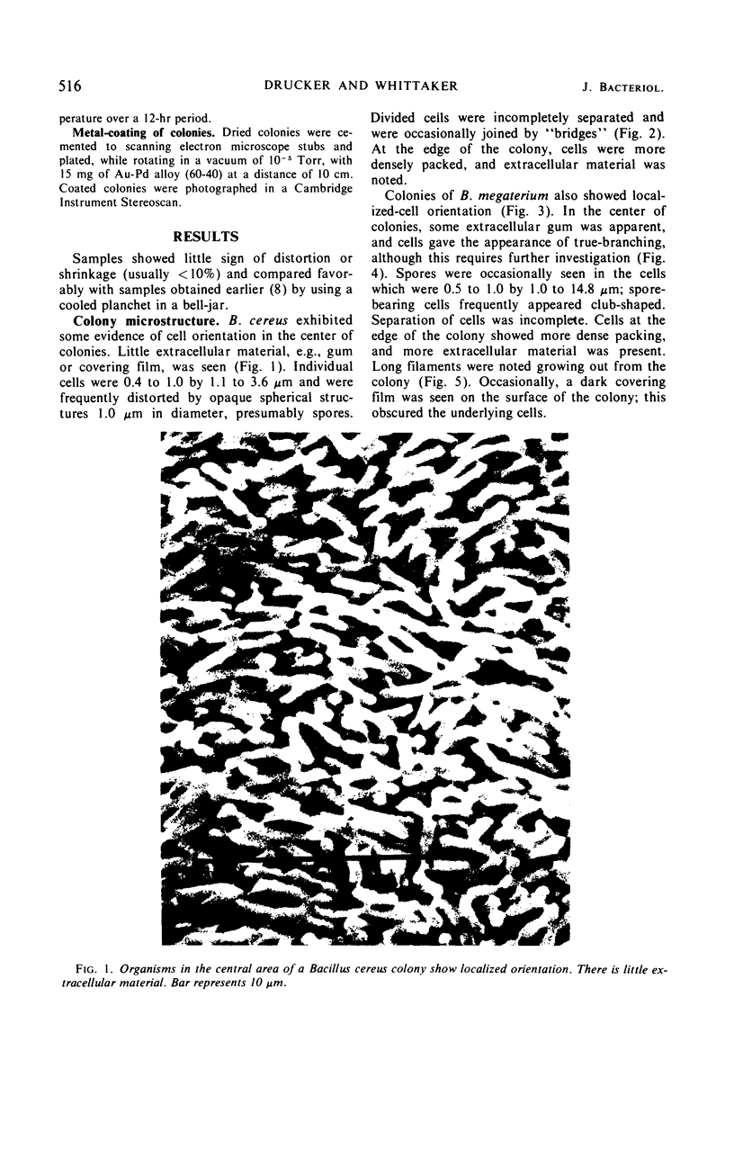
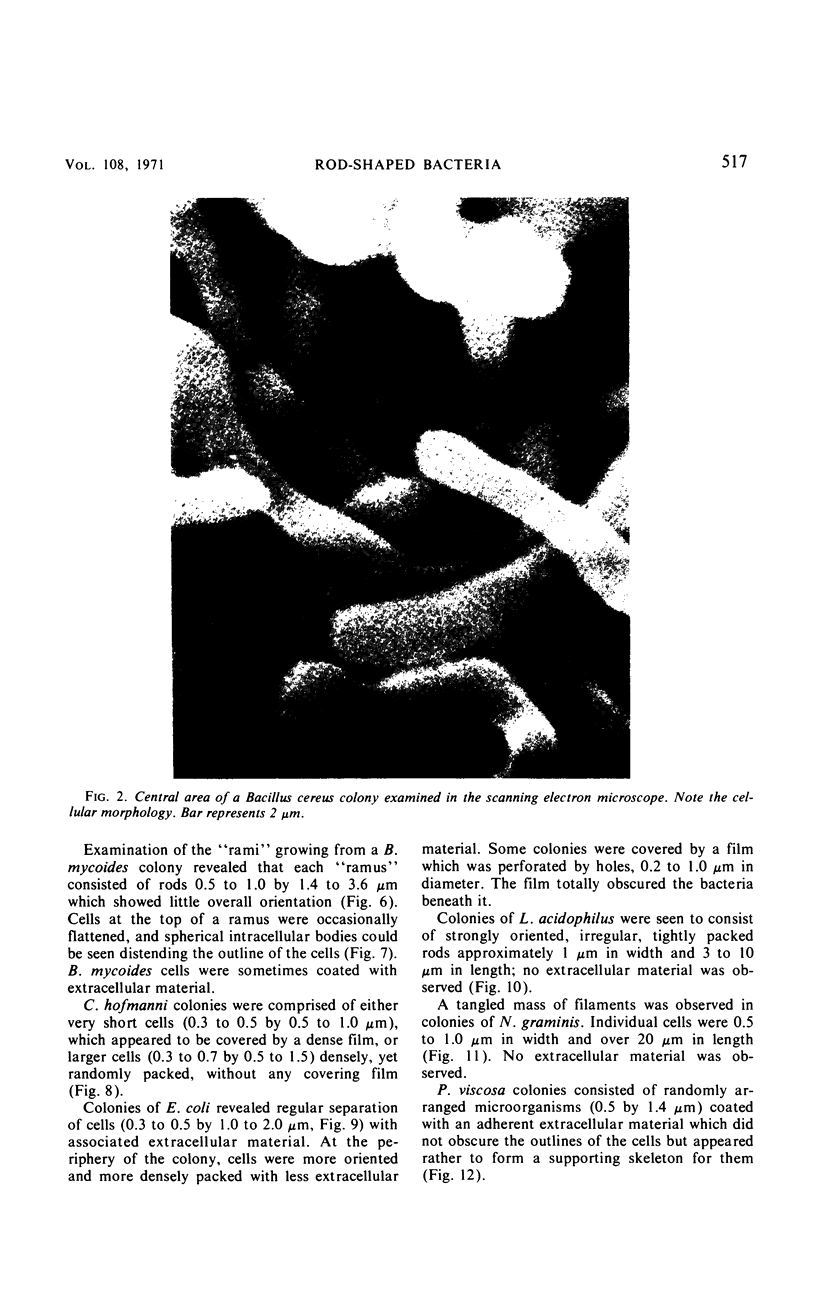
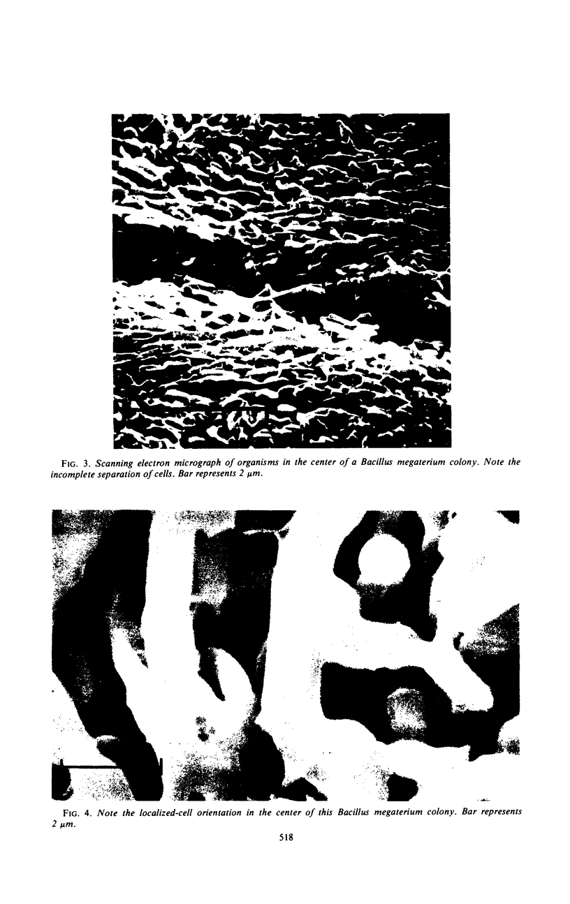
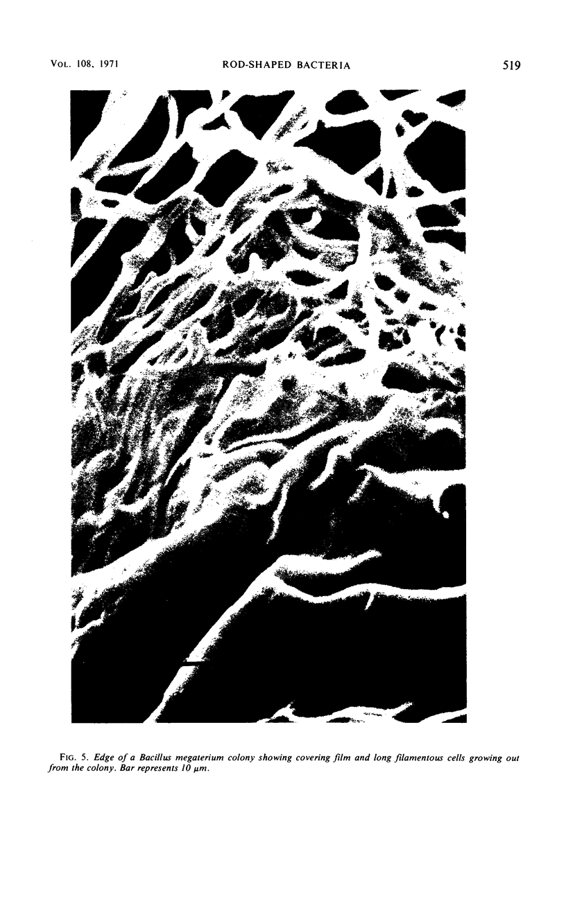
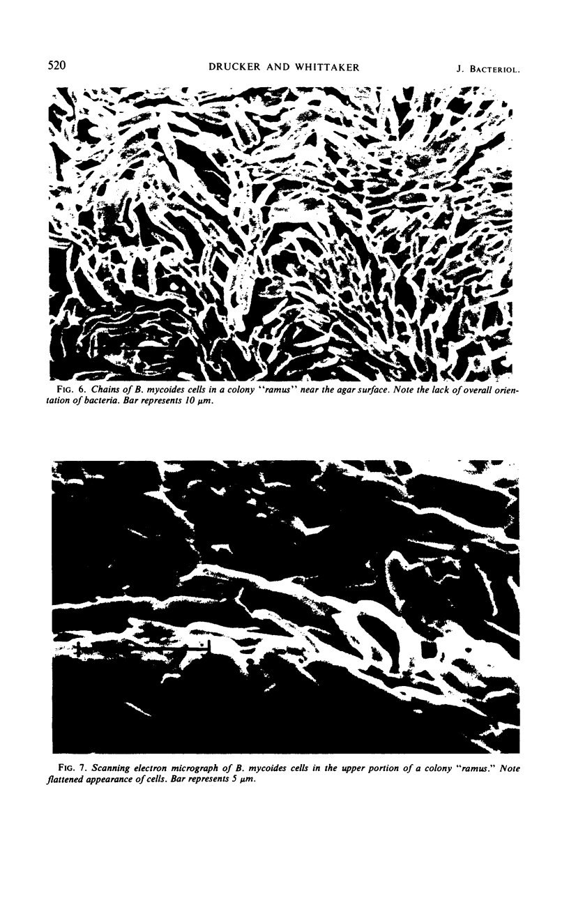
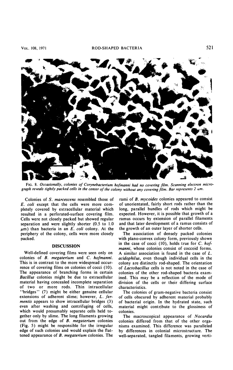
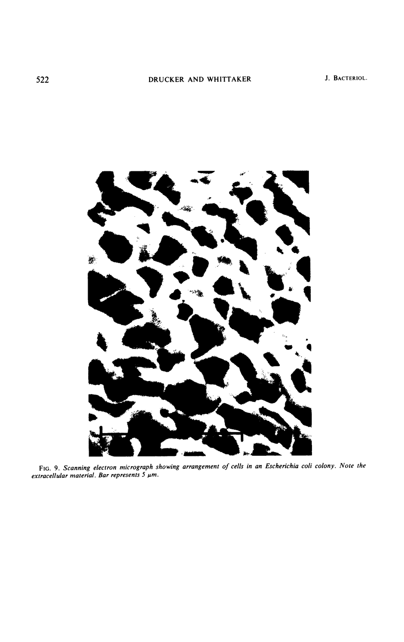
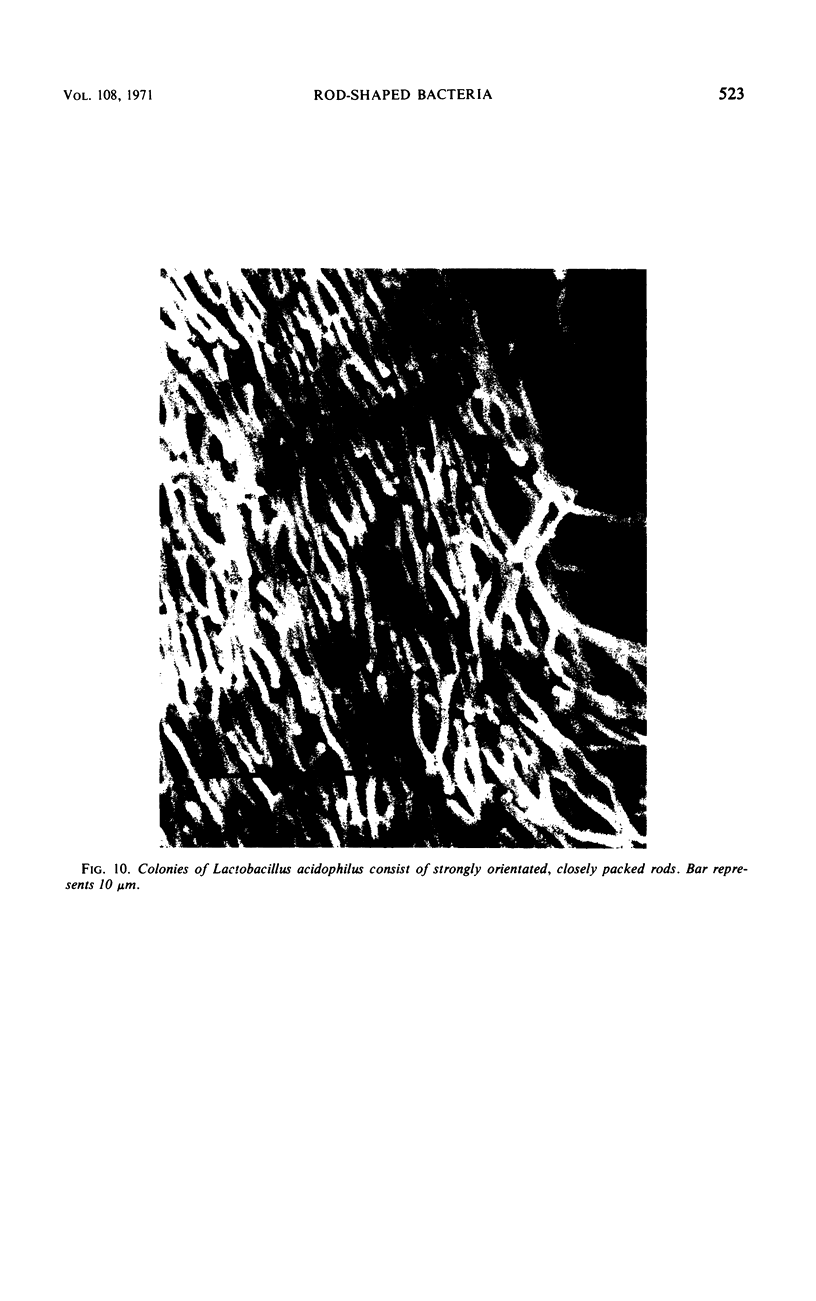
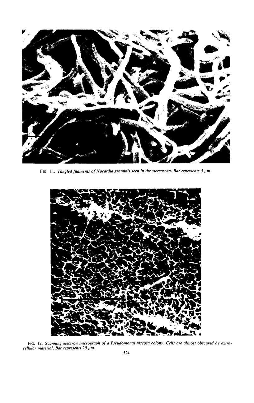
Whole colonies of Bacillus cereus, B. megaterium, B. mycoides CN2495, Corynebacterium hofmanni NCTC1938, Escherichia coli, Lactobacillus acidophilus NCIB1899, Nocardia graminis NCTC4728, Pseudomonas viscosa, and Serratia marcescens were prepared for scanning electron microscopic examination by freeze-drying and metal-coating. The arrangement of individual cells within colonies could be seen. Cells of Bacillus colonies tended to be longer than in liquid culture and irregular in shape and to give the appearance of branching. B. megaterium colonies frequently had a dense covering film. Colonies of gram-negative bacteria consisted of fairly short rods covered by much adherent extracellular material. L. acidophilus had colonies comprised of densely packed, well-oriented rods. C. hofmanni colonies contained coccobacilli, packed together. Correlations were observed between plano-convex colony form and densely packed cells, rough colony form and random arrangement of well-separated microorganisms, and irregular colony edge and tendency of cells to grow out from the colony in filaments.
Full text
PDF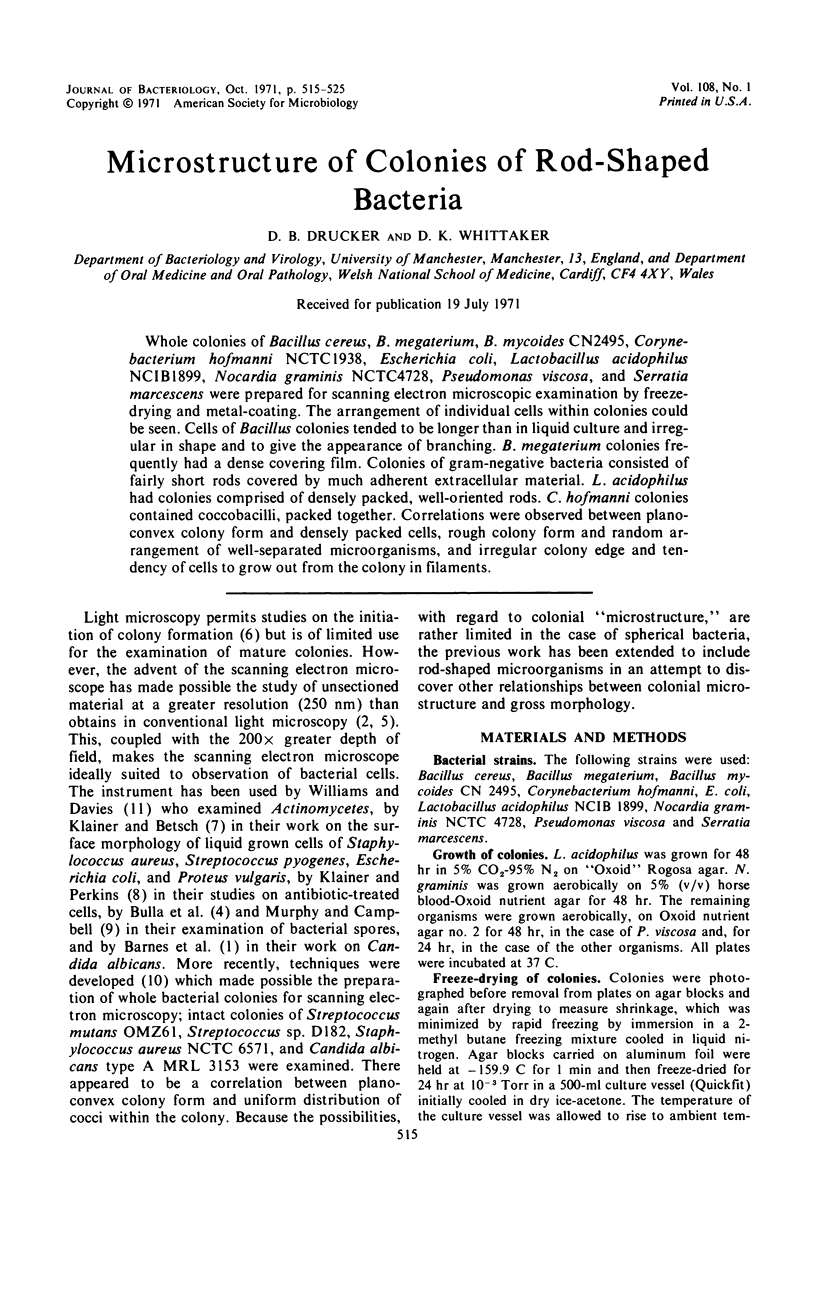
Images in this article
Selected References
These references are in PubMed. This may not be the complete list of references from this article.
- Barnes W. G., Flesher A., Berger A. E., Arnold J. D. Scanning electron microscopic studies of Candida albicans. J Bacteriol. 1971 Apr;106(1):276–280. doi: 10.1128/jb.106.1.276-280.1971. [DOI] [PMC free article] [PubMed] [Google Scholar]
- Boyde A., Knight P. J. The use of scanning electron microscopy in clinical dental research. Br Dent J. 1969 Oct 7;127(7):313–322. [PubMed] [Google Scholar]
- Boyde A., Williams R. A. Estimation of the volumes of bacterial cells by scanning electron microscopy. Arch Oral Biol. 1971 Mar;16(3):259–267. doi: 10.1016/0003-9969(71)90019-7. [DOI] [PubMed] [Google Scholar]
- Crewe A. V., Wall J. A scanning microscope with 5 A resolution. J Mol Biol. 1970 Mar;48(3):375–393. doi: 10.1016/0022-2836(70)90052-5. [DOI] [PubMed] [Google Scholar]
- Driedger A. A. The ordered growth pattern of microcolonies of Micrococcus radiodurans: first generation sectoring of induced lethal mutations. Can J Microbiol. 1970 Nov;16(11):1133–1135. doi: 10.1139/m70-191. [DOI] [PubMed] [Google Scholar]
- Klainer A. S., Betsch C. J. Scanning-beam electron microscopy of selected microorganisms. J Infect Dis. 1970 Mar;121(3):339–343. doi: 10.1093/infdis/121.3.339. [DOI] [PubMed] [Google Scholar]
- Klainer A. S., Perkins R. L. Antibiotic-induced alterations in the surface morphology of bacterial cells: a scanning-beam electron miscroscopy study. J Infect Dis. 1970 Oct;122(4):323–328. doi: 10.1093/infdis/122.4.323. [DOI] [PubMed] [Google Scholar]
- Murphy J. A., Campbell L. L. Surface features of Bacillus polymyxa spores as revealed by scanning electron microscopy. J Bacteriol. 1969 May;98(2):737–743. doi: 10.1128/jb.98.2.737-743.1969. [DOI] [PMC free article] [PubMed] [Google Scholar]
- Whittaker D. K., Drucker D. B. Scanning electron microscopy of intact colonies of microorganisms. J Bacteriol. 1970 Nov;104(2):902–909. doi: 10.1128/jb.104.2.902-909.1970. [DOI] [PMC free article] [PubMed] [Google Scholar]
- Williams S. T., Davies F. L. Use of scanning electron microscope for the examination of actinomycetes. J Gen Microbiol. 1967 Aug;48(2):171–177. doi: 10.1099/00221287-48-2-171. [DOI] [PubMed] [Google Scholar]