

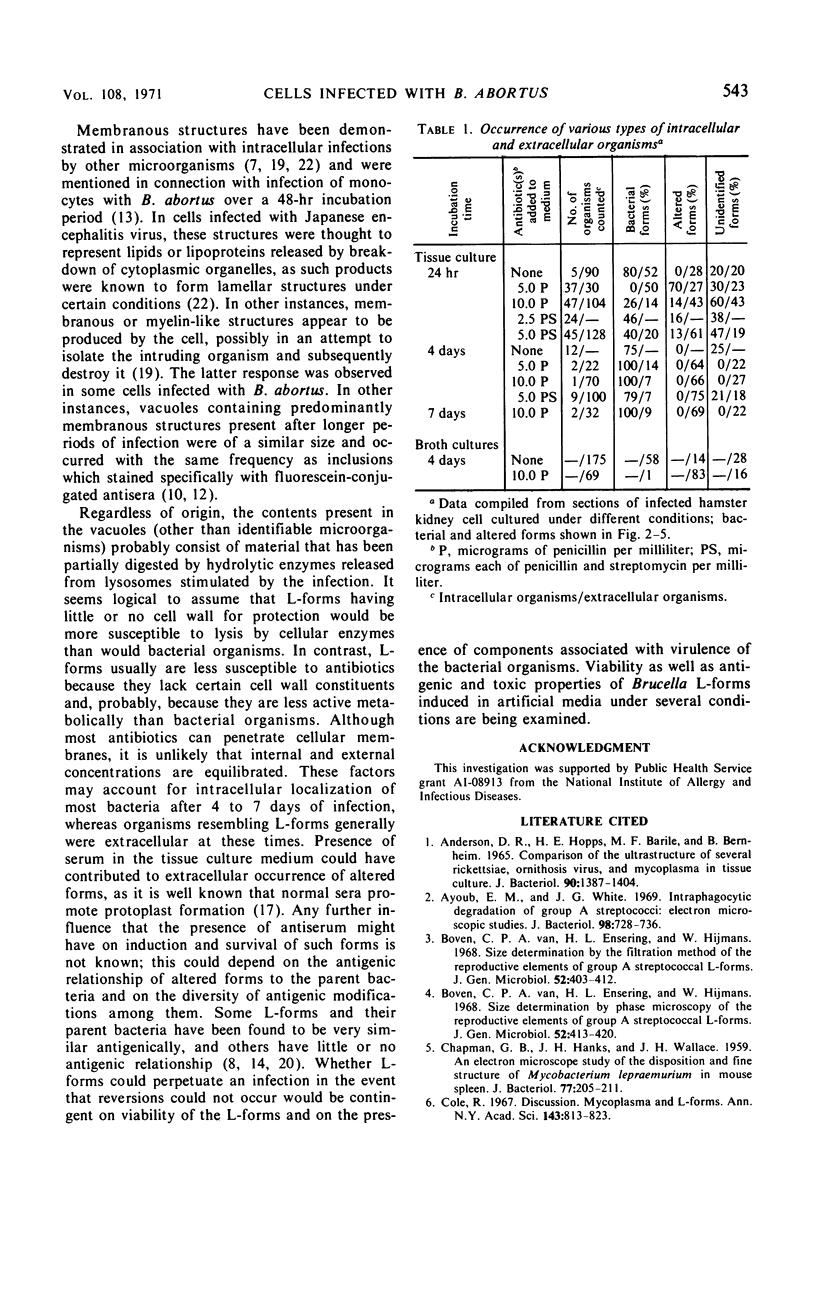
Abstract
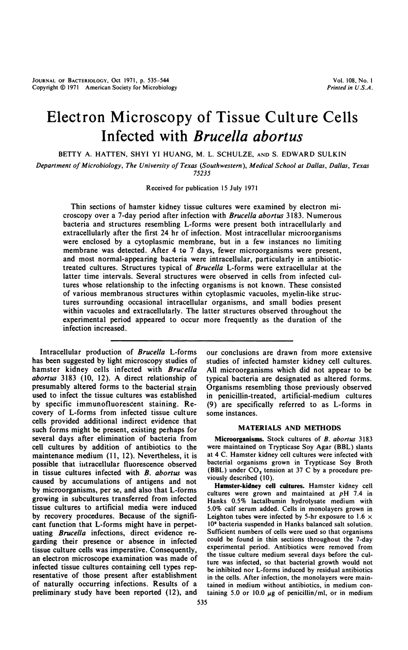
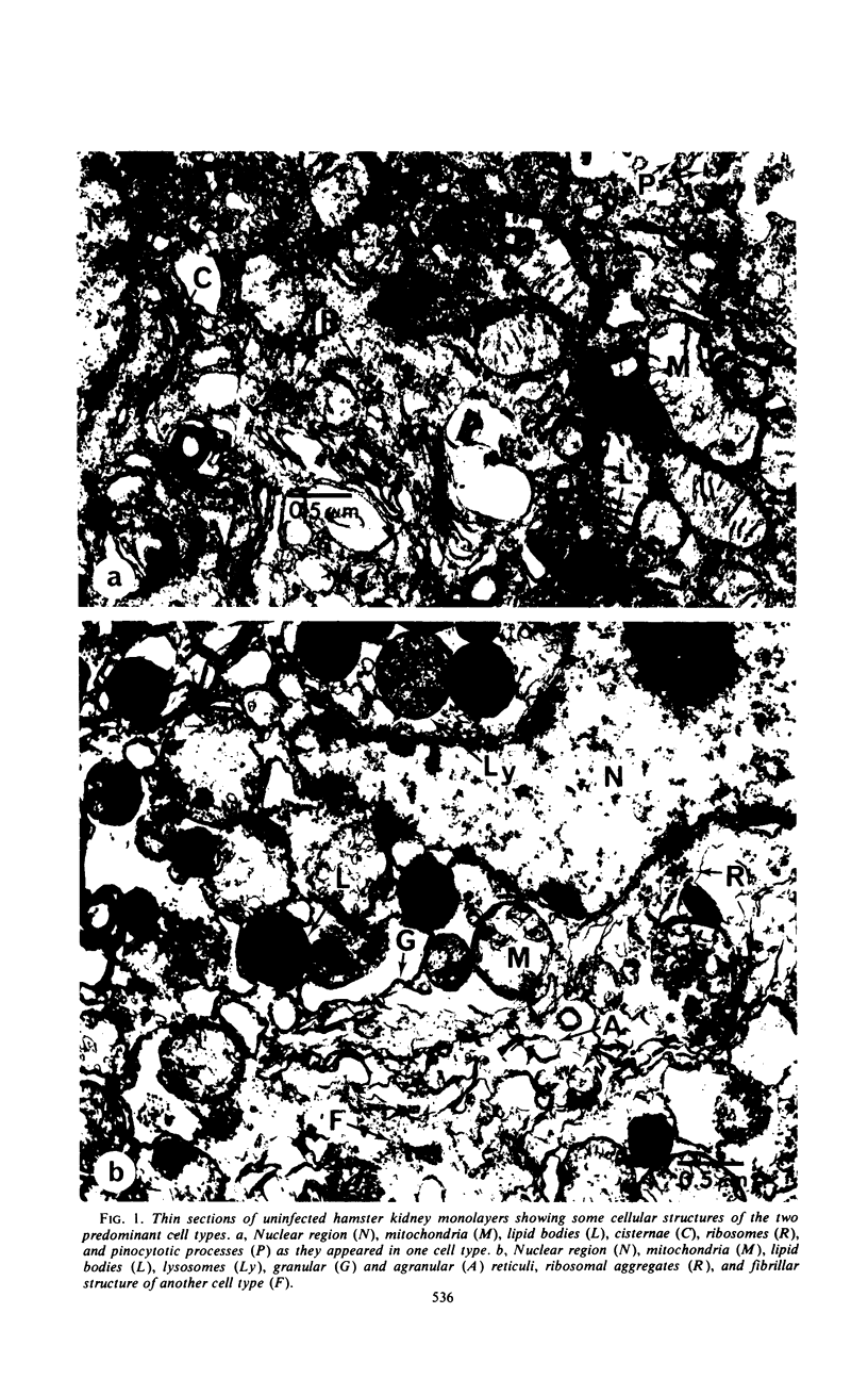
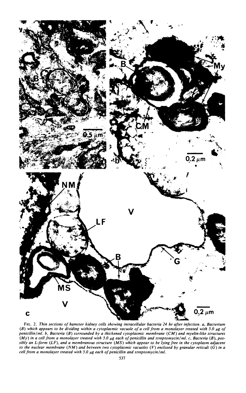
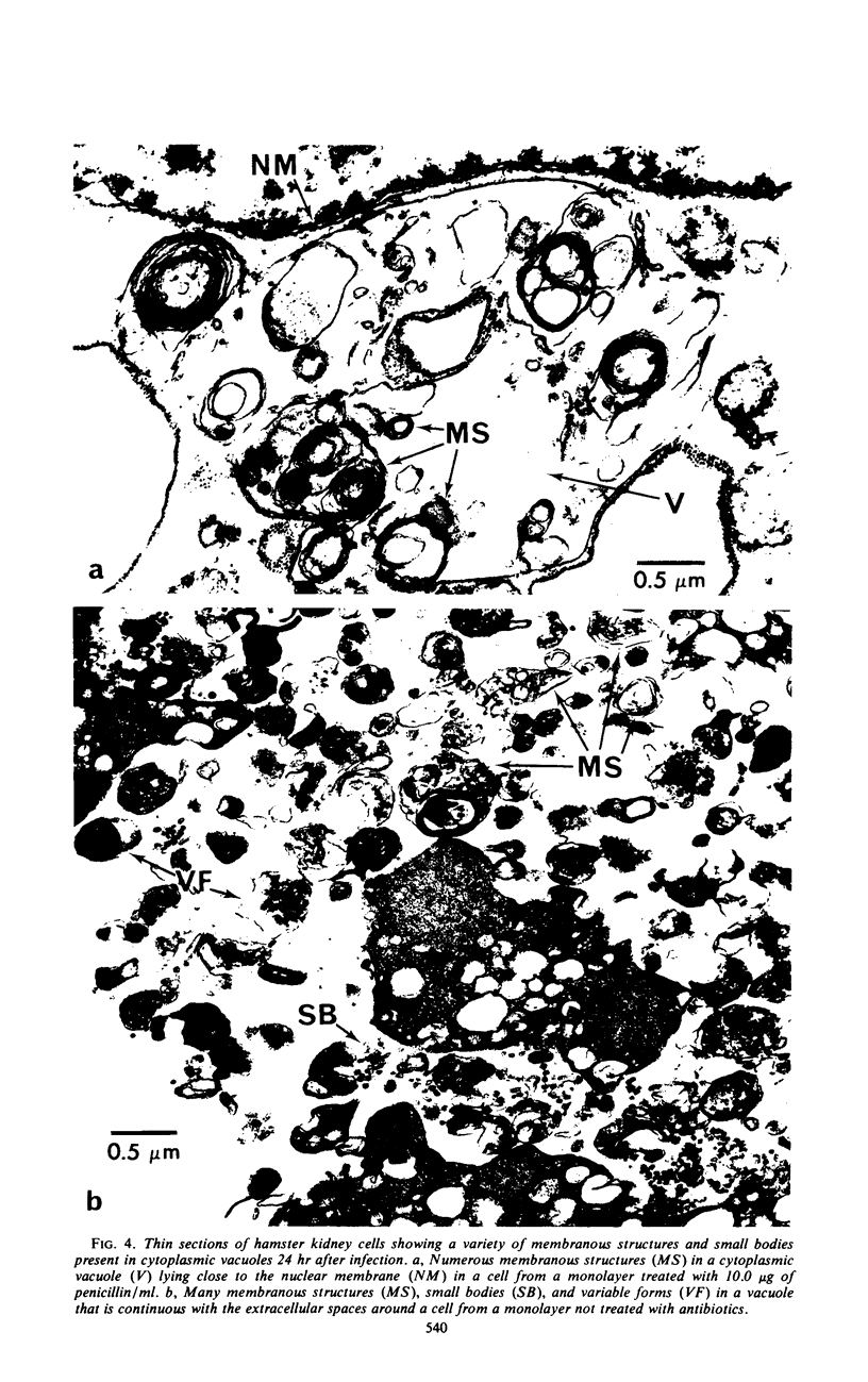
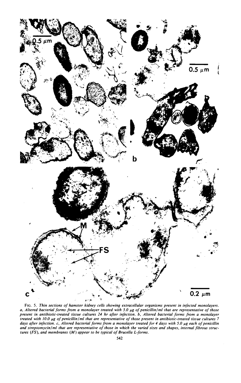
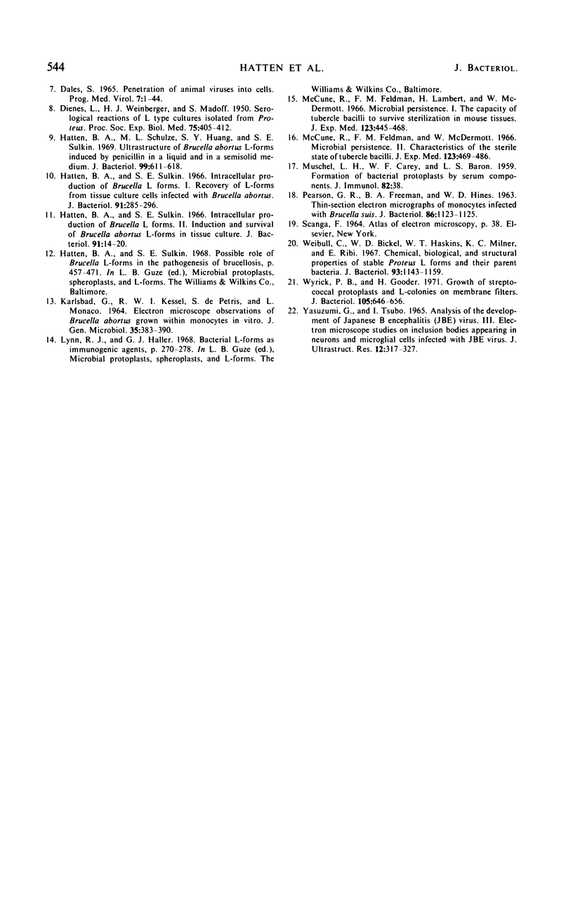
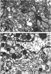
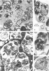
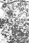
Thin sections of hamster kidney tissue cultures were examined by electron microscopy over a 7-day period after infection with Brucella abortus 3183. Numerous bacteria and structures resembling L-forms were present both intracellularly and extracellularly after the first 24 hr of infection. Most intracellular microorganisms were enclosed by a cytoplasmic membrane, but in a few instances no limiting membrane was detected. After 4 to 7 days, fewer microorganisms were present, and most normal-appearing bacteria were intracellular, particularly in antibiotic-treated cultures. Structures typical of Brucella L-forms were extracellular at the latter time intervals. Several structures were observed in cells from infected cultures whose relationship to the infecting organisms is not known. These consisted of various membranous structures within cytoplasmic vacuoles, myelin-like structures surrounding occasional intracellular organisms, and small bodies present within vacuoles and extracellularly. The latter structures observed throughout the experimental period appeared to occur more frequently as the duration of the infection increased.
Full text
PDF
Images in this article
Selected References
These references are in PubMed. This may not be the complete list of references from this article.
- Anderson D. R., Hopps H. E., Barile M. F., Bernheim B. C. Comparison of the ultrastructure of several rickettsiae, ornithosis virus, and Mycoplasma in tissue culture. J Bacteriol. 1965 Nov;90(5):1387–1404. doi: 10.1128/jb.90.5.1387-1404.1965. [DOI] [PMC free article] [PubMed] [Google Scholar]
- Ayoub E. M., White J. G. Intraphagocytic degradation of group A streptococci: electron microsopic studies. J Bacteriol. 1969 May;98(2):728–736. doi: 10.1128/jb.98.2.728-736.1969. [DOI] [PMC free article] [PubMed] [Google Scholar]
- CHAPMAN G. B., HANKS J. H., WALLACE J. H. An electron microscope study of the disposition and fine structure of Mycobacterium lepraemurium in mouse spleen. J Bacteriol. 1959 Feb;77(2):205–211. doi: 10.1128/jb.77.2.205-211.1959. [DOI] [PMC free article] [PubMed] [Google Scholar]
- Cole R. Discussion. Mycoplasma and L forms. Ann N Y Acad Sci. 1967 Jul 28;143(1):813–823. doi: 10.1111/j.1749-6632.1967.tb27729.x. [DOI] [PubMed] [Google Scholar]
- DIENES L., WEINBERGER H. J., MADOFF S. Serological reactions of L type cultures isolated from proteus. Proc Soc Exp Biol Med. 1950 Nov;75(2):409–412. doi: 10.3181/00379727-75-18213. [DOI] [PubMed] [Google Scholar]
- Dales S. Pentration of animal viruses into cells. Prog Med Virol. 1965;7:1–43. [PubMed] [Google Scholar]
- Hatten B. A., Schulze M. L., Huang S. Y., Sulkin S. E. Ultrastructure of Brucella abortus L-forms induced by penicillin in a liquid and in a semisolid medium. J Bacteriol. 1969 Aug;99(2):611–618. doi: 10.1128/jb.99.2.611-618.1969. [DOI] [PMC free article] [PubMed] [Google Scholar]
- Hatten B. A., Sulkin S. E. Intracellular Production of Brucella L Forms I. Recovery of L Forms from Tissue Culture Cells Infected with Brucella abortus. J Bacteriol. 1966 Jan;91(1):285–296. doi: 10.1128/jb.91.1.285-296.1966. [DOI] [PMC free article] [PubMed] [Google Scholar]
- Hatten B. A., Sulkin S. E. Intracellular production of Brucella L forms . II. Induction and survival of Brucella abortus L forms in tissue culture. J Bacteriol. 1966 Jan;91(1):14–20. doi: 10.1128/jb.91.1.14-20.1966. [DOI] [PMC free article] [PubMed] [Google Scholar]
- KARLSBAD G., KESSEL R. W., DE PETRIS S., MONACO L. ELECTRON MICROSCOPE OBSERVATIONS OF BRUCELLA ABORTUS GROWN WITHIN MONOCYTES IN VITRO. J Gen Microbiol. 1964 Jun;35:383–390. doi: 10.1099/00221287-35-3-383. [DOI] [PubMed] [Google Scholar]
- MUSCHEL L. H., CAREY W. F., BARON L. S. Formation of bacterial protoplasts by serum components. J Immunol. 1959 Jan;82(1):38–42. [PubMed] [Google Scholar]
- McCune R. M., Feldmann F. M., Lambert H. P., McDermott W. Microbial persistence. I. The capacity of tubercle bacilli to survive sterilization in mouse tissues. J Exp Med. 1966 Mar 1;123(3):445–468. doi: 10.1084/jem.123.3.445. [DOI] [PMC free article] [PubMed] [Google Scholar]
- McCune R. M., Feldmann F. M., McDermott W. Microbial persistence. II. Characteristics of the sterile state of tubercle bacilli. J Exp Med. 1966 Mar 1;123(3):469–486. doi: 10.1084/jem.123.3.469. [DOI] [PMC free article] [PubMed] [Google Scholar]
- PEARSON G. R., FREEMAN B. A., HINES W. D. THIN-SECTION ELECTRON MICROGRAPHS OF MONOCYTES INFECTED WITH BRUCELLA SUIS. J Bacteriol. 1963 Nov;86:1123–1125. doi: 10.1128/jb.86.5.1123-1125.1963. [DOI] [PMC free article] [PubMed] [Google Scholar]
- Weibull C., Bickel W. D., Haskins W. T., Milner K. C., Ribi E. Chemical, biological, and structural properties of stable Proteus L forms and their parent bacteria. J Bacteriol. 1967 Mar;93(3):1143–1159. doi: 10.1128/jb.93.3.1143-1159.1967. [DOI] [PMC free article] [PubMed] [Google Scholar]
- Wyrick P. B., Gooder H. Growth of streptococcal protoplasts and L-colonies on membrane filters. J Bacteriol. 1971 Feb;105(2):646–656. doi: 10.1128/jb.105.2.646-656.1971. [DOI] [PMC free article] [PubMed] [Google Scholar]
- YASUZUMI G., TSUBO I. ANALYSIS OF THE DEVELOPMENT OF JAPANESE B ENCEPHALITIS (JBE) VIRUS. 3. ELECTRON MICROSCOPE STUDIES ON INCLUSION BODIES APPEARING IN NEURONS AND MICROGLIAL CELLS INFECTED WITH JBE VIRUS. J Ultrastruct Res. 1965 Apr;12:317–327. doi: 10.1016/s0022-5320(65)80102-2. [DOI] [PubMed] [Google Scholar]