

Abstract
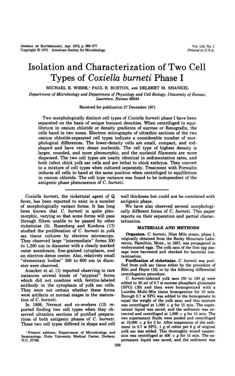
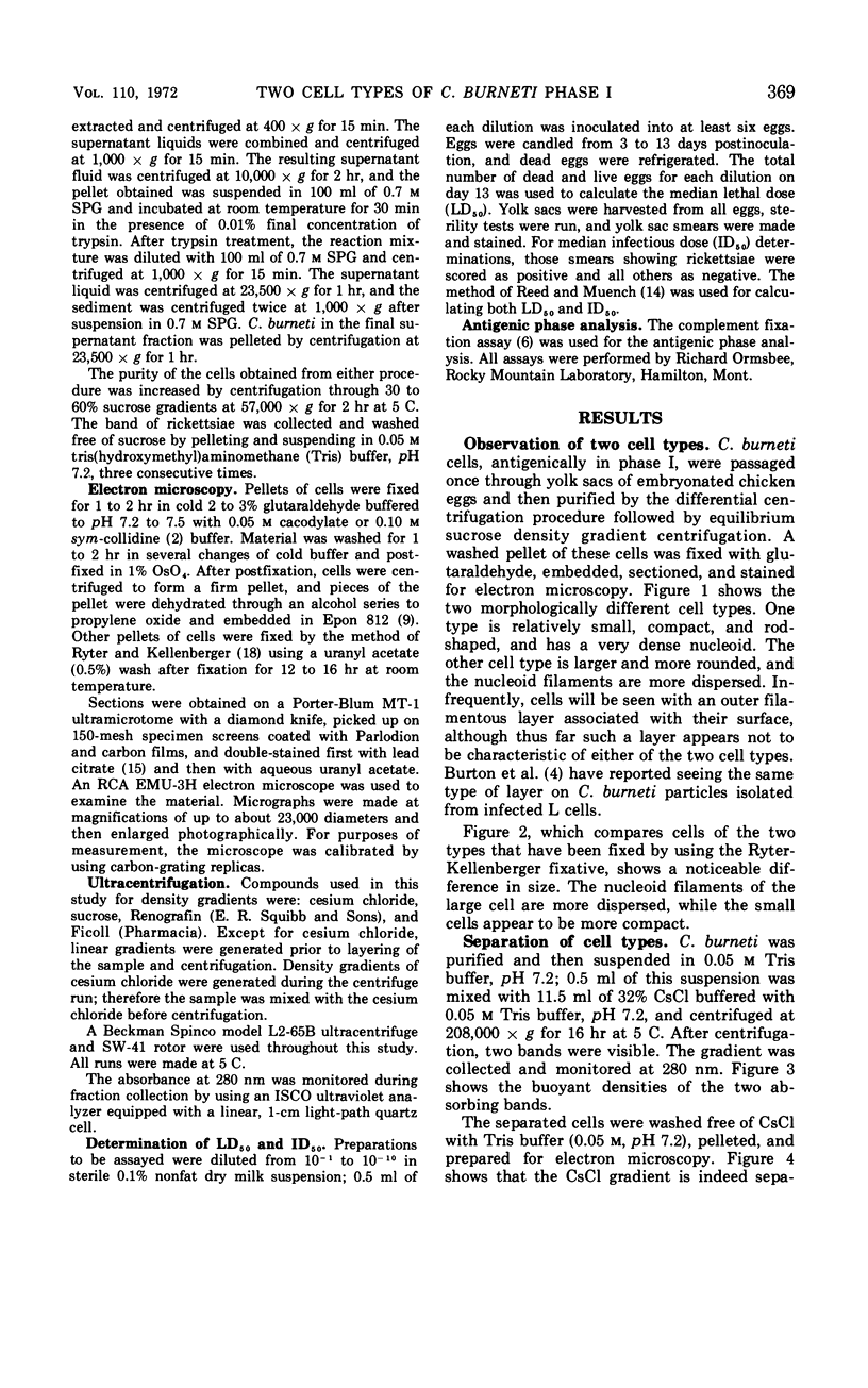
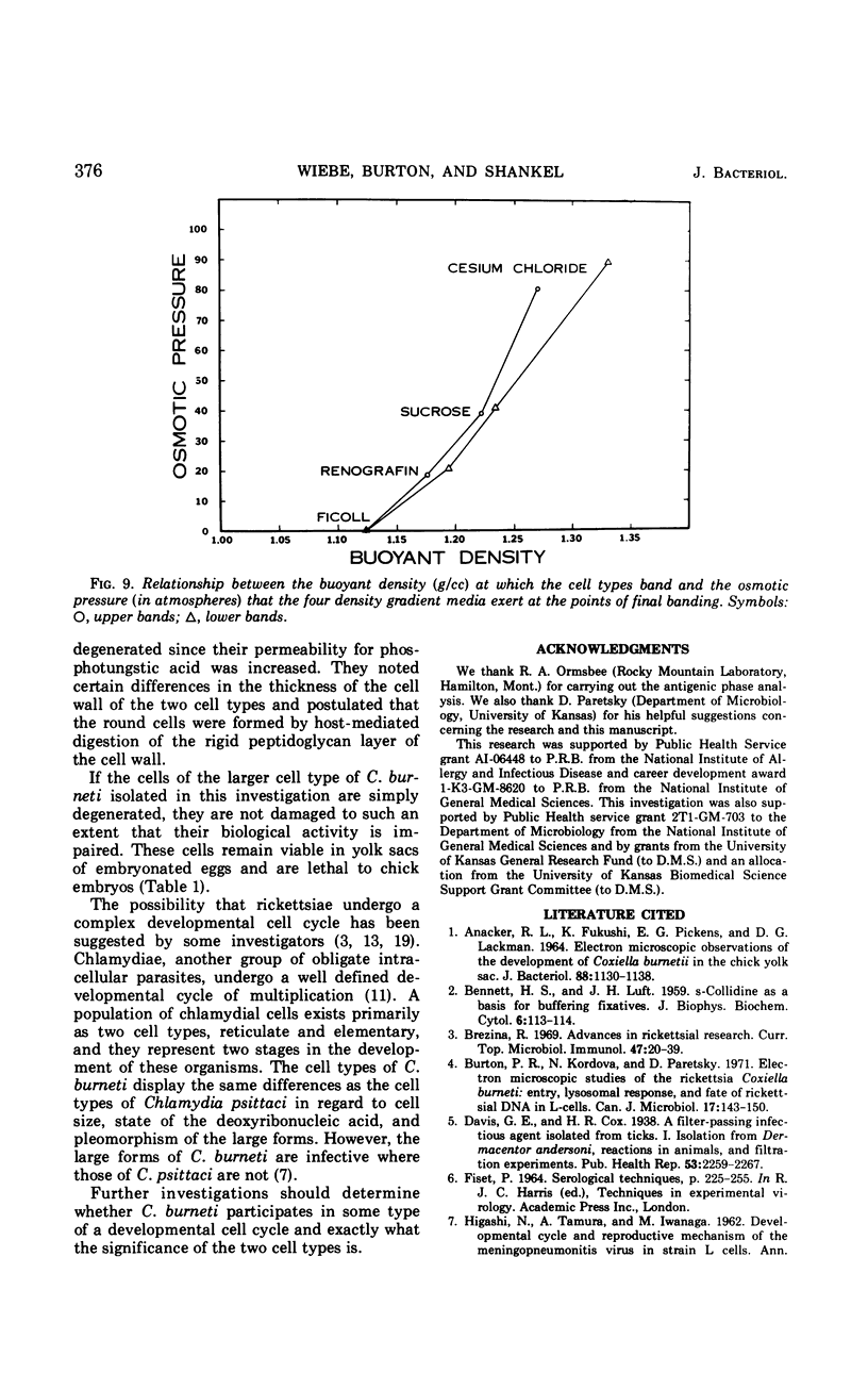
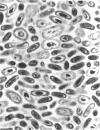
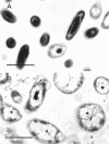
Two morphologically distinct cell types of Coxiella burneti phase I have been separated on the basis of unique buoyant densities. When centrifuged to equilibrium in cesium chloride or density gradients of sucrose or Renografin, the cells band in two zones. Electron micrographs of ultrathin sections of the two cesium chloride-separated cell types indicate a considerable number of morphological differences. The lower-density cells are small, compact, and rodshaped and have very dense nucleoids. The cell type of highest density is larger, rounded, and more pleomorphic, and the nucleoid filaments are more dispersed. The two cell types are nearly identical in sedimentation rates, and both infect chick yolk sac cells and are lethal to chick embryos. They convert to a mixture of cell types when cultured separately. Treatment with Formalin induces all cells to band at the same position when centrifuged to equilibrium in cesium chloride. The cell type variance was found to be independent of the antigenic phase phenomenon of C. burneti.
Full text
PDF
Images in this article
Selected References
These references are in PubMed. This may not be the complete list of references from this article.
- ANACKER R. L., FUKUSHI K., PICKENS E. G., LACKMAN D. B. ELECTRON MICROSCOPIC OBSERVATIONS OF THE DEVELOPMENT OF COXIELLA BURNETII IN THE CHICK YOLK SAC. J Bacteriol. 1964 Oct;88:1130–1138. doi: 10.1128/jb.88.4.1130-1138.1964. [DOI] [PMC free article] [PubMed] [Google Scholar]
- BENNETT H. S., LUFT J. H. zeta-Collidine as a basis for buffering fixatives. J Biophys Biochem Cytol. 1959 Aug;6(1):113–114. doi: 10.1083/jcb.6.1.113. [DOI] [PMC free article] [PubMed] [Google Scholar]
- Brezina R. Advances in rickettsial research. Curr Top Microbiol Immunol. 1969;47:20–39. doi: 10.1007/978-3-642-46160-6_2. [DOI] [PubMed] [Google Scholar]
- Burton P. R., Kordová N., Paretsky D. Electron microscopic studies of the rickettsia Coxiella burneti: entry, lysosomal response, and fate of rickettsial DNA in L-cells. Can J Microbiol. 1971 Feb;17(2):143–150. doi: 10.1139/m71-025. [DOI] [PubMed] [Google Scholar]
- HIGASHI N., TAMURA A., IWANAGA M. Developmental cycle and reproductive mechanism of the meningopneumonitis virus in strain L cells. Ann N Y Acad Sci. 1962 Mar 5;98:100–121. doi: 10.1111/j.1749-6632.1962.tb30536.x. [DOI] [PubMed] [Google Scholar]
- HOYER B. H., ORMSBEE R. A., FISET P., LACKMAN D. B. Differentiation of Phase I and Phase II Coxiella burnetti by equilibrium density gradient sedimentation. Nature. 1963 Feb 9;197:573–574. doi: 10.1038/197573a0. [DOI] [PubMed] [Google Scholar]
- LUFT J. H. Improvements in epoxy resin embedding methods. J Biophys Biochem Cytol. 1961 Feb;9:409–414. doi: 10.1083/jcb.9.2.409. [DOI] [PMC free article] [PubMed] [Google Scholar]
- McEwen C. R. Tables for estimating sedimentation through linear concentration gradients of sucrose solution. Anal Biochem. 1967 Jul;20(1):114–149. doi: 10.1016/0003-2697(67)90271-0. [DOI] [PubMed] [Google Scholar]
- Moulder J. W. The contribution of model systems to the understanding of infectious diseases. Perspect Biol Med. 1971 Spring;14(3):486–502. doi: 10.1353/pbm.1971.0024. [DOI] [PubMed] [Google Scholar]
- Nermut M. V., Schramek S., Brezina R. Electron microscopy of Coxiella burneti phase I and II. Acta Virol. 1968 Sep;12(5):446–452. [PubMed] [Google Scholar]
- Ormsbee R. A. Rickettsiae (as organisms). Annu Rev Microbiol. 1969;23:275–292. doi: 10.1146/annurev.mi.23.100169.001423. [DOI] [PubMed] [Google Scholar]
- REYNOLDS E. S. The use of lead citrate at high pH as an electron-opaque stain in electron microscopy. J Cell Biol. 1963 Apr;17:208–212. doi: 10.1083/jcb.17.1.208. [DOI] [PMC free article] [PubMed] [Google Scholar]
- ROSENBERG M., KORDOVA N. Study of intracellular forms of Coxiella burneti in the electron microscope. Acta Virol. 1960 Jan;4:52–55. [PubMed] [Google Scholar]
- RYTER A., KELLENBERGER E., BIRCHANDERSEN A., MAALOE O. Etude au microscope électronique de plasmas contenant de l'acide désoxyribonucliéique. I. Les nucléoides des bactéries en croissance active. Z Naturforsch B. 1958 Sep;13B(9):597–605. [PubMed] [Google Scholar]
- WISSEMAN C. L., Jr, HAHN F. E., JACKSON E. B., BOZEMAN M. F., SMADEL J. E. Metabolic studies of rickettsiae. II. Studies on the pathway of glutamate oxidation by purified suspensions of Rickettsia mooseri. J Immunol. 1952 Mar;68(3):251–264. [PubMed] [Google Scholar]
- Wisseman C. L., Jr Some biological properties of rickettsiae pathogenic for man. Zentralbl Bakteriol Orig. 1968 Apr;206(3):299–313. [PubMed] [Google Scholar]