Abstract
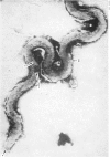
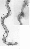
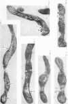
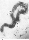
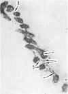
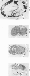
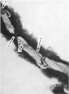
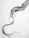
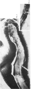
In recent years many investigations have been carried out on the morphology of Treponema pallidum by means of the electron microscope, and the use of ultra-thin sections has shown up a number of structural details. However, there is still need for much more evidence before the internal structure of treponemes can be elucidated fully and the functions of the structures interpreted. To provide such evidence, the authors have examined under the electron microscope negative-stained treponemes and ultra-thin sections, using both cultivated strains and treponemes obtained direct from syphilids in people suffering from fresh secondary syphilis. It has been shown that treponemes have a complex structure. T. pallidum has a two-layered outer wall, a cytoplasmic membrane proper, cytoplasm and a bunch of fibrils following a different path in different places on the treponeme. The sites of insertion of the fibrils (the basal granules) were investigated; structures similar to mesosomes and nucleoids were found. Cysts and granular forms are described.
Full text
PDF


Images in this article
Selected References
These references are in PubMed. This may not be the complete list of references from this article.
- Bladen H. A., Hampp E. G. Ultrastructure of Treponema microdentium and Borrelia vincentii. J Bacteriol. 1964 May;87(5):1180–1191. doi: 10.1128/jb.87.5.1180-1191.1964. [DOI] [PMC free article] [PubMed] [Google Scholar]
- CHRISTIANSEN S. Protective layer covering pathogenic treponemata. Lancet. 1963 Feb 23;1(7278):423–425. doi: 10.1016/s0140-6736(63)92309-2. [DOI] [PubMed] [Google Scholar]
- GREIFELT A., MOLBERT E. Elektronenmikroskopische Untersuchungen zur Morphologie des Treponema pallidum. Hautarzt. 1955 Jan;6(1):17–20. [PubMed] [Google Scholar]
- HARDY P. H., Jr, NELL E. E. Influence of osmotic pressure on the morphology of the Reiter treponeme. J Bacteriol. 1961 Dec;82:967–978. doi: 10.1128/jb.82.6.967-978.1961. [DOI] [PMC free article] [PubMed] [Google Scholar]
- LISTGARTEN M. A., LOESCHE W. J., SOCRANSKY S. S. MORPHOLOGY OF TREPONEMA MICRODENTIUM AS REVEALED BY ELECTRON MICROSCOPY OF ULTRATHIN SECTIONS. J Bacteriol. 1963 Apr;85:932–939. doi: 10.1128/jb.85.4.932-939.1963. [DOI] [PMC free article] [PubMed] [Google Scholar]
- LISTGARTEN M. A., SOCRANSKY S. S. ELECTRON MICROSCOPY OF AXIAL FIBRILS, OUTER ENVELOPE, AND CELL DIVISION OF CERTAIN ORAL SPIROCHETES. J Bacteriol. 1964 Oct;88:1087–1103. doi: 10.1128/jb.88.4.1087-1103.1964. [DOI] [PMC free article] [PubMed] [Google Scholar]
- MORTON H. E., FORD W. T. Preliminary observations of the action of penicillin on Treponema pallidum in vivo. Am J Syph Gonorrhea Vener Dis. 1953 Nov;37(6):529–535. [PubMed] [Google Scholar]
- MORTON H. E., OSKAY J. Electron microscope studies of treponemes; the effect of penicillin on the Nichols strain of Treponema pallidum. Am J Syph Gonorrhea Vener Dis. 1950 Jan;34(1):34-9, illust. [PubMed] [Google Scholar]
- MORTON H. E., RAKE G., ROSE N. R. Electron microscope studies of treponemes. III. Flagella. Am J Syph Gonorrhea Vener Dis. 1951 Nov;35(6):503–516. [PubMed] [Google Scholar]
- Mudd S., Polevitzky K., Anderson T. F. Bacterial Morphology as shown by the Electron Microscope: V. Treponema pallidum, T. macrodentium and T. microdentium. J Bacteriol. 1943 Jul;46(1):15–24. doi: 10.1128/jb.46.1.15-24.1943. [DOI] [PMC free article] [PubMed] [Google Scholar]
- OVCHINNIKOV N. M., ZELIKOVA R. L. Deistvie penitsillina na blednuiu spirokhetu v probirke (elektronnomikrospipicheskoe issledovanie). Vestn Venerol Dermatol. 1951 Jan-Feb;1:18–20. [PubMed] [Google Scholar]
- RYTER A., PILLOT J. [Electron microscope study of the external and internal structure of the Reiter treponema]. Ann Inst Pasteur (Paris) 1963 Apr;104:496–501. [PubMed] [Google Scholar]
- SWAIN R. H. Electron microscopic studies of the morphology of pathogenic spirochaetes. J Pathol Bacteriol. 1955 Jan-Apr;69(1-2):117–128. doi: 10.1002/path.1700690117. [DOI] [PubMed] [Google Scholar]
- WATSON J. H. L., ANGULO J. J., LEON-BLANCO F., VARELA G., WEDDERBURN C. C. Electron microscopic observations of flagellation in some species of the genus Treponema Schaudinn. J Bacteriol. 1951 Apr;61(4):455–461. doi: 10.1128/jb.61.4.455-461.1951. [DOI] [PMC free article] [PubMed] [Google Scholar]


