

Abstract
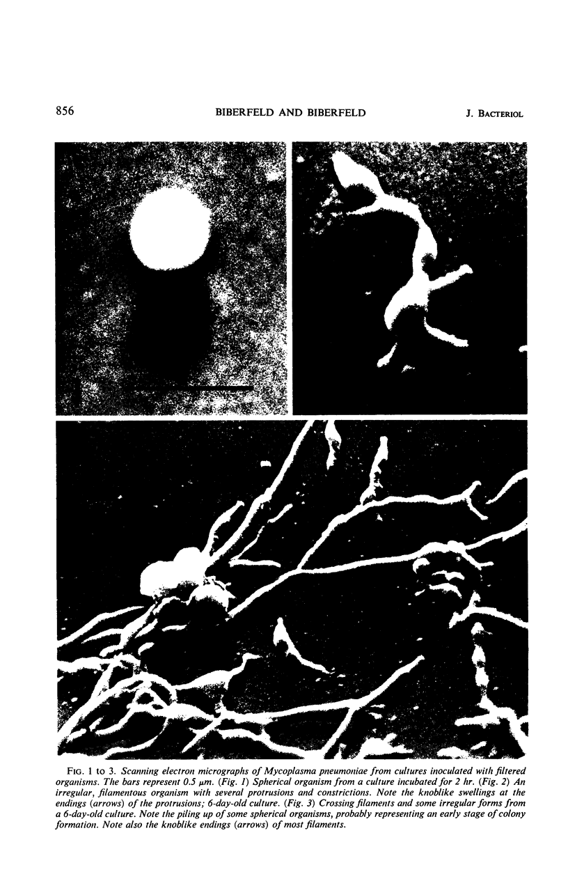
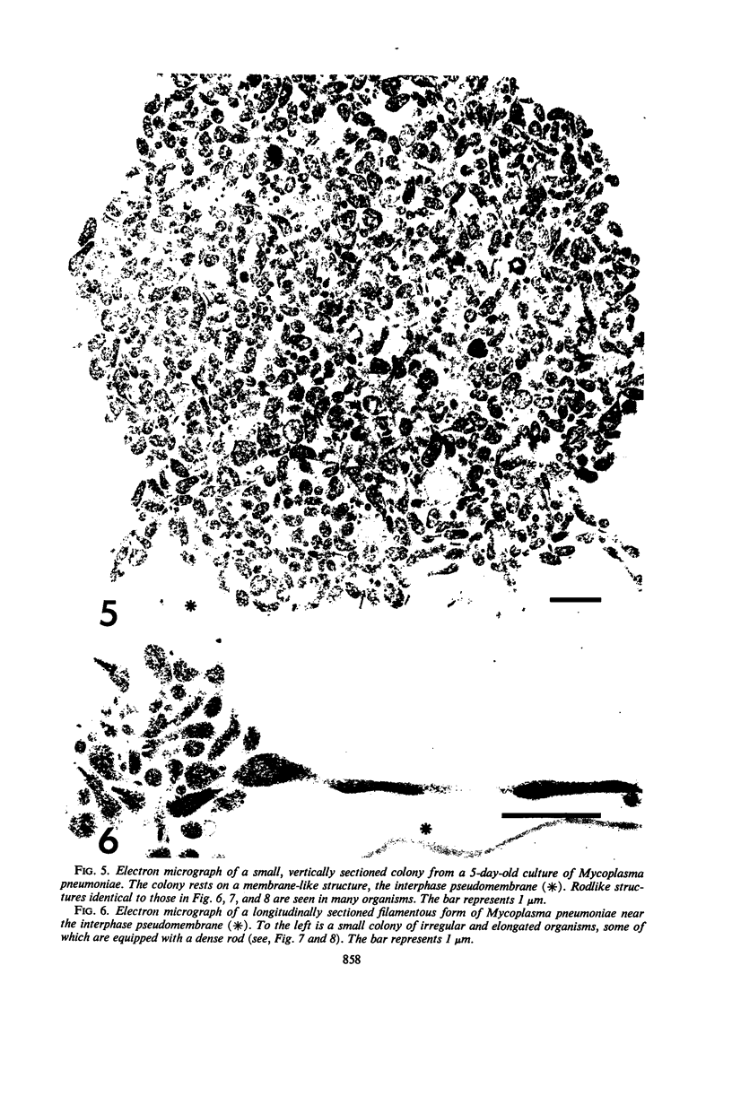
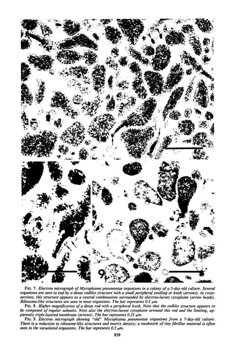
The ultrastructure of Mycoplasma pneumoniae cultivated in broth on glass and plastic surfaces was studied by scanning and transmission electron microscopy. The organisms grew as filaments, which by over-crossing eventually formed a dense network on the surface and in colonies composed mainly of rounded and elongated forms. The filaments were usually thinner at the ends and terminated with a knob-like structure. Some filaments possessed short ramifications which also ended with a knob, and others showed constrictions. Sectioned organisms were seen to contain ribosome-like structures. Many organisms had a specialized structure at their thinner end, which consisted of a dense rod surrounded by electron-lucent cytoplasm and ending with a platelike thickening.
Full text
PDF


Images in this article


Selected References
These references are in PubMed. This may not be the complete list of references from this article.
- Biberfeld P., Holm G., Perlmann P. Morphological observations on lymphocyte peripolesis and cytotoxic action in vitro. Exp Cell Res. 1968 Oct;52(2):672–677. doi: 10.1016/0014-4827(68)90508-9. [DOI] [PubMed] [Google Scholar]
- Bredt W. Growth morphology of Mycoplasma pneumoniae strain FH on glass surface. Proc Soc Exp Biol Med. 1968 Jun;128(2):338–340. doi: 10.3181/00379727-128-33009. [DOI] [PubMed] [Google Scholar]
- Bredt W. Motility and multiplication of Mycoplasma pneumoniae. A phase contrast study. Pathol Microbiol (Basel) 1968;32(6):321–326. doi: 10.1159/000162074. [DOI] [PubMed] [Google Scholar]
- Bredt W. Phasenkontrastmikroskopische Untersuchungen zu Morphologie und Vermehrung von Mycoplasma pneumoniae an Glas. Zentralbl Bakteriol Orig. 1968;208(4):549–562. [PubMed] [Google Scholar]
- DOMERMUTH C. H., NIELSEN M. H., FREUNDT E. A., BIRCH-ANDERSEN A. ULTRASTRUCTURE OF MYCOPLASMA SPECIES. J Bacteriol. 1964 Sep;88:727–744. doi: 10.1128/jb.88.3.727-744.1964. [DOI] [PMC free article] [PubMed] [Google Scholar]
- Furness G., Pipes F. J., McMurtrey M. J. Analysis of the life cycle of Mycoplasma pneumoniae by synchronized division and by ultraviolet and x irradiations. J Infect Dis. 1968 Feb;118(1):7–13. doi: 10.1093/infdis/118.1.7. [DOI] [PubMed] [Google Scholar]
- HAYFLICK L., CHANOCK R. M. MYCOPLASMA SPECIES OF MAN. Bacteriol Rev. 1965 Jun;29:185–221. doi: 10.1128/br.29.2.185-221.1965. [DOI] [PMC free article] [PubMed] [Google Scholar]
- Kim K. S., Clyde W. A., Jr, Denny F. W. Physical properties of human Mycoplasma species. J Bacteriol. 1966 Jul;92(1):214–219. doi: 10.1128/jb.92.1.214-219.1966. [DOI] [PMC free article] [PubMed] [Google Scholar]
- MOLLENHAUER H. H. PLASTIC EMBEDDING MIXTURES FOR USE IN ELECTRON MICROSCOPY. Stain Technol. 1964 Mar;39:111–114. [PubMed] [Google Scholar]
- Nelson J. B., Lyons M. J. Phase-contrast and electron microscopy of murine strains of Mycoplasma. J Bacteriol. 1965 Dec;90(6):1750–1763. doi: 10.1128/jb.90.6.1750-1763.1965. [DOI] [PMC free article] [PubMed] [Google Scholar]
- STEMPAK J. G., WARD R. T. AN IMPROVED STAINING METHOD FOR ELECTRON MICROSCOPY. J Cell Biol. 1964 Sep;22:697–701. doi: 10.1083/jcb.22.3.697. [DOI] [PMC free article] [PubMed] [Google Scholar]
- Somerson N. L., James W. D., Walls B. E., Chanock R. M. Growth of Mycoplasma pneumoniae on a glass surface. Ann N Y Acad Sci. 1967 Jul 28;143(1):384–389. doi: 10.1111/j.1749-6632.1967.tb27680.x. [DOI] [PubMed] [Google Scholar]
- Taylor-Robinson D., Manchee R. J. Adherence of mycoplasmas to glass and plastic. J Bacteriol. 1967 Nov;94(5):1781–1782. doi: 10.1128/jb.94.5.1781-1782.1967. [DOI] [PMC free article] [PubMed] [Google Scholar]
- VENABLE J. H., COGGESHALL R. A SIMPLIFIED LEAD CITRATE STAIN FOR USE IN ELECTRON MICROSCOPY. J Cell Biol. 1965 May;25:407–408. doi: 10.1083/jcb.25.2.407. [DOI] [PMC free article] [PubMed] [Google Scholar]
- Zucker-Franklin D., Davidson M., Thomas L. The interaction of mycoplasmas with mammalian cells. I. HeLa cells, neutrophils, and eosinophils. J Exp Med. 1966 Sep 1;124(3):521–532. doi: 10.1084/jem.124.3.521. [DOI] [PMC free article] [PubMed] [Google Scholar]