Summary
Progesterone is an ovarian steroid hormone that is essential for normal breast development during puberty and in preparation for lactation. The actions of progesterone are primarily mediated by its high affinity receptors, including the classical progesterone receptor (PR) -A and -B isoforms, located in diverse tissues such as the brain where progesterone controls reproductive behavior, and the breast and reproductive organs. Progestins are frequently prescribed as contraceptives or to alleviate menopausal symptoms, wherein progestin is combined with estrogen as a means to block estrogen-induced endometrial growth. Estrogen is undisputed as a potent breast mitogen, and inhibitors of the estrogen receptor (ER) and estrogen producing enzymes (aromatases) are effective first-line cancer therapies. However, PR action in breast cancer remains controversial. Herein, we review existing evidence from in vitro and in vivo models, and discuss the challenges to defining a role for progesterone in breast cancer.
Keywords: progesterone, progesterone receptor, breast cancer, estrogen receptor, growth factor, protein kinase, steroid hormone, hormone replacement therapy
Introduction: Alterations between the normal and neoplastic breast
Complex factors contribute to the challenge of demonstrating a clear role for progesterone in breast cancer. First, progesterone is difficult to study in isolation from other hormones (i.e. growth factors, prolactin) that also contribute to breast cancer biology. Second, progesterone receptor (PR) isoforms are expressed in response to estrogen receptor-alpha (ER) mediated transcriptional events, but can also occur independently of ER [1]. The subset of mammary epithelial cells (MECs) in the breast that express both PR-A and PR-B also express ER, and estrogen is usually required in order to induce the robust expression of PR in these ER+ cells. As estrogen is also a potent breast mitogen, this makes difficult to separate the effects of progesterone alone from those of estrogen. Indeed, PR isoforms are grossly understudied relative to ER in both the normal and neoplastic breast.
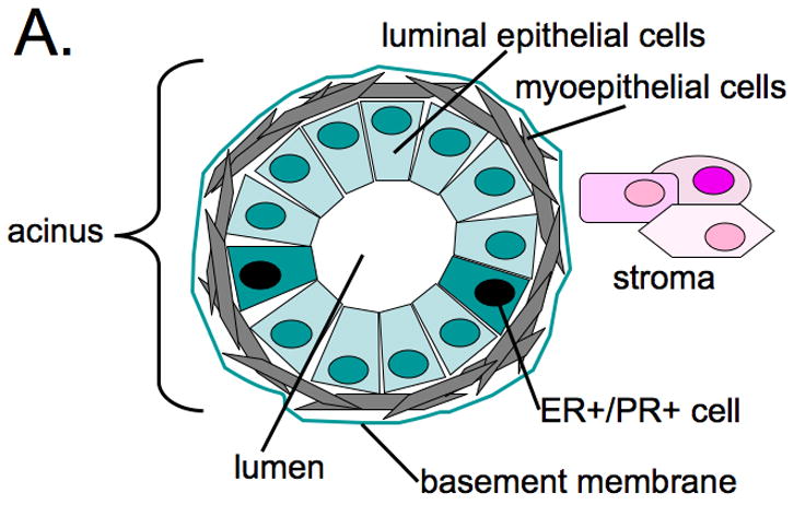
Studies in steroid hormone receptor knock-out mice have revealed that the concerted actions of estrogen and progesterone are required for normal mammary gland development [2,3]; estrogen/ER promotes the growth of ducts that invade the mammary fat pad emanating from the nipple, while estrogen/ER and progesterone/PR are required for the development of the terminal end-buds (TEBs) or acini located at the ends of ducts that later become the milk producing structures in the lactating mammary gland (Fig. 1). Additional required hormones, known as epidermal growth factor (EGF) and insulin-like growth factor (IGF-1) augment the proliferation of terminal end-buds during normal breast development, and promote ductal outgrowth and side branching induced by estrogen plus progesterone [4,5]. In fact, PR isoform expression in response to estrogen requires the presence of EGF [6], suggesting the existence of important cross talk between EGF receptors (EGFR) and/or family members (erbB2) and both steroid hormone receptors.
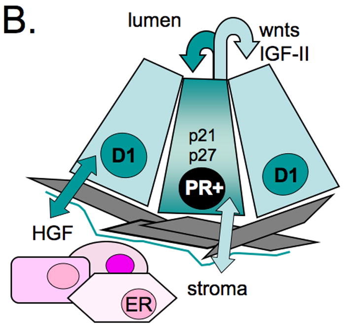
Figure 1. Mammary gland structure.


A. Acini, located at the ends of ducts in the adult mammary gland, are the functional units of the lactating mammary gland. Luminal epithelial cells (apical) exist as polar cells in contact with myoepithelial cells (basal). Epithelial cell populations are separated from the stroma by a basement membrane. B. Steroid hormone receptor positive (ER+/PR+) cells occur adjacent to proliferating cells in the normal mammary gland. Communication (paracrine signaling) between the epithelial and stromal compartments mediates proliferation of ER/PR negative cells. Early events during breast cancer development may mediate switching from paracrine to autocrine mechanisms of proliferation in ER+/PR+ cells.
Another limitation to deciphering a role for progesterone/PR action in breast cancer development is that normal proliferating breast epithelial cells are steroid hormone receptor negative [7]. In the normal adult mammary gland, ER+/PR+ cells represent only about 7–10% of the luminal epithelial cell population; these cells are most often non-dividing, but usually lie adjacent to proliferating cells (Fig. 1). The most current information suggests that ER+/PR+ cells are capable of proliferating, but are growth-arrested by the expression of inhibitory molecules, such as TGF-beta or high levels of p21 and p27, the endogenous inhibitors of cell-cycle-dependent protein kinases (CDKs). Communication between the breast epithelial and stromal compartments mediates the proliferation of nearby or adjacent cells by expression and secretion of locally active pro-proliferative molecules such as wnts, IGF-II [7], or stroma-derived hepatocyte growth factor (HGF) [8]. Recent evidence suggests that ER+/PR+ cells may act as “feeder cells” by providing growth-promoting substances (i.e. wnts) to nearby progenitor or stem cell populations [9].
In contrast to the normal breast, where proliferating cells are most often devoid of steroid hormone receptors, the majority of breast cancers (~70%) express ER and PR at the time of diagnosis. Although steroid hormone receptor-positive tumors are most often slower growing relative to receptor-negative tumors [10], ER+/PR+ breast epithelial cells may undergo an early switch to autocrine or paracrine signaling mechanisms whereby negative controls on proliferation are somehow lifted. Another setting where PR-containing cells clearly divide is in the pregnant mammary gland, where PR-B colocalizes with cyclin D1 in BrdU-stained (dividing) cells [11]. Thus, pathways involved in normal mammary gland growth and development may inappropriately “re-assert” themselves during breast cancer progression. Experimental evidence in model organisms (primates, mice, rats) and humans suggests a pro-proliferative role for progestins [12–14]. Herein, we review the status of progesterone/PR action in breast cancer models, and suggest a potential for future development of PR antagonists as part of combined breast cancer therapies.
Integration of PR classical and membrane-associated rapid signaling
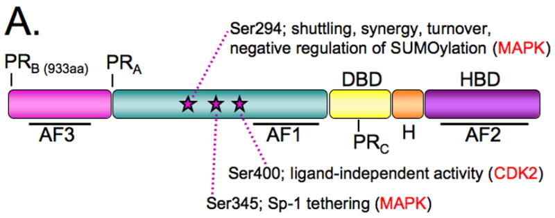
PR isoforms are classically defined as ligand-activated transcription factors and members of a large family of related steroid hormone receptors (that includes ER, androgen receptor (AR), glucocorticoid receptor (GR), and mineralocorticoid receptor). PRs are activated upon binding the naturally occurring ovarian steroid hormone, progesterone, or via binding to synthetic ligands (progestins) and regulate gene expression by binding directly or indirectly to specific sites in DNA (Fig. 2). Three PR isoforms (Fig. 2A) are the distinct protein products of a single gene located on chromosome 11 at q22-23. Transcription of PR isoforms is governed by the use of “distal” and “proximal” promoter regions [15]. The presence of internal translational start sites within common mRNAs results in the creation of three protein isoforms that consist of the full length PR-B (116 kDa), N-terminally-truncated PR-A (94 kDa), and PR-C-isoforms (60 kDa). PR-positive cells most often co-express PR-A and PR-B isoforms; these receptors exhibit different transcriptional activities within the same promoter context, but can also recognize entirely different gene promoters [16,17]. PR-B is essential for normal mammary gland development [18], while PR-A is required for uterine development and reproductive function [19]. PR-C is devoid of transcriptional activity, but when expressed, can enhance PR activity in breast cancer cells [20] or function as a dominant inhibitor of PR-B in the uterus [21].
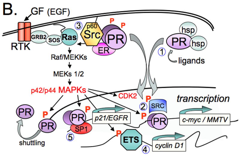
Figure 2. Progesterone receptor-dependent integrated actions.


A. Ligand activated PR-B and PR-A transcription factors contain a hormone binding domain (HBD), hinge region (H), DNA-binding domain (DBD), and amino terminus. Activation functions (AFs) represent the sites of co-regulator interaction required for transcription. Serines 294, 345, and 400 are regulatory sites that are phosphorlyated in response to progestins and/or mitogenic signaling pathways that modify PR function. B. Phosphorylation (P) of specific sites in PR couples multiple receptor functions. 1. Ligand-binding mediates dissociation of heat-shock proteins (hsps) and nuclear accumulation of PR. 2. Nuclear PRs regulate gene expression via the classical (PRE-dependent) pathway; phosphorylated PR recruit regulatory molecules that are phospho-proteins, and may function in one or more inter-connected processes (transcription, localization, and turnover). 3. PR and growth factors activate MAPKs via a c-Src kinase-dependent pathway, and this may result in positive regulation of PR transcriptional activity via “feed-back” regulation (i.e. direct phosphorylation of liganded PR or co-activators), occurring in both the absence and presence of ligands and on PRE-containing or other PR-regulated gene promoters. 4. Activation of MAPKs by PR provides for regulation of genes whose promoters do not contain PREs and are otherwise independent of PR-transcriptional activities but utilize PR-activated MAPKs. 5. In response to progestins, c-Src and MAPK-dependent phosphorylation of PR Ser345 mediates tethering to Sp1 and selective regulation of growth promoting genes via Sp1 sites (p21, EGFR).
Unliganded PRs are complexed with chaperone molecules including heat shock proteins (hsps); these interactions allow proper protein folding and assembly of stable PR molecules competent to bind hormone [22]. Hsps also mediate important aspects of PR protein trafficking. After binding to progesterone, receptor conformational changes induce dimerization and hsp dissociation (Fig. 2). Activated receptors associate with co-regulators, including steroid receptor coactivators (SRCs 1–3), are withheld in the nucleus, and bind directly to specific progesterone response elements (PREs) and PRE-like sequences in the promoter regions of target genes such as c-myc [23], fatty acid synthetase [24], and MMTV [25]. Treatment with progestin also results in the upregulation of genes without canonical PREs in their proximal promoter regions, such as Epidermal Growth Factor Receptor [26], c-fos [27], p21 [28], IRS-2 [29], and cyclin D1 [30]. Regulation of genes without PREs, PRE half-sites, or PRE-like sequences can occur through PR tethering to other DNA-binding transcription factors, such as Specificity protein 1 [28], Activating Protein 1 [31] or Signal Transducers and Activators of Transcription (Stats) [32,33].
The genomic or classical actions of steroid hormone treatment are delayed by several minutes to hours, dictated by the time required for transcription and translation of target genes. Recently, however, rapidly occurring (within a few minutes) extranuclear or non-genomic effects of cell membrane-localized steroid hormone receptors have entered the forefront. For example, progestin treatment of breast cancer cells causes a rapid and transient (2–15 mins) activation of cytoplasmic protein kinases, including mitogen-activated protein kinase (MAPK), PI3K, and p60-Src kinase [34–36]. Similar activities have been reported for membrane-associated ERalpha and AR [37]. These effects are mediated by direct binding of steroid hormone receptors to protein-protein interaction domains of signaling molecules located in or near the plasma membrane, in close proximity to growth factor receptors and their immediate effectors. Human PR contains an N-terminal proline-rich (PXXP) motif that mediates direct binding to the Src-homology three (SH3) domains of signaling molecules in the p60-Src kinase family in a ligand-dependent manner [34]. In vitro experiments demonstrate that progestin-bound, purified PR-A and PR-B directly activate the c-Src-related protein kinase, Hck; PR-B but not PR-A activates c-Src and MAPKs in vivo. Mutation of the PXXP sequence in PR-B disrupts the c-Src/PR interaction and blocks progestin-induced activation of c-Src (or Hck) and p42/p44 MAPKs. Furthermore, mutation of the PR-B DNA-binding domain (DBD) abolished PR transcriptional activity without blocking progestin-induced c-Src or MAP kinase activation. Thus, non-genomic MAPK activation by progestin/PR-B/c-Src complexes most likely occurs by way of a c-Src-dependent mechanism involving Ras activation of the Raf/MEK/MAPK module (Fig. 2B). ER in association with other signaling and adaptor molecules is suspected to reside in similar cytoplasmic signaling complexes, possibly in association with PR and c-Src [37].
In studies using human breast or prostate cancer cell lines, the rapid signaling actions of membrane associated AR, PR and/or ER have been shown to contribute to the regulation of cell proliferation in response to their respective hormone ligands [38–40]. While potential roles in human physiology (i.e. whole organisms) are less clear, steroid hormone receptor-mediated activation of cytoplasmic signaling molecules may primarily serve to potentiate the nuclear functions of these receptors (Fig. 2). For example, amplification of PR nuclear functions likely occurs through rapid, direct phosphorylation of PR proteins and/or receptor co-regulators in response to activation of PR-induced cytoplasmic pathways that are mechanistically coupled to ligand binding. Thus, appropriately phosphorylated and activated receptor complexes are efficiently directed to selected target genes. Clearly, such positive feedback explains the dramatic influence of activated signaling pathways on PR nuclear function. Indeed, several progestin/PR-dependent events are MAPK or c-Src-dependent, including upregulation of cyclins D1 and E, CDK2 activation, S-phase entry, and anchorage-independent cell growth in soft-agar [26,41,42]. C-Src- and MAPK-dependent direct phosphorylation of PR Ser345 is required for PR tethering to Sp1 transcription factors bound to the p21 and EGFR promoters [43]. PR/Sp1 tethering upon c-Src/MAPK pathway activation is predicted to alter PR promoter selectivity, favoring the use of Sp1-driven promoters within PR-target genes. Kinases also confer hyperactivity and ligand-independence to phosphorylated PR-B [42,44,45]. For example, MAPKs mediate PR hypersensitivity to ligand by phosphorylation of PR Ser294, an event that derepresses receptor activity by preventing PR sumoylation [46]. Activated CDK2 or loss of p27 induces PR ligand-independent activity via Ser400 phosphorylation [42]. Although more studies are needed, it is becoming clear that activation of cytoplasmic protein kinases is an integral feature of PR nuclear action (i.e. phosphorylation events are required for gene regulation leading to changes in cell biology). Thus, rapid phosphorylation events may primarily act to alter PR transcriptional activity, but clearly also mediate promoter selectivity [47].
How might the membrane-associated signaling actions of steroid hormone receptors, including PR, contribute to deregulated breast cancer cell growth and/or increased breast cancer risk? Perhaps by linking steroid hormone action to the expression of MAPK-regulated genes (i.e. the endpoint of MAPK signaling is the phosphorylation of transcription factors). In support of this concept, the extranuclear actions of liganded ERs induce a state of “adaptive hypersensitivity” during endocrine therapy in which growth factor signaling pathways are coopted by upregulated ERs [48]. In this model of ER-dependent MAPK activation, liganded ERs localized at the cell membrane interact with the adapter protein Shc and induce its phosphorylation, leading to recruitment of adaptor molecules and activation of Ras and the Raf-1/MEK/MAPK module. MAPK then regulates genes via direct phosphorylation of Ets factors and/or AP1 components (i.e. independently of ER transcriptional activity). ER activation of MAPK explains why many tumors respond well to aromatase inhibitors, yet fail to respond to selective estrogen receptor modulators (SERMS) designed to inhibit ER transcriptional activity in the nucleus, but not ER-dependent MAPK activation in the cytoplasm. Breast cancers often exhibit heightened c-Src and MAPK activities [49,50] and elevated cyclin D1, an AP1 target gene whose expression is sensitive to multiple kinase inputs [51–53]. Steroid hormone receptors including PRs may contribute to the constitutive signaling of cytoplasmic mitogenic protein kinases via their membrane-associated activities, thereby circumventing endocrine-based (i.e. antiestrogen) therapies (Fig. 2).
Probing PR action in animal models
Studies in rodents demonstrate that PR-A and PR-B are differentially expressed during mammary gland development, with PR-A predominantly expressed during ductal sidebranching, while PR-B expression coincides with the formation of alveoli [54,55]. PR-B but not PR-A is expressed in proliferating cells. Some but not all proliferating cells in both compartments are PR-B+, suggesting that progesterone may induce proliferation through either direct or paracrine mechanisms. In contrast, cells in adult virgin glands are PR and cyclin D1 positive, but fail to proliferate, possibly due to high levels of the cyclin-dependent protein kinase inhibitors, p21 and p27 [55]. During the menstrual cycle, MEC undergo sequential waves of proliferation and apoptosis. Notably, in primates (macaques and humans) increased terminal duct lobular unit cell proliferation coincides with the peak of serum progesterone that occurs during the luteal phase [13,56–58], again suggesting a paracrine mechanism for this hormone in adult tissues. Upregulation of local IGF-1 may be a cooperating factor in this regard [59]. In animal models of postmenopausal hormone replacement therapy, both parous and nulliparous early and late postmenopausal mice were subjected to estrogen alone (E) or estrogen plus progestin (E+P); E+P produced a greater proliferative response relative to E alone regardless of parity or treatment time. E+P was also shown to act directly on the mammary gland, rather than via systemic effects [60]. Similar results occurred in surgically postmenopausal macaques [61] and in postmenopausal humans [62]. Although breast cancer development was not modeled in the above animal studies, the results (i.e. increased proliferation) are consistent with human clinical data [63,64], which revealed increased tumor number and size in women taking E+P, while E alone did not significantly alter breast cancer risk or tumor size.
Progesterone may act via proto-oncogenes and growth factors to affect breast cell proliferation and breast cancer etiology. As the majority of early breast cancer lesions express both ER and PR and these receptors remain high in at least 60% of advanced decease, early events may include a switch in the ability of normally quiescent ER+/PR+ cells to respond directly to steroid hormones and proliferate (Fig. 1). Notably, deregulation of the cell cycle is a hallmark of breast cancer. Up to 40% of breast cancers overexpress cyclin D1, while at least 30% have lost p27 or p21 and/or contain activated CDK2 [65,66]. Mutation or loss of p53 is also a frequent occurrence [67]. Numerous in vitro studies have demonstrated linkage of PR action to cell cycle regulation [42,68–70]. Namely, PR interacts directly with cyclins A or E and CDK2 [69,70]. PR activity is highest in the DNA synthesis (S) phase of the cell cycle, when CDK2 activity peaks [68,69]. Furthermore, PR transcriptional activity becomes ligand-independent and CDK2-driven upon loss of p27 [42]. Progestins, acting through PR-B-dependent transcription, induce cyclin D1 expression and cell cycle re-entry in antiestrogen-arrested breast cancer cells [71]. This suggests that in the breast, progesterone/PR action is tightly coupled to mechanisms of cell cycle control. In breast cancer, the mitogenic potential of activated PRs may manifest particularly during loss of checkpoint control and/or elevation of CDK or other mitogenic kinase activities. For example, TGF-□ (an EGFR ligand) transgenic mice develop ER+/PR+ proliferative hyperplasias (early lesions) that rapidly progress to ER+/PR- tumors [72]. Recent in vitro studies demonstrate that BRCA-1 knock-down enhances progestin-induced PR transcriptional activity, while progestin-induced MEC proliferation is increased in genetically engineered mice lacking BRCA-1 in the mammary gland [73]. Related to this finding, in recent studies using mice lacking mammary gland expression of both BRCA-1 and p53, PR protein levels were dramatically increased, and the development of aggressive ER+/PR+ tumors in virgin mice was completely blocked by antiprogestins [74]. In future studies, it will be important to define how negative regulation of cell proliferation in ER+/PR+ normal MECs is somehow lifted to allow progression of early lesions to malignant cancer, and if early events include loss of checkpoint control or alteration of DNA damage and repair pathways in PR+ cells.
PR action in human breast cancer cell models
The biochemistry of PR action is well characterized, having been largely defined using PR+ human breast cancer cell lines, or PR-null cells into which wild-type or modified PRs has been re-expressed. Numerous studies have focused on PR interactions with regulatory proteins, changes in PR subcellular localization, or post-translational modifications of PRs (i.e. phosphorylation, ubiquitinylation, or sumoylation) or other conditions that affect PR transcriptional activities, usually measured on artificial gene promoters (reporter genes) containing one or more tandem PRE sites [75]. Growth factors, including EGF or heregulin, promote transcriptional synergy in the presence of progestins on PR-target genes [45,76,77]. As discussed above, phosphorylation events primarily serve to augment PR action in a promoter selective manner [46]. Despite this depth of basic understanding, the details of gene regulation and the associated changes in cell biology in response to PR activation remain elusive. Only a handful of endogenous progesterone-responsive genes have been described in detail [23,29,78]. The majority of genes regulated in response to progesterone lack PR-binding consensus sequences or progesterone responsive elements (PREs), and the presence of one or more PREs or PRE half-sites fails to accurately predict progesterone-responsive regulation [17]. Many genes are regulated upon PR expression, but independently of progesterone [16,79]. Furthermore, several genes are downregulated in response to progesterone/PR-dependent transcriptional repression, largely by unknown mechanisms [16,17]. In most cases, the regulation of specific genes in response to progesterone/PR is only loosely tied (by correlation) to changes in cell or tumor biology. For example, many PR-regulated genes are associated with aspects of tumor progression towards aggressive tumor phenotype. In addition, the PR-A to PR-B ratio is frequently altered (i.e. away from 1:1) in breast tumors relative to normal tissue [80], a condition predicted to dramatically alter the genetic program [16,81].
Confounding the role of progesterone in breast cancer is the finding that progesterone has biphasic effects on the proliferation of breast cancer cell lines grown in vitro (cells grown in plastic culture dishes supplemented with progestin-containing media). Cultured cell lines undergo an initial burst of proliferation characterized by increased S-phase entry that peaks at 18 hrs of progesterone treatment [30,82,83]. Cell cycle progression is driven by successive upregulation of G1/S and G2 cyclins, p21, and elevated CDK2 activity. This is followed 24–48 hours (one to three cell cycles) later by cell growth inhibition in which p27 is upregulated and the cells ultimately arrest at the G1/S boundary. Thus, the response of cultured breast cancer cells to progesterone is both proliferative and inhibitory, in contrast to the clear mitogenic effects of estrogens in the same cell line models. For this reason, it has been suggested that progesterone acts primarily as a priming agent, with growth promoting activity dependent upon cellular context and/or the presence of secondary agents [84]. For example, progestins upregulate selected components of growth factor-initiated signaling pathways, including IRS-2 [29], and EGFR family members and their ligands; progestin-treated breast cancer cells are more responsive to EGF-induced proliferation than are progestin-naïve cells [30]. Thus, progesterone may act in part by sensitizing breast cancer cells to growth factor and cytokine signals [85].
Of note is that progesterone treatment of PR+ breast cancer cells growing in culture has also been implicated in pro-survival (resistance to chemotherapy-induced apoptosis [86]) and tumor cell differentiation (from luminal to myoepithelial phenotype) with minimal effects on tumor growth [87]; this transition is associated with poor clinical prognosis. Similarly, epithelial to mesenchymal transition (EMT) is an early event that precedes tumor cell invasion and metastasis, and may occur independently of changes in proliferation. During EMT, stationary epithelial cells become fibroblast-like and acquire the ability to migrate and invade locally. Interestingly, many progesterone regulated genes encode molecules involved in signal transduction and cell adhesion to extracellular matrix (ECM) or other basement membrane components [16,17]. IRS-2, a PR-B-regulated gene, is a mediator of increased cell motility [47]. The effects of progestins have recently been evaluated in soft-agar, a type of 3D culture system that allows breast cancer cells to grow as anchorage-independent colonies more similar to the organization of acini found in vivo. In contrast to mono-layer cultures, progestins are clearly mitogenic in this system, wherein PR-B induces transcriptional upregulation of wnt-1, leading to sustained MAPK activity, upregulation of cyclin D1, and the formation of abundant large colonies [41]. This suggests that the mitogenic actions of progesterone require the establishment of cell polarity, a property that is not supported in 2D culture systems. In addition to gaining further insight into the role of what appears to be excessive cross talk between progesterone/PR and signaling pathway components, a clear definition of the specific actions of progesterone/PR that are relevant to more advanced breast cancer cell biology (i.e. tumor progression to metastasis, including EMT) is needed. This may require study conditions that mimic or preserve breast epithelial cell architecture in which PR+ luminal epithelial cells are polarized, and in contact with basement membrane components (Fig. 1). In support of this concept, normal MECs respond differently to estrogen and progesterone when cultured in the presence of variable ECM components (i.e. collagen type I, fibronectin, laminin) [8]; ECM proteins increase the expression of EGF and IGF receptors. In this context, estrogen plus progestin inhibit EGF/IGF-induced MEC proliferation in an ECM-dependent manner [88]. Clearly, ER and PR interactions with ECM are complex [8]. Deregulation of ECM protein expression and/or integrin signaling (i.e. early events in cancer progression) is likely to dramatically alter hormone responsiveness.
Future Perspective
Many aspects of PR action originally discovered in animal or cell line models of breast cancer have not been established in humans. However, a direct role for PRs in breast cancer is illustrated by the clinical findings of the Women’s Health Initiative (WHI) and Million Women Study, demonstrating that women taking a progestin in combination with estrogen as part of hormone replacement therapy (EPT), experienced a greater breast cancer risk relative to estrogen alone; tumors were larger and of higher grade [63,64]. The Million Women Study also found that women were more likely to die of breast cancer if they were taking EPT at the time of diagnosis. Thus, while substantial preclinical data suggest an important role for PR function in modulating breast cancer biology, validation of these findings are dependent on a clinical strategy to disrupt PR function in human breast cancers. It will then be important to decipher the contribution of both nuclear and membrane PR activities, and target them appropriately with selective PR modulators, in addition to targeting the relevant kinases (c-Src, MAPKs, and CDK2) required for steroid hormone receptor action. We suggest that PR activities be routinely targeted as part of combination therapies aimed at blocking both ER-□ and PR-B, along with the associated essential protein kinases.
Acknowledgments
Studies on the role of progesterone and breast cancer were funded by NIH/NCI grants (to C.A. Lange) R01 CA123763-10A1 (formerly R01 DK53825) and R21 CA116790-01A1. We thank Andrea Daniel and Douglas Yee for helpful comments.
Footnotes
Publisher's Disclaimer: This is a PDF file of an unedited manuscript that has been accepted for publication. As a service to our customers we are providing this early version of the manuscript. The manuscript will undergo copyediting, typesetting, and review of the resulting proof before it is published in its final citable form. Please note that during the production process errors may be discovered which could affect the content, and all legal disclaimers that apply to the journal pertain.
References
- 1.Hewitt SC, Korach KS. Progesterone action and responses in the alphaERKO mouse. Steroids. 2000;65(10–11):551–557. doi: 10.1016/s0039-128x(00)00113-6. [DOI] [PubMed] [Google Scholar]
- 2.Feng Y, Manka D, Wagner KU, Khan SA. Estrogen receptor-alpha expression in the mammary epithelium is required for ductal and alveolar morphogenesis in mice. Proc Natl Acad Sci U S A. 2007;104(37):14718–14723. doi: 10.1073/pnas.0706933104. [DOI] [PMC free article] [PubMed] [Google Scholar]
- 3.Mulac-Jericevic B, Conneely OM. Reproductive tissue selective actions of progesterone receptors. Reproduction. 2004;128(2):139–146. doi: 10.1530/rep.1.00189. [DOI] [PubMed] [Google Scholar]
- 4.Haslam SZ, Counterman LJ, Nummy KA. Effects of epidermal growth factor, estrogen, and progestin on DNA synthesis in mammary cells in vivo are determined by the developmental state of the gland. J Cell Physiol. 1993;155(1):72–78. doi: 10.1002/jcp.1041550110. [DOI] [PubMed] [Google Scholar]
- 5.Ruan W, Monaco ME, Kleinberg DL. Progesterone stimulates mammary gland ductal morphogenesis by synergizing with and enhancing insulin-like growth factor-I action. Endocrinology. 2005;146(3):1170–1178. doi: 10.1210/en.2004-1360. [DOI] [PubMed] [Google Scholar]
- 6.Ankrapp DP, Bennett JM, Haslam SZ. Role of epidermal growth factor in the acquisition of ovarian steroid hormone responsiveness in the normal mouse mammary gland. J Cell Physiol. 1998;174(2):251–260. doi: 10.1002/(SICI)1097-4652(199802)174:2<251::AID-JCP12>3.0.CO;2-F. [DOI] [PubMed] [Google Scholar]
- 7.Robinson GW, Hennighausen L, Johnson PF. Side-branching in the mammary gland: the progesterone-Wnt connection. Genes Dev. 2000;14(8):889–894. [PubMed] [Google Scholar]
- 8.Haslam SZ, Woodward TL. Host microenvironment in breast cancer development: Epithelial-cell-stromal-cell interactions and steroid hormone action in normal and cancerous mammary gland. Breast Cancer Res. 2003;5(4):208–215. doi: 10.1186/bcr615. [DOI] [PMC free article] [PubMed] [Google Scholar]
- 9.Li Y, Rosen JM. Stem/progenitor cells in mouse mammary gland development and breast cancer. J Mammary Gland Biol Neoplasia. 2005;10(1):17–24. doi: 10.1007/s10911-005-2537-2. [DOI] [PubMed] [Google Scholar]
- 10.Wenger CR, Clark GM. S-phase fraction and breast cancer--a decade of experience. Breast Cancer Res Treat. 1998;51(3):255–265. doi: 10.1023/a:1006188512927. [DOI] [PubMed] [Google Scholar]
- 11.Aupperlee MD, Smith KT, Kariagina A, Haslam SZ. Progesterone receptor isoforms A and B: temporal and spatial differences in expression during murine mammary gland development. Endocrinology. 2005;146(8):3577–3588. doi: 10.1210/en.2005-0346. [DOI] [PubMed] [Google Scholar]
- 12.Chlebowski RT, Hendrix SL, Langer RD, Stefanick ML, Gass M, Lane D, Rodabough RJ, Gilligan MA, Cyr MG, Thomson CA, Khandekar J, Petrovitch H, McTiernan A. Influence of estrogen plus progestin on breast cancer and mammography in healthy postmenopausal women: the Women’s Health Initiative Randomized Trial. Jama. 2003;289(24):3243–3253. doi: 10.1001/jama.289.24.3243. [DOI] [PubMed] [Google Scholar]
- 13.Graham JD, Clarke CL. Physiological action of progesterone in target tissues. Endocr Rev. 1997;18(4):502–519. doi: 10.1210/edrv.18.4.0308. [DOI] [PubMed] [Google Scholar]
- 14.Haslam SZ, Osuch JR, Raafat AM, Hofseth LJ. Postmenopausal hormone replacement therapy: effects on normal mammary gland in humans and in a mouse postmenopausal model. J Mammary Gland Biol Neoplasia. 2002;7(1):93–105. doi: 10.1023/a:1015726608146. [DOI] [PubMed] [Google Scholar]
- 15.Kraus WL, Montano MM, Katzenellenbogen BS. Cloning of the rat progesterone receptor gene 5′-region and identification of two functionally distinct promoters. Mol Endocrinol. 1993;7(12):1603–1616. doi: 10.1210/mend.7.12.8145766. [DOI] [PubMed] [Google Scholar]
- 16.Jacobsen BM, Richer JK, Schittone SA, Horwitz KB. New human breast cancer cells to study progesterone receptor isoform ratio effects and ligand-independent gene regulation. J Biol Chem. 2002;277(31):27793–27800. doi: 10.1074/jbc.M202584200. [DOI] [PubMed] [Google Scholar]
- 17.Richer JK, Jacobsen BM, Manning NG, Abel MG, Wolf DM, Horwitz KB. Differential gene regulation by the two progesterone receptor isoforms in human breast cancer cells. J Biol Chem. 2002;277(7):5209–5218. doi: 10.1074/jbc.M110090200. [DOI] [PubMed] [Google Scholar]
- 18.Mulac-Jericevic B, Lydon JP, DeMayo FJ, Conneely OM. Defective mammary gland morphogenesis in mice lacking the progesterone receptor B isoform. Proc Natl Acad Sci U S A. 2003;100(17):9744–9749. doi: 10.1073/pnas.1732707100. [DOI] [PMC free article] [PubMed] [Google Scholar]
- 19.Mulac-Jericevic B, Mullinax RA, DeMayo FJ, Lydon JP, Conneely OM. Subgroup of reproductive functions of progesterone mediated by progesterone receptor-B isoform. Science. 2000;289(5485):1751–1754. doi: 10.1126/science.289.5485.1751. [DOI] [PubMed] [Google Scholar]
- 20.Wei LL, Norris BM, Baker CJ. An N-terminally truncated third progesterone receptor protein, PR(C), forms heterodimers with PR(B) but interferes in PR(B)-DNA binding. J Steroid Biochem Mol Biol. 1997;62(4):287–297. doi: 10.1016/s0960-0760(97)00044-7. [DOI] [PubMed] [Google Scholar]
- 21.Condon JC, Hardy DB, Kovaric K, Mendelson CR. Up-regulation of the progesterone receptor (PR)-C isoform in laboring myometrium by activation of nuclear factor-kappaB may contribute to the onset of labor through inhibition of PR function. Mol Endocrinol. 2006;20(4):764–775. doi: 10.1210/me.2005-0242. [DOI] [PubMed] [Google Scholar]
- 22.Pratt WB, Toft DO. Regulation of signaling protein function and trafficking by the hsp90/hsp70-based chaperone machinery. Exp Biol Med (Maywood) 2003;228(2):111–133. doi: 10.1177/153537020322800201. [DOI] [PubMed] [Google Scholar]
- 23.Moore MR, Zhou JL, Blankenship KA, Strobl JS, Edwards DP, Gentry RN. A sequence in the 5′ flanking region confers progestin responsiveness on the human c-myc gene. J Steroid Biochem Mol Biol. 1997;62(4):243–252. doi: 10.1016/s0960-0760(97)00036-8. [DOI] [PubMed] [Google Scholar]
- 24.Chalbos D, Chambon M, Ailhaud G, Rochefort H. Fatty acid synthetase and its mRNA are induced by progestins in breast cancer cells. J Biol Chem. 1987;262(21):9923–9926. [PubMed] [Google Scholar]
- 25.Krusekopf S, Chauchereau A, Milgrom E, Henderson D, Cato AC. Co-operation of progestational steroids with epidermal growth factor in activation of gene expression in mammary tumor cells. J Steroid Biochem Mol Biol. 1991;40(1–3):239–245. doi: 10.1016/0960-0760(91)90188-b. [DOI] [PubMed] [Google Scholar]
- 26.Lange CA, Richer JK, Shen T, Horwitz KB. Convergence of progesterone and epidermal growth factor signaling in breast cancer. Potentiation of mitogen-activated protein kinase pathways. J Biol Chem. 1998;273(47):31308–31316. doi: 10.1074/jbc.273.47.31308. [DOI] [PubMed] [Google Scholar]
- 27.Richer JK, Lange CA, Wierman AM, Brooks KM, Tung L, Takimoto GS, Horwitz KB. Progesterone receptor variants found in breast cells repress transcription by wild-type receptors. Breast Cancer Res Treat. 1998;48(3):231–241. doi: 10.1023/a:1005941117247. [DOI] [PubMed] [Google Scholar]
- 28.Owen GI, Richer JK, Tung L, Takimoto G, Horwitz KB. Progesterone regulates transcription of the p21(WAF1) cyclin- dependent kinase inhibitor gene through Sp1 and CBP/p300. J Biol Chem. 1998;273(17):10696–10701. doi: 10.1074/jbc.273.17.10696. [DOI] [PubMed] [Google Scholar]
- 29.Cui X, Lazard Z, Zhang P, Hopp TA, Lee AV. Progesterone crosstalks with insulin-like growth factor signaling in breast cancer cells via induction of insulin receptor substrate-2. Oncogene. 2003;22(44):6937–6941. doi: 10.1038/sj.onc.1206803. [DOI] [PubMed] [Google Scholar]
- 30.Groshong SD, Owen GI, Grimison B, Schauer IE, Todd MC, Langan TA, Sclafani RA, Lange CA, Horwitz KB. Biphasic regulation of breast cancer cell growth by progesterone: role of the cyclin-dependent kinase inhibitors, p21 and p27(Kip1) Mol Endocrinol. 1997;11(11):1593–1607. doi: 10.1210/mend.11.11.0006. [DOI] [PubMed] [Google Scholar]
- 31.Tseng L, Tang M, Wang Z, Mazella J. Progesterone receptor (hPR) upregulates the fibronectin promoter activity in human decidual fibroblasts. DNA Cell Biol. 2003;22(10):633–640. doi: 10.1089/104454903770238102. [DOI] [PubMed] [Google Scholar]
- 32.Proietti C, Salatino M, Rosemblit C, Carnevale R, Pecci A, Kornblihtt AR, Molinolo AA, Frahm I, Charreau EH, Schillaci R, Elizalde PV. Progestins induce transcriptional activation of signal transducer and activator of transcription 3 (Stat3) via a Jak- and Src-dependent mechanism in breast cancer cells. Mol Cell Biol. 2005;25(12):4826–4840. doi: 10.1128/MCB.25.12.4826-4840.2005. [DOI] [PMC free article] [PubMed] [Google Scholar]
- 33.Richer JK, Lange CA, Manning NG, Owen G, Powell R, Horwitz KB. Convergence of progesterone with growth factor and cytokine signaling in breast cancer. Progesterone receptors regulate signal transducers and activators of transcription expression and activity. J Biol Chem. 1998;273(47):31317–31326. doi: 10.1074/jbc.273.47.31317. [DOI] [PubMed] [Google Scholar]
- 34.Boonyaratanakornkit V, Scott MP, Ribon V, Sherman L, Anderson SM, Maller JL, Miller WT, Edwards DP. Progesterone receptor contains a proline-rich motif that directly interacts with SH3 domains and activates c-Src family tyrosine kinases. Mol Cell. 2001;8(2):269–280. doi: 10.1016/s1097-2765(01)00304-5. [DOI] [PubMed] [Google Scholar]
- 35.Migliaccio A, Piccolo D, Castoria G, Di Domenico M, Bilancio A, Lombardi M, Gong W, Beato M, Auricchio F. Activation of the Src/p21ras/Erk pathway by progesterone receptor via cross-talk with estrogen receptor. Embo J. 1998;17(7):2008–2018. doi: 10.1093/emboj/17.7.2008. [DOI] [PMC free article] [PubMed] [Google Scholar]
- 36.Saitoh M, Ohmichi M, Takahashi K, Kawagoe J, Ohta T, Doshida M, Takahashi T, Igarashi H, Mori-Abe A, Du B, Tsutsumi S, Kurachi H. Medroxyprogesterone acetate induces cell proliferation through up-regulation of cyclin D1 expression via phosphatidylinositol 3-kinase/Akt/nuclear factor-kappaB cascade in human breast cancer cells. Endocrinology. 2005;146(11):4917–4925. doi: 10.1210/en.2004-1535. [DOI] [PubMed] [Google Scholar]
- 37.Shupnik MA. Crosstalk between steroid receptors and the c-Src-receptor tyrosine kinase pathways: implications for cell proliferation. Oncogene. 2004;23(48):7979–7989. doi: 10.1038/sj.onc.1208076. [DOI] [PubMed] [Google Scholar]
- 38.Migliaccio A, Castoria G, Di Domenico M, Ballare C, Beato M, Auricchio F. The progesterone receptor/estradiol receptor association and the progestin-triggered S-phase entry. Ernst Schering Res Found Workshop. 2005;52:39–54. doi: 10.1007/3-540-27147-3_3. [DOI] [PubMed] [Google Scholar]
- 39.Migliaccio A, Castoria G, Di Domenico M, de Falco A, Bilancio A, Lombardi M, Barone MV, Ametrano D, Zannini MS, Abbondanza C, Auricchio F. Steroid-induced androgen receptor-oestradiol receptor beta-Src complex triggers prostate cancer cell proliferation. Embo J. 2000;19(20):5406–5417. doi: 10.1093/emboj/19.20.5406. [DOI] [PMC free article] [PubMed] [Google Scholar]
- 40.Skildum A, Faivre E, Lange CA. Progesterone receptors induce cell cycle progression via activation of mitogen-activated protein kinases. Mol Endocrinol. 2005;19(2):327–339. doi: 10.1210/me.2004-0306. [DOI] [PubMed] [Google Scholar]
- 41.Faivre E, Lange C. Progesterone receptors upregulate Wnt-1 to induce EGFR transactivation and c-Src-dependent sustained activation of Erk1/2 MAP Kinase in breast cancer cells. Mol Cell Biol. 2006 doi: 10.1128/MCB.01539-06. [DOI] [PMC free article] [PubMed] [Google Scholar]
- 42.Pierson-Mullany LK, Lange CA. Phosphorylation of progesterone receptor serine 400 mediates ligand-independent transcriptional activity in response to activation of cyclin-dependent protein kinase 2. Mol Cell Biol. 2004;24(24):10542–10557. doi: 10.1128/MCB.24.24.10542-10557.2004. [DOI] [PMC free article] [PubMed] [Google Scholar]
- 43.Faivre E, Daniel AR, Hillard C, Lange CA. Progesterone Receptor Rapid Signaling Mediates Ser345 Phosphorylation and tethering to Sp1 Transcription Factors. Mol Endo. doi: 10.1210/me.2007-0437. In Press. [DOI] [PMC free article] [PubMed] [Google Scholar]
- 44.Labriola L, Salatino M, Proietti CJ, Pecci A, Coso OA, Kornblihtt AR, Charreau EH, Elizalde PV. Heregulin induces transcriptional activation of the progesterone receptor by a mechanism that requires functional ErbB-2 and mitogen-activated protein kinase activation in breast cancer cells. Mol Cell Biol. 2003;23(3):1095–1111. doi: 10.1128/MCB.23.3.1095-1111.2003. [DOI] [PMC free article] [PubMed] [Google Scholar]
- 45.Qiu M, Lange CA. MAP kinases couple multiple functions of human progesterone receptors: degradation, transcriptional synergy, and nuclear association. J Steroid Biochem Mol Biol. 2003;85:147–157. doi: 10.1016/s0960-0760(03)00221-8. [DOI] [PubMed] [Google Scholar]
- 46.Daniel AR, Faivre EJ, Lange CA. Phosphorylation-dependent Antagonism of Sumoylation De-represses Progesterone Receptor Action in Breast Cancer Cells. Mol Endocrinol. 2007 doi: 10.1210/me.2007-0248. [DOI] [PubMed] [Google Scholar]
- 47.Byron SA, Horwitz KB, Richer JK, Lange CA, Zhang X, Yee D. Insulin receptor substrates mediate distinct biological responses to insulin-like growth factor receptor activation in breast cancer cells. Br J Cancer. 2006;95(9):1220–1228. doi: 10.1038/sj.bjc.6603354. [DOI] [PMC free article] [PubMed] [Google Scholar]
- 48.Santen R, Jeng MH, Wang JP, Song R, Masamura S, McPherson R, Santner S, Yue W, Shim WS. Adaptive hypersensitivity to estradiol: potential mechanism for secondary hormonal responses in breast cancer patients. J Steroid Biochem Mol Biol. 2001;79(1–5):115–125. doi: 10.1016/s0960-0760(01)00151-0. [DOI] [PubMed] [Google Scholar]
- 49.Gee JM, Robertson JF, Ellis IO, Nicholson RI. Phosphorylation of ERK1/2 mitogen-activated protein kinase is associated with poor response to anti-hormonal therapy and decreased patient survival in clinical breast cancer. Int J Cancer. 2001;95(4):247–254. doi: 10.1002/1097-0215(20010720)95:4<247::aid-ijc1042>3.0.co;2-s. [DOI] [PubMed] [Google Scholar]
- 50.Wilson GR, Cramer A, Welman A, Knox F, Swindell R, Kawakatsu H, Clarke RB, Dive C, Bundred NJ. Activated c-SRC in ductal carcinoma in situ correlates with high tumour grade, high proliferation and HER2 positivity. Br J Cancer. 2006;95(10):1410–1414. doi: 10.1038/sj.bjc.6603444. [DOI] [PMC free article] [PubMed] [Google Scholar]
- 51.Acosta JJ, Munoz RM, Gonzalez L, Subtil-Rodriguez A, Dominguez-Caceres MA, Garcia-Martinez JM, Calcabrini A, Lazaro-Trueba I, Martin-Perez J. Src mediates prolactin-dependent proliferation of T47D and MCF7 cells via the activation of focal adhesion kinase/Erk1/2 and phosphatidylinositol 3-kinase pathways. Mol Endocrinol. 2003;17(11):2268–2282. doi: 10.1210/me.2002-0422. [DOI] [PubMed] [Google Scholar]
- 52.Mulloy R, Salinas S, Philips A, Hipskind RA. Activation of cyclin D1 expression by the ERK5 cascade. Oncogene. 2003;22(35):5387–5398. doi: 10.1038/sj.onc.1206839. [DOI] [PubMed] [Google Scholar]
- 53.Sakamoto K, Creamer BA, Triplett AA, Wagner KU. The Janus kinase 2 is required for expression and nuclear accumulation of cyclin D1 in proliferating mammary epithelial cells. Mol Endocrinol. 2007;21(8):1877–1892. doi: 10.1210/me.2006-0316. [DOI] [PubMed] [Google Scholar]
- 54.Aupperlee MD, Haslam SZ. Differential hormonal regulation and function of progesterone receptor isoforms in normal adult mouse mammary gland. Endocrinology. 2007;148(5):2290–2300. doi: 10.1210/en.2006-1721. [DOI] [PubMed] [Google Scholar]
- 55.Kariagina A, Aupperlee MD, Haslam SZ. Progesterone receptor isoforms and proliferation in the rat mammary gland during development. Endocrinology. 2007;148(6):2723–2736. doi: 10.1210/en.2006-1493. [DOI] [PubMed] [Google Scholar]
- 56.Navarrete MA, Maier CM, Falzoni R, Quadros LG, Lima GR, Baracat EC, Nazario AC. Assessment of the proliferative, apoptotic and cellular renovation indices of the human mammary epithelium during the follicular and luteal phases of the menstrual cycle. Breast Cancer Res. 2005;7(3):R306–313. doi: 10.1186/bcr994. [DOI] [PMC free article] [PubMed] [Google Scholar]
- 57.Soderqvist G, Isaksson E, von Schoultz B, Carlstrom K, Tani E, Skoog L. Proliferation of breast epithelial cells in healthy women during the menstrual cycle. Am J Obstet Gynecol. 1997;176(1 Pt 1):123–128. doi: 10.1016/s0002-9378(97)80024-5. [DOI] [PubMed] [Google Scholar]
- 58.Stute P, Wood CE, Kaplan JR, Cline JM. Cyclic changes in the mammary gland of cynomolgus macaques. Fertil Steril. 2004;82(Suppl 3):1160–1170. doi: 10.1016/j.fertnstert.2004.04.035. [DOI] [PubMed] [Google Scholar]
- 59.Dabrosin C. Increase of free insulin-like growth factor-1 in normal human breast in vivo late in the menstrual cycle. Breast Cancer Res Treat. 2003;80(2):193–198. doi: 10.1023/A:1024575103524. [DOI] [PubMed] [Google Scholar]
- 60.Haslam SZ. Experimental mouse model of hormonal therapy effects on the postmenopausal mammary gland. Breast Dis. 2005;24:71–78. doi: 10.3233/bd-2006-24106. [DOI] [PubMed] [Google Scholar]
- 61.Soderqvist G. Effects of sex steroids on proliferation in normal mammary tissue. Ann Med. 1998;30(6):511–524. [PubMed] [Google Scholar]
- 62.Hofseth LJ, Raafat AM, Osuch JR, Pathak DR, Slomski CA, Haslam SZ. Hormone replacement therapy with estrogen or estrogen plus medroxyprogesterone acetate is associated with increased epithelial proliferation in the normal postmenopausal breast. J Clin Endocrinol Metab. 1999;84(12):4559–4565. doi: 10.1210/jcem.84.12.6194. [DOI] [PubMed] [Google Scholar]
- 63.Anderson GL, Limacher M, Assaf AR, Bassford T, Beresford SA, Black H, Bonds D, Brunner R, Brzyski R, Caan B, Chlebowski R, Curb D, Gass M, Hays J, Heiss G, Hendrix S, Howard BV, Hsia J, Hubbell A, Jackson R, Johnson KC, Judd H, Kotchen JM, Kuller L, LaCroix AZ, Lane D, Langer RD, Lasser N, Lewis CE, Manson J, Margolis K, Ockene J, O’Sullivan MJ, Phillips L, Prentice RL, Ritenbaugh C, Robbins J, Rossouw JE, Sarto G, Stefanick ML, Van Horn L, Wactawski-Wende J, Wallace R, Wassertheil-Smoller S. Effects of conjugated equine estrogen in postmenopausal women with hysterectomy: the Women’s Health Initiative randomized controlled trial. Jama. 2004;291(14):1701–1712. doi: 10.1001/jama.291.14.1701. [DOI] [PubMed] [Google Scholar]
- 64.Beral V. Breast cancer and hormone-replacement therapy in the Million Women Study. Lancet. 2003;362(9382):419–427. doi: 10.1016/s0140-6736(03)14065-2. [DOI] [PubMed] [Google Scholar]
- 65.Alkarain A, Jordan R, Slingerland J. p27 deregulation in breast cancer: prognostic significance and implications for therapy. J Mammary Gland Biol Neoplasia. 2004;9(1):67–80. doi: 10.1023/B:JOMG.0000023589.00994.5e. [DOI] [PubMed] [Google Scholar]
- 66.Steeg PS, Zhou Q. Cyclins and breast cancer. Breast Cancer Res Treat. 1998;52(1–3):17–28. doi: 10.1023/a:1006102916060. [DOI] [PubMed] [Google Scholar]
- 67.Kumar S, Walia V, Ray M, Elble RC. p53 in breast cancer: mutation and countermeasures. Front Biosci. 2007;12:4168–4178. doi: 10.2741/2378. [DOI] [PubMed] [Google Scholar]
- 68.Narayanan R, Adigun AA, Edwards DP, Weigel NL. Cyclin-dependent kinase activity is required for progesterone receptor function: novel role for cyclin A/Cdk2 as a progesterone receptor coactivator. Mol Cell Biol. 2005;25(1):264–277. doi: 10.1128/MCB.25.1.264-277.2005. [DOI] [PMC free article] [PubMed] [Google Scholar]
- 69.Narayanan R, Edwards DP, Weigel NL. Human progesterone receptor displays cell cycle-dependent changes in transcriptional activity. Mol Cell Biol. 2005;25(8):2885–2898. doi: 10.1128/MCB.25.8.2885-2898.2005. [DOI] [PMC free article] [PubMed] [Google Scholar]
- 70.Pierson-Mullany LK, Skildum A, Faivre E, Lange CA. Cross-talk between growth factor and progesterone receptor signaling pathways: implications for breast cancer cell growth. Breast Dis. 2003;18:21–31. doi: 10.3233/bd-2003-18104. [DOI] [PubMed] [Google Scholar]
- 71.McGowan EM, Russell AJ, Boonyaratanakornkit V, Saunders DN, Lehrbach GM, Sergio CM, Musgrove EA, Edwards DP, Sutherland RL. Progestins reinitiate cell cycle progression in antiestrogen-arrested breast cancer cells through the B-isoform of progesterone receptor. Cancer Res. 2007;67(18):8942–8951. doi: 10.1158/0008-5472.CAN-07-1255. [DOI] [PubMed] [Google Scholar]
- 72.Rose-Hellekant TA, Sandgren EP. Transforming growth factor alpha- and c-myc-induced mammary carcinogenesis in transgenic mice. Oncogene. 2000;19(8):1092–1096. doi: 10.1038/sj.onc.1203350. [DOI] [PubMed] [Google Scholar]
- 73.Ma Y, Katiyar P, Jones LP, Fan S, Zhang Y, Furth PA, Rosen EM. The breast cancer susceptibility gene BRCA1 regulates progesterone receptor signaling in mammary epithelial cells. Mol Endocrinol. 2006;20(1):14–34. doi: 10.1210/me.2004-0488. [DOI] [PMC free article] [PubMed] [Google Scholar]
- 74.Poole AJ, Li Y, Kim Y, Lin SC, Lee WH, Lee EY. Prevention of Brca1-mediated mammary tumorigenesis in mice by a progesterone antagonist. Science. 2006;314(5804):1467–1470. doi: 10.1126/science.1130471. [DOI] [PubMed] [Google Scholar]
- 75.Lange CA, Gioeli D, Hammes SR, Marker PC. Integration of rapid signaling events with steroid hormone receptor action in breast and prostate cancer. Annu Rev Physiol. 2007;69:171–199. doi: 10.1146/annurev.physiol.69.031905.160319. [DOI] [PubMed] [Google Scholar]
- 76.Daniel AR, Qiu M, Faivre EJ, Ostrander JH, Skildum A, Lange CA. Linkage of progestin and epidermal growth factor signaling: phosphorylation of progesterone receptors mediates transcriptional hypersensitivity and increased ligand-independent breast cancer cell growth. Steroids. 2007;72(2):188–201. doi: 10.1016/j.steroids.2006.11.009. [DOI] [PMC free article] [PubMed] [Google Scholar]
- 77.Shen T, Horwitz KB, Lange CA. Transcriptional hyperactivity of human progesterone receptors is coupled to their ligand-dependent down-regulation by mitogen-activated protein kinase-dependent phosphorylation of serine 294. Mol Cell Biol. 2001;21(18):6122–6131. doi: 10.1128/MCB.21.18.6122-6131.2001. [DOI] [PMC free article] [PubMed] [Google Scholar]
- 78.McGowan EM, Clarke CL. Effect of overexpression of progesterone receptor A on endogenous progestin-sensitive endpoints in breast cancer cells. Mol Endocrinol. 1999;13(10):1657–1671. doi: 10.1210/mend.13.10.0356. [DOI] [PubMed] [Google Scholar]
- 79.Jacobsen BM, Schittone SA, Richer JK, Horwitz KB. Progesterone-independent effects of human progesterone receptors (PRs) in estrogen receptor-positive breast cancer: PR isoform-specific gene regulation and tumor biology. Mol Endocrinol. 2005;19(3):574–587. doi: 10.1210/me.2004-0287. [DOI] [PubMed] [Google Scholar]
- 80.Graham JD, Yeates C, Balleine RL, Harvey SS, Milliken JS, Bilous AM, Clarke CL. Characterization of progesterone receptor A and B expression in human breast cancer. Cancer Res. 1995;55(21):5063–5068. [PubMed] [Google Scholar]
- 81.Jacobsen BM, Richer JK, Sartorius CA, Horwitz KB. Expression profiling of human breast cancers and gene regulation by progesterone receptors. J Mammary Gland Biol Neoplasia. 2003;8(3):257–268. doi: 10.1023/b:jomg.0000010028.48159.84. [DOI] [PubMed] [Google Scholar]
- 82.Hissom JR, Moore MR. Progestin effects on growth in the human breast cancer cell line T-47D--possible therapeutic implications. Biochemical and Biophysical Research Communications. 1987;145(2):706–711. doi: 10.1016/0006-291x(87)91022-9. [DOI] [PubMed] [Google Scholar]
- 83.Musgrove EA, Lee CS, Sutherland RL. Progestins both stimulate and inhibit breast cancer cell cycle progression while increasing expression of transforming growth factor alpha, epidermal growth factor receptor, c-fos, and c-myc genes. Mol Cell Biol. 1991;11(10):5032–5043. doi: 10.1128/mcb.11.10.5032. [DOI] [PMC free article] [PubMed] [Google Scholar]
- 84.Lange CA, Richer JK, Horwitz KB. Hypothesis: Progesterone primes breast cancer cells for cross-talk with proliferative or antiproliferative signals. Mol Endocrinol. 1999;13(6):829–836. doi: 10.1210/mend.13.6.0290. [DOI] [PubMed] [Google Scholar]
- 85.Lange CA. Making sense of cross-talk between steroid hormone receptors and intracellular signaling pathways: who will have the last word? Mol Endocrinol. 2004;18(2):269–278. doi: 10.1210/me.2003-0331. [DOI] [PubMed] [Google Scholar]
- 86.Moore MR, Spence JB, Kiningham KK, Dillon JL. Progestin inhibition of cell death in human breast cancer cell lines. J Steroid Biochem Mol Biol. 2006;98(4–5):218–227. doi: 10.1016/j.jsbmb.2005.09.008. [DOI] [PubMed] [Google Scholar]
- 87.Sartorius CA, Harvell DM, Shen T, Horwitz KB. Progestins initiate a luminal to myoepithelial switch in estrogen-dependent human breast tumors without altering growth. Cancer Res. 2005;65(21):9779–9788. doi: 10.1158/0008-5472.CAN-05-0505. [DOI] [PubMed] [Google Scholar]
- 88.Woodward TL, Lu H, Haslam SZ. Laminin inhibits estrogen action in human breast cancer cells. Endocrinology. 2000;141(8):2814–2821. doi: 10.1210/endo.141.8.7607. [DOI] [PubMed] [Google Scholar]


