Abstract
Vitamin A is required for reproduction and normal embryonic development. We have determined that all-trans-retinoic acid (atRA) can support development of the mammalian embryo to parturition in vitamin A-deficient (VAD) rats. At embryonic day (E) 0.5, VAD dams were fed purified diets containing either 12 μg of atRA per g of diet (230 μg per rat per day) or 250 μg of atRA per g of diet (4.5 mg per rat per day) or were fed the purified diet supplemented with a source of retinol (100 units of retinyl palmitate per day). An additional group was fed both 250 μg of atRA per g of diet in combination with retinyl palmitate. Embryonic survival to E12.5 was similar for all groups. However, embryonic development in the group fed 12 μg of atRA per g of diet was grossly abnormal. The most notable defects were in the region of the hindbrain, which included a loss of posterior cranial nerves (IX, X, XI, and XII) and postotic pharyngeal arches as well as the presence of ectopic otic vesicles and a swollen anterior cardinal vein. All embryonic abnormalities at E12.5 were prevented by feeding pharmacological amounts of atRA (250 μg/g diet) or by supplementation with retinyl palmitate. Embryos from VAD dams receiving 12 μg of atRA per g of diet were resorbed by E18.5, whereas those in the group fed 250 μg of atRA per g of diet survived to parturition but died shortly thereafter. Equivalent results were obtained by using commercial grade atRA or atRA that had been purified to eliminate any potential contamination by neutral retinoids, such as retinol. Thus, 250 μg of atRA per g of diet fed to VAD dams (≈4.5 mg per rat per day) can prevent the death of embryos at midgestation and prevents the early embryonic abnormalities that arise when VAD dams are fed insufficient amounts of atRA.
Vitamin A (retinol, ROL) is a nutrient that is essential for reproduction. Female rats severely deficient in vitamin A do not become pregnant when mated with normal males (1, 2). ROL supports all known functions of the vitamin including reproduction, whereas the vitamin A metabolite, retinoic acid (RA), does not (2). Vitamin A-deficient (VAD) dams supplemented with 10–100 μg of RA per g of diet can become pregnant (2), but embryos die in utero by E17.5¶ unless ROL is given to the dam on or before E9.5 (3). To date, attempts to support embryonic development to parturition in VAD dams with exogenous RA alone have failed.
A specific requirement for RA during embryonic development in the mammal has been inferred from studies of transgenic mice with null mutations for various retinoic acid receptors (RARs) and/or retinoid X receptors (RXRs). Mice with mutations in more than one RAR subtype (4) exhibit many of the same defects described in early studies of embryos from dams maintained on marginal vitamin A intakes (5). RA or the ability to synthesize RA has also been identified in the mammalian embryo at several stages of development (6–9). Thus, it is perplexing that the highest affinity endogenous ligand for the RARs, all-trans-retinoic acid (atRA), has not been shown to fully support female reproduction.
In this study, we found that supplementation of VAD dams with 250 μg of atRA per g of diet (≈4.5 mg of atRA per rat per day) supports the survival of rat embryos to parturition. Furthermore, we determined that 200–230 μg of atRA per rat per day either administered as an oral bolus dose or mixed into the diet (12 μg of atRA per g of diet) is unable to support normal embryonic development to E12.5 and results in defects that may account for the embryo lethality that occurs at midgestation.
MATERIALS AND METHODS
Generation of Rat Embryos.
Weanling female rats (Harlan–Sprague–Dawley) were fed a purified diet devoid of vitamin A activity (10) and supplemented with vitamins D, E, and K (11). When symptoms of vitamin A deficiency appeared (body weight plateau and xerophthalmia), animals received the same diet containing 12 μg of atRA per g of diet for a minimum of 2 weeks. atRA was obtained from Sigma and was either used directly or purified first by using a solvent partition extraction procedure (12). Plasma ROL levels were analyzed by HPLC in a random sample of animals to verify deficiency (<2 μg of ROL per 100 ml). The remaining animals were mated with normal males between 6 and 10 p.m. Noon of the day vaginal sperm was detected was designated E0.5. Mating took place between postnatal days 105 and 165. During gestation, VAD dams received purified diets containing either atRA (12 or 250 μg/g diet), retinyl palmitate (RP, 100 units/day), or RP in combination with atRA (250 μg/g diet). Dams were killed on E12.5 or E18.5 or allowed to give birth. A second group of VAD animals was given a bolus dose supplement of 50 μg of atRA per rat per day by oral gavage before mating, 50 μg per rat per day from E0.5 to E5.5, and 200 μg of atRA per rat per day from E5.5 to E12.5 in two divided doses 12 hours apart as described (13). In a subset of these animals, atRA supplementation was discontinued at E11.5. Dams in these groups were killed at E12.5 and E13.5.
At E12.5 and E13.5, dams were examined for resorptions and dead and live embryos. A live embryo was defined as having a vascularized yolk sac and heart beat. Embryos were fixed in 4% paraformaldehyde or Dent’s fixative (dimethyl sulfoxide/methanol, 1:4, vol/vol) overnight at 4°C, dehydrated to 100% methanol, and stored at −20°C until use. Live embryos were scored for developmental stage (14), and a subset of 4% paraformaldehyde-fixed embryos was picked at random for analysis by using fluorescent microscopy as described (15). A subset of embryos preserved with Dent’s fixative was analyzed for neurofilament antibody staining (2H3) in whole mount (16). The 2H3 hybridoma was obtained from the Developmental Studies Hybridoma Bank (Department of Biological Sciences, The University of Iowa, Iowa City, IA).
At E18.5, dams were examined for resorptions and dead and live fetuses. A live fetus was defined as having a vascularized yolk sac, a heart beat, and a response to external stimuli. Fetuses were fixed in 4% paraformaldehyde or 95% ethanol and held at 4°C until use. A subset of fetuses fixed in 4% paraformaldehyde was analyzed for gross external morphology and growth, whereas a subset of fetuses fixed in 95% ethanol was analyzed for skeletal morphology by using alcian blue and alizarin red (Sigma), which stain cartilage and ossified tissue, respectively (17).
When labor commenced (E22.5–E24.5), dams were observed every 2–3 h to accurately record the delivery of live and/or dead neonates. Surviving neonates were allowed to grow until weaning before being killed.
Retinoid Measurement in Plasma and Diet.
Retinoids were extracted from plasma and diet samples according to the method of Meyer et al. (18) and analyzed by reverse-phase HPLC according to the method of MacCrehan and Schönberger (19). Extraction efficiency and HPLC column recoveries were monitored by the addition of a known amount of [11,12-3H(N)]atRA (49.3 Ci/mmol; 1 Ci = 37 GBq) or [11,12-3H(N)]all-trans-retinol (atROL; 37.1 Ci/mmol) from New England Nuclear (Dupont) to diet or plasma samples before extraction. The extraction efficiency of atROL from plasma was 73 ± 5% and that of atRA from purified diet was 91 ± 4%. atRA was stable in the diet for at least 2 weeks when stored at 4°C, but it degraded at a rate of 10% per day in the animal cage at room temperature. For this reason, animals were provided with fresh diet daily, and diet was not stored for longer than 2 weeks at 4°C.
Statistical Analysis.
Maternal and embryonic data were analyzed by diet group by using a two-way ANOVA followed by a least significant difference test (Fisher’s least significant difference comparisons) when appropriate (P < 0.05). All numerical values are presented as means ± SE.
RESULTS
Embryonic Survival.
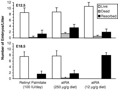
At E12.5, the number of live, dead, and resorbed embryos did not differ between VAD dams maintained on diets containing either the lower (12 μg/g diet) or higher (250 μg/g diet) level of atRA and did not differ from the control dams receiving a source of ROL (Fig. 1). However, by E18.5, all of the fetuses in the group fed 12 μg of atRA per g of diet were completely resorbed (Fig. 1). In contrast, when VAD dams were provided with 250 μg of atRA per g of diet from E0.5 to E18.5 of pregnancy, the majority of fetuses were alive and did not differ in number from the group receiving a source of ROL alone. Food intake was measured daily and showed that VAD dams fed 250 μg of atRA per g of diet ingested 4.5 ± 0.2 mg of atRA per rat per day, whereas those receiving 12 μg of atRA per g of diet consumed 230 ± 9 μg of atRA per rat per day. VAD dams fed the higher level of atRA delivered live pups; however, all of the pups died within 60 min after birth and none appeared to have milk in their stomachs. Neonatal mortality occurred in this group regardless of whether the dams received supplemental RP, demonstrating that mortality was caused by atRA excess rather than vitamin A deficiency. Pups from VAD dams fed a source of ROL alone were normal in all respects and survived to weaning (P21).
Figure 1.
Embryo/fetal survival at E12.5 and E18.5. The number of live, dead, and resorbed embryos and fetuses were assessed at the time of dissection. At E12.5 and E18.5, the number of litters studied equaled five for the RP group, six and four, respectively, for the group fed 250 μg of atRA per g of diet, and seven and six, respectively, for the group fed 12 μg of atRA per g of diet group.
General Embryonic Morphology at E12.5.
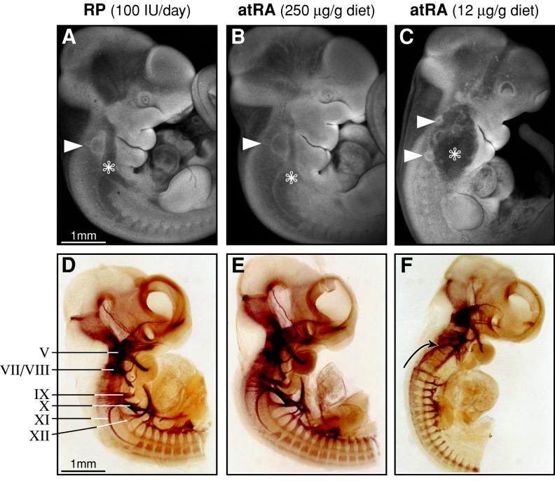
External morphology was normal at E12.5 in all embryos from VAD dams that were provided with a source of ROL alone or in combination with 250 μg of atRA per of g diet. Likewise, embryos from the group fed 250 μg of atRA per g of diet group were morphologically normal and indistinguishable from those given a source of ROL (Fig. 2 and data not shown). In contrast, embryos in the group fed 12 μg of atRA per g of diet exhibited numerous defects, including incomplete spiral torsion of the tail (43%), reduction of forelimb size (91%), defects in eye development (100%), and dysmorphogenesis in the region of the developing hindbrain. The most striking abnormality in this region was the appearance of multiple immature otic vesicles, which were present in 93% of the embryos (52 of 56 embryos, Fig. 2). The most rostral otic vesicle appeared to be positioned normally, dorsal to the second pharyngeal arch and lateral to the hindbrain, whereas the second (ectopic) vesicle was located posterior to the first. All otic vesicles were internalized but immature in appearance, and in a few cases, multiple otic vesicles appeared unilaterally, with the opposite side exhibiting a single immature vesicle. Additional defects present in 100% of embryos examined from the group fed 12 μg of atRA per g of diet group included an enlarged anterior cardinal vein and the absence of postotic pharyngeal arches (Fig. 2). The maxillary and mandibular processes (first pharyngeal arch) and the hyoid arch (second pharyngeal arch) were also less well developed in this group.
Figure 2.
External morphology of the hindbrain region and cranial nerve staining of embryos at E12.5. Embryonic morphology of the otic vesicle (solid arrowhead) and anterior cardinal vein (asterisk) was normal in all embryos from VAD dams supplemented with RP (A) or fed 250 μg of atRA per g of diet (B). The results shown are representative of 31 embryos selected at random from VAD dams fed RP (n = 5 litters) and 39 embryos from the group fed 250 μg of atRA per g of diet (n = 6 litters). All embryos from VAD dams fed 12 μg of atRA per g of diet were abnormal (C; 56 embryos from seven litters). Embryos had immature otic vesicles with the majority (93%) having two or more externally visible otic vesicles (solid arrowheads). Pharyngeal arches 1 and 2 were reduced in size and arches 3 and 4 were absent. Enlargement of the anterior cardinal vein (asterisk) was observed in 100% of embryos from the group fed 12 μg of atRA per g of diet. The normal complement of cranial nerves was readily identified by neurofilament antibody staining in randomly selected embryos from VAD dams supplemented with RP (D; n = 8 embryos) or receiving 250 μg of atRA per g of diet (E; n = 8 embryos). No 2H3 antibody staining was evident in peripheral regions normally innervated by cranial nerves IX, X, XI, and XII in embryos from VAD dams fed 12 μg of atRA per g of diet (F; n = 13 embryos). Axons in the facial nerve and those exiting from the posterior hindbrain appear to abnormally project axons in a more rostral direction (arrow). Nonspecific antibody staining was assessed by using ascites containing an antibody that does not bind to rat proteins (data not shown). Cranial nerves: V, trigeminal; VII, facial; VIII, vestibulo-cochlear; IX, glossopharyngeal; X, vagus; XI, accessory; XII, hypoglossal.
Cranial Nerve Morphogenesis.
The presence of external abnormalities in the hindbrain region of embryos in the group fed 12 μg of atRA per g of diet prompted us to evaluate cranial nervous system development at E12.5. Cranial nerve development was normal in all embryos from VAD dams given a source of ROL during pregnancy (Fig. 2). Likewise, the high atRA-containing diet (250 μg/g diet = 4.5 mg per rat per day) supported normal cranial nerve morphogenesis in both the absence and presence of exogenous ROL. In contrast, all embryos (100% penetrance) from dams fed the lower level of dietary atRA (12 μg of atRA per g of diet = 230 μg per rat per day) lacked cranial nerves IX (glossopharyngeal), X (vagus), XI (accessory), and XII (hypoglossal). In addition, the proximal (superior and jugular) and distal (petrosal and nodose) cranial ganglia of nerves IX and X, respectively, were missing. The few branchiomotor neuronal rootlets exiting the postotic hindbrain extended their axons abnormally in a rostral direction toward the facial nerve (VII). The facial nerve, although present, appeared to exit the hindbrain caudal to its normal position, and neurite outgrowth was less organized than normal. Thus, 230 μg of atRA per rat per day did not support normal development of the hindbrain, whereas an intake of 4.5 mg per rat per day yielded apparently normal embryos at E12.5.
Oral Bolus Administration of atRA and Embryonic Development to E12.5.
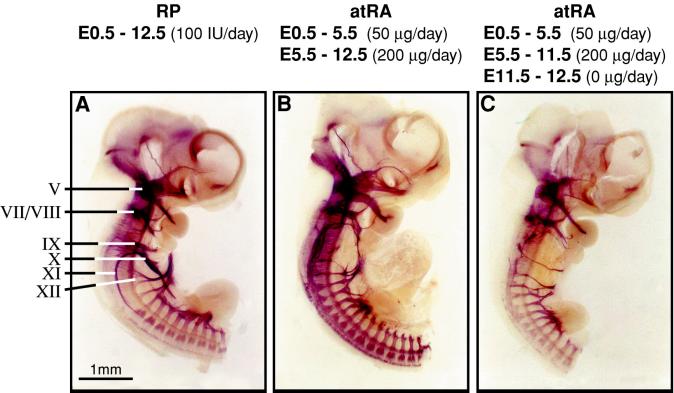
Dickman et al. (13) recently reported that embryos from VAD dams that received supplementation of atRA (50 μg of atRA per day from E0.5 to E5.5 and 200 μg of atRA per day in two divided doses from E5.5 to E13.5) with oral bolus doses are RA-sufficient and normal in all respects. This result contrasts with our finding that embryonic development is abnormal when VAD dams are maintained on a synthetic atRA-containing diet providing a slightly higher amount of the vitamin (230 μg per rat per day). To test whether mode of vitamin administration (oral bolus dosing vs. incorporation into the diet) could explain these disparate results, we provided oral bolus doses of atRA to VAD dams to yield RA-sufficient embryos according to the protocol of Dickman et al. (13). We found that the resulting embryos were abnormal at both E12.5 (Fig. 3B, 31 of 31 embryos) and E13.5 (data not shown, 24 of 24 embryos) and were phenotypically indistinguishable from embryos from dams receiving 230 μg of atRA per rat per day mixed into the diet (Fig. 2F). Embryos from dams receiving oral bolus administration of atRA exhibited a reduction of forelimb size, defects in eye development, and malformations in the region of the developing hindbrain, including the presence of ectopic otic vesicles, enlargement of the anterior cardinal vein, a lack of postotic pharyngeal arches, and a loss of cranial nerves IX, X, XI, and XII (Fig. 3 and data not shown). Dickman et al. (13) failed to observe any abnormalities in embryos generated in this fashion and reported that cranial nerve X was deleted only if oral bolus doses of atRA were eliminated from E11.5 to E13.5. In contrast, we found that cranial nerve X, together with cranial nerves IX, XI, and XII, was absent from embryos regardless of whether atRA was deleted from the regimen or not (Fig. 3 C and B, respectively).
Figure 3.
Oral bolus administration of atRA (200 μg per rat per day) is ineffective in preventing cranial nerve abnormalities at E12.5. Cranial nerve morphology was normal in VAD dams supplemented with RP (A). VAD dams supplemented with oral bolus doses of atRA from E0.5 to E12.5 (B) as described in the text exhibited abnormal posterior cranial nerve development and were morphologically indistinguishable from embryos obtained from VAD dams fed 12 μg of atRA per g of diet (230 μg per rat per day; see Fig. 2). Embryos from dams receiving no supplemental atRA after E11.5 exhibited a similar loss of cranial nerves IX, X, XI, and XII (C).
Embryonic Morphology at E18.5.
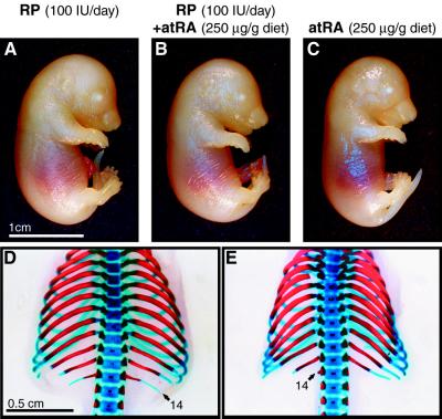
The external morphology of fetuses from dams fed the VAD diet and a source of ROL during gestation was normal. Fetuses from dams fed 250 μg of atRA per g of diet (4.5 mg of atRA per rat per day) with or without additional ROL appeared morphologically similar to the control group (Fig. 4). Despite the rather high intake of atRA during pregnancy by these dams, none of the resulting embryos exhibited any central nervous system (exencephaly and spina bifida) or craniofacial (protruding tongue and cleft palate) abnormalities that are characteristic of vitamin A toxicity (20, 21). However, when skeletal development was studied, 54% of the embryos from the high RA-containing diet group exhibited an extranumerary (14th) lumbar rib. In all but one case, the malformation was bilateral. This type of abnormality was also observed at the same frequency in the group receiving both a source of ROL and the high RA-containing diet, indicating that the skeletal malformation resulted from retinoid toxicity rather than deficiency. Thus, 4.5 mg of atRA per rat per day can support the development of embryos to E18.5; however, the embryos exhibit at least one sign of retinoid toxicity.
Figure 4.
External morphology and skeletal staining of embryos at E18.5. External fetal morphology from VAD dams supplemented with RP (A; representative of 38 fetuses from five litters), RP in combination with 250 μg of atRA per g of diet (B; representative of 48 fetuses from five litters), or fed 250 μg of atRA per g of diet alone (C; representative of 31 fetuses from four litters) was indistinguishable between the different diet groups. None of the embryos exhibited external malformations associated with retinoid toxicity (20). An extranumerary lumbar (14th) rib was present in 10 of 16 fetuses examined from VAD dams fed RP in combination with 250 μg of atRA per g and in 7 of 13 fetuses from VAD dams fed 250 μg of atRA per g of diet alone. An example of the range of skeletal malformations that were observed is shown in D (full 14th rib) and E (rudiment of a 14th rib).
Analysis of Embryos at E12.5 and E18.5 from Dams Maintained on Purified atRA.
Because a large amount of atRA was required to normalize development at E12.5 and to prevent embryo lethality in utero, we wanted to verify that the commercial batch of atRA did not contain any neutral retinoid contaminant (e.g., ROL) that could be responsible for rescuing the VAD embryos. Analysis of 8 μg of the commercial atRA preparation by HPLC and diode array detection revealed no absorbance in the region of authentic atROL (maximum absorbance = 325 nm, detection limit = 2 ng). However, to ensure that the preparation was free of ROL, the commercial atRA was purified according to the method of Napoli (12). This step involved partitioning less polar contaminants into hexane while retaining the atRA salt in the ethanol/water layer (pH 11.0). We verified that this method would remove all nonacid forms of retinoid by adding radiolabeled ROL to the original atRA preparation, and after completion of the partitioning, no radiolabeled ROL could be found in the atRA-containing fraction.
VAD dams were fed diets containing either 12 or 250 μg of purified atRA per g of diet, and a second set of animals was fed atRA obtained directly from the commercial vendor (three to four animals per diet group; 30–40 embryos examined per diet group and embryonic day). Embryos and fetuses from VAD dams fed 250 μg of purified atRA per g of diet did not differ in external embryonic morphology at either E12.5 or E18.5 of development from animals receiving atRA that had not been subjected to additional purification (data not shown). Likewise, all of the embryos from dams fed 12 μg of atRA per g of diet exhibited defects in external morphology and cranial nerve defects, regardless of whether they were fed purified atRA (data not shown). These results demonstrate that dietary atRA and not an ROL-like impurity prevents embryonic abnormalities at E12.5 and supports embryonic survival to E18.5.
DISCUSSION
In the present study, we demonstrated that atRA provided in the diet of the pregnant VAD dam at 250 μg/g diet or the equivalent of 4.5 mg per rat per day supports the survival of embryos to parturition. Before this report, it had been shown that up to 100 μg of atRA per g of diet did not support pregnancy in full (2) and that a source of ROL was needed on or before E9.5 to prevent embryonic resorption (3). The ability of atRA to support embryonic development to term suggests that the RAR- and RXR-mediated signaling pathways mediate most, if not all, of the actions of vitamin A during pregnancy in the female.
The amount of atRA that is required to enable VAD dams to carry fetuses to term is similar to that which has been shown to induce teratogenesis in vitamin A-sufficient rodents. Despite this fact, embryos from dams receiving 250 μg of atRA per g of diet appear normal at E12.5 and show only minor differences from the RP (a source of ROL)-supplemented control group at E18.5. Previous studies of the teratogenic effects of the vitamin showed that 15 mg of atRA per kg per day administered as an oral bolus dose to pregnant rats between E7.5 and E15.5 results in exencephaly, protruding tongue, and cleft palate in 50–60% of fetuses, whereas 6 mg of atRA per kg per day given between E6.5 and E15.5 leads to the generation of only a supernumerary (14th) lumbar rib in about 42% of the fetuses (20). Fetuses from VAD dams fed 250 μg of atRA per g of diet (16–18 mg of atRA per kg per day) exhibited no evidence of any central nervous system or craniofacial defects at E18.5; however, an additional lumbar (14th) rib was present in 54% of the fetuses examined at E18.5 (7 of 13). It is likely that the consumption of atRA throughout the day resulted in a pharmacokinetic profile that is less teratogenic than bolus administration. Although the high atRA-containing diet (250 μg of atRA per g) is able to support embryonic development to term, the pups die shortly after delivery because of vitamin A toxicity. It is possible that elevated levels of atRA are needed only at critical stages of embryonic development and that toxicity could be avoided by increasing the maternal intake of atRA only at those times when it is specifically needed.
The demand for atRA during pregnancy certainly seems high relative to the 47 μg of atRA per rat per day that is needed to support normal growth and cellular differentiation in the adult rat (22). It is possible that maternal atRA catabolism has been induced because of continuous exposure to dietary atRA and that this may be contributing to the high requirement for atRA in VAD dams. It should be noted that atRA is not normally found in the diet but, instead, is synthesized in vivo from ROL. During critical stages of development, the embryo may need to generate locally high concentrations of atRA, which cannot be mimicked by feeding 12 μg of atRA per g of diet to the VAD dam. Although embryonic tissue needs can be met by feeding 250 μg of atRA per g of diet to the VAD dam, this may lead to toxicity in tissues with a lesser requirement.
When atRA is provided at less than optimal levels during gestation (12 μg of atRA per g of diet = 230 μg of atRA per rat per day), the resulting embryos are abnormal at E12.5 and are completely resorbed by E18.5. We were unable to reproduce the work of Dickman et al. (13) who stated that embryos from VAD rats fed 140–200 μg of atRA per rat per day were completely normal. We showed that 200–230 μg of atRA per rat per day was insufficient to support normal embryonic development regardless of whether it was incorporated into the diet or given as a bolus oral dose. Embryos from VAD dams fed this level of atRA exhibited severe developmental abnormalities (ectopic otic vesicles, enlarged anterior cardinal vein, and loss of postotic pharyngeal arches and cranial nerves). These defects can be directly attributed to insufficient atRA, because they are prevented by providing dams with higher levels of dietary atRA during gestation (250 μg of atRA per g of diet = 4.5 mg of atRA per rat per day). Thus, embryos from VAD dams receiving 200 μg of atRA per rat per day are also VAD at E12.5 or E13.5 of development and should not be referred to as RA sufficient. We cannot fully explain the divergent results of our work and those of Dickman et al., although it is possible that this group might not have fully depleted their animals of ROL or their diet contained trace amounts of ROL. Based on the work presented here, we question the validity of the model put forth by Dickman et al. (13) to study vitamin A and embryonic development.
There is some question regarding whether embryo lethality occurring at midgestation in VAD dams supplemented with insufficient atRA results from a primary defect in the embryonic compartment or results from placental necrosis. Howell et al. (23) indicated that the earliest detectable lesion was necrosis of cells in the placental labyrinth and junctional zone, which could be detected histologically by day 14.5 to 15.5 of pregnancy. However, subsequent work showed that by E14.5 of development a reduction in total protein, DNA, and RNA could be detected in embryos from VAD dams supplemented with 12 μg of atRA per g of diet compared with ROL-supplemented controls (24). In the present report, we demonstrate that defects in the cranial nervous system and vascular development are present in embryos at least 2 days before the time when histological changes are reported to occur in the placenta. Thus, our results suggest that death of embryos occurring at midgestation in VAD dams maintained on 12 μg of atRA per g of diet is caused by a primary defect in embryonic rather than placental development.
The present study emphasizes the importance of vitamin A in the normal development of the posterior hindbrain including posterior cranial nerves and sensory ganglia. The cranial motor nerves IX–XII, which are missing in rat embryos receiving insufficient atRA, arise from the posterior hindbrain. In VAD quail embryos, the posterior hindbrain is reportedly deleted (25). In contrast, embryos from VAD pregnant rats receiving insufficient atRA (12 μg of atRA per g of diet = 230 μg per rat per day) possess a posterior hindbrain, but this region develops abnormally (J.C.W. and M.C.-D., unpublished data). The neurons of the proximal ganglia of cranial nerves IX and X originate from hindbrain-derived neural crest cells, whereas the neurons of the distal ganglia arise from ectodermal placode cells (26). The absence of these ganglia suggests that atRA is necessary for the development of these neurogenic precursor cells. A similar effect on neurogenic neural crest cells has not been reported in studies of RAR mutant fetuses and may reflect the ability of remaining RARs to substitute functionally for deleted receptor subtypes (4, 27).
A number of the defects in cranial nerves observed in this study are reminiscent of defects observed in transgenic Hoxa-1 (28, 29), Hoxa-1 3′-RA response element (30) and Hoxa-1/Hoxb-1 null mutant mice (31). Mice carrying a null mutation for Hoxa-1 exhibit a loss of the proximal portion of cranial nerves IX and X, a reduction in the size of the corresponding proximal ganglia (superior and jugular), and no connection of the distal sensory ganglia IX and X (petrosal and nodose) with the central nervous system. Mice (13%) with a mutation in the 3′-RA response element of Hoxa-1 also exhibit a loss unilaterally of the proximal portion of cranial nerve IX. Double null mutant mice for Hoxa-1 (+/− or −/−) and Hoxb-1 (−/−) also exhibit cranial nerve abnormalities that include a loss unilaterally of the proximal part of the glossopharyngeal (IX) and vagus (X) nerves, loss of the accessory (XI) nerve, and a reduction in the distal ganglia of cranial nerves IX and X. These cranial nerves are also affected in embryos from VAD dams receiving insufficient atRA; however, the effects of vitamin A deficiency are more severe and 100% penetrant. Thus, a limited portion of the vitamin A deficiency phenotype described here may be explained by a loss of class 1 Hox gene expression that is under retinoid control. However, the effect of vitamin A deficiency clearly extends beyond a simple loss of Hoxa-1 and/or Hoxb-1 function.
In this report we demonstrate that normal otic vesicle formation is under the regulation of atRA. Otic placodes are believed to be induced by secreted hindbrain factors at the level of the presumptive rhombomere 5/6 border that act on adjacent ectoderm (32, 33). In the absence of sufficient atRA, the adjacent postotic neuroepithelium may retain a more anterior-like fate, thus inducing ectopic otic vesicles dorsolateral to the posterior hindbrain. In agreement with this hypothesis, atRA has been shown to transform neural tissue from an anterior baseline fate to one that is posterior in character (34–37). The presence of ectopic otic vesicles in embryos from VAD dams receiving insufficient atRA combined with the ability of higher amounts of atRA (250 μg/g diet) to restore the normal pattern of otic vesicle formation shows that atRA plays a role both in restricting otic vesicle induction to its proper location and in otic vesicle maturation.
In conclusion, atRA can support the development of embryos to parturition, obviating the need for ROL before midgestation. Furthermore, these studies emphasize the critical importance of vitamin A in the normal development of the otic vesicle, postotic cranial nerves, and pharyngeal arches. This work provides the basis of a model system that can now be exploited to study the effects of vitamin A deficiency on both the early and late stages of embryonic development in the rat.
Acknowledgments
We thank the University of Wisconsin-Madison College of Agricultural and Life Sciences Statistical Consulting Group for their statistical assistance and the University of Wisconsin-Madison Biochemistry Media Lab for graphic design. This work was supported by the National Institutes of Health (Grant DK-14881).
ABBREVIATIONS
- RA
retinoic acid
- atRA
all-trans-retinoic acid
- VAD
vitamin A deficient
- RP
retinyl palmitate
- ROL
retinol
- atROL
all-trans-retinol
- E
embryonic day
- RAR
retinoic acid receptor
Footnotes
The numbering of embryonic days (E) for literature cited has been adjusted so that 12:00 p.m. (noon) of the first day of pregnancy is designated E0.5.
References
- 1.Evans H M. J Biol Chem. 1928;77:651–654. [Google Scholar]
- 2.Thompson J N, Howell J M, Pitt G A J. Proc R Soc London B Biol Sci. 1964;159:510–535. doi: 10.1098/rspb.1964.0017. [DOI] [PubMed] [Google Scholar]
- 3.Wellik D M, DeLuca H F. Biol Reprod. 1995;53:1392–1397. doi: 10.1095/biolreprod53.6.1392. [DOI] [PubMed] [Google Scholar]
- 4.Kastner P, Mark M, Chambon P. Cell. 1995;83:859–869. doi: 10.1016/0092-8674(95)90202-3. [DOI] [PubMed] [Google Scholar]
- 5.Wilson J G, Roth C B, Warkany J. Am J Anat. 1953;92:189–217. doi: 10.1002/aja.1000920202. [DOI] [PubMed] [Google Scholar]
- 6.Hogan B L M, Thaller C, Eichele G. Nature (London) 1992;359:237–241. doi: 10.1038/359237a0. [DOI] [PubMed] [Google Scholar]
- 7.Creech Kraft J, Shepard T, Juchau M R. Reprod Toxicol. 1993;7:11–15. doi: 10.1016/0890-6238(93)90004-q. [DOI] [PubMed] [Google Scholar]
- 8.McCaffery P, Dräger U C. Proc Natl Acad Sci USA. 1994;91:7194–7197. doi: 10.1073/pnas.91.15.7194. [DOI] [PMC free article] [PubMed] [Google Scholar]
- 9.Horton C, Maden M. Dev Dyn. 1995;202:312–323. doi: 10.1002/aja.1002020310. [DOI] [PubMed] [Google Scholar]
- 10.Suda T, DeLuca H F, Tanaka Y. J Nutr. 1970;100:1049–1052. doi: 10.1093/jn/100.9.1049. [DOI] [PubMed] [Google Scholar]
- 11.National Research Council. Nutrient Requirements of Laboratory Animals. 4th Rev. Ed. Washington, DC: National Academy Press; 1995. pp. 11–79. [Google Scholar]
- 12.Napoli J L. Methods Enzymol. 1986;123:112–124. doi: 10.1016/s0076-6879(86)23015-3. [DOI] [PubMed] [Google Scholar]
- 13.Dickman E D, Thaller C, Smith S M. Development (Cambridge, UK) 1997;124:3111–3121. doi: 10.1242/dev.124.16.3111. [DOI] [PubMed] [Google Scholar]
- 14.Brown N A, Fabro S. Teratology. 1981;24:65–78. doi: 10.1002/tera.1420240108. [DOI] [PubMed] [Google Scholar]
- 15.Zucker R M, Elstein K H, Shuey D L, Ebron-McCoy M, Rogers J M. Teratology. 1995;51:430–434. doi: 10.1002/tera.1420510608. [DOI] [PubMed] [Google Scholar]
- 16.Wall N A, Jones C M, Hogan B L M, Wright C V E. Mech Dev. 1992;37:111–120. doi: 10.1016/0925-4773(92)90073-s. [DOI] [PubMed] [Google Scholar]
- 17.Lufkin T, Mark M, Hart C P, Dollé P, LeMeur M, Chambon P. Nature (London) 1992;359:835–841. doi: 10.1038/359835a0. [DOI] [PubMed] [Google Scholar]
- 18.Meyer E, Lambert W E, De Leenheer A P. Clin Chem. 1994;40:48–50. [PubMed] [Google Scholar]
- 19.MacCrehan W A, Schönberger E. J Chromatogr. 1987;417:65–78. doi: 10.1016/0378-4347(87)80092-0. [DOI] [PubMed] [Google Scholar]
- 20.Collins M D, Tzimas G, Hummler H, Bürgin H, Nau H. Toxicol Appl Pharmacol. 1994;127:132–144. doi: 10.1006/taap.1994.1147. [DOI] [PubMed] [Google Scholar]
- 21.Geelen J A G. CRC Crit Rev Toxicol. 1979;6:351–376. doi: 10.3109/10408447909043651. [DOI] [PubMed] [Google Scholar]
- 22.Zile M, DeLuca H F. J Nutr. 1968;94:302–308. doi: 10.1093/jn/94.3.302. [DOI] [PubMed] [Google Scholar]
- 23.Howell J M, Thompson J N, Pitt G A J. J Reprod Fertil. 1964;7:251–258. doi: 10.1530/jrf.0.0070251. [DOI] [PubMed] [Google Scholar]
- 24.Takahashi Y I, Smith J E, Winick M, Goodman D S. J Nutr. 1975;105:1299–1310. doi: 10.1093/jn/105.10.1299. [DOI] [PubMed] [Google Scholar]
- 25.Maden M, Gale E, Kostetskii I, Zile M. Curr Biol. 1996;6:417–426. doi: 10.1016/s0960-9822(02)00509-2. [DOI] [PubMed] [Google Scholar]
- 26.D’Amico-Martel A, Noden D M. Am J Anat. 1983;166:445–468. doi: 10.1002/aja.1001660406. [DOI] [PubMed] [Google Scholar]
- 27.Lohnes D, Mark M, Mendelsohn C, Dollé P, Dierich A, Gorry P, Gansmuller A, Chambon P. Development (Cambridge, UK) 1994;120:2723–2748. doi: 10.1242/dev.120.10.2723. [DOI] [PubMed] [Google Scholar]
- 28.Chisaka O, Musci T S, Capecchi M R. Nature (London) 1992;355:516–520. doi: 10.1038/355516a0. [DOI] [PubMed] [Google Scholar]
- 29.Mark M, Lufkin T, Vonesch J-L, Ruberte E, Olivo J-C, Dollé P, Gorry P, Lumsden A, Chambon P. Development (Cambridge, UK) 1993;119:319–338. doi: 10.1242/dev.119.2.319. [DOI] [PubMed] [Google Scholar]
- 30.Dupé V, Davenne M, Brocard J, Dollé P, Mark M, Dierich A, Chambon P, Rijli F M. Development (Cambridge, UK) 1997;124:399–410. doi: 10.1242/dev.124.2.399. [DOI] [PubMed] [Google Scholar]
- 31.Gavalas A, Studer M, Lumsden A, Rijli R M, Krumlauf R, Chambon P. Development (Cambridge, UK) 1998;125:1123–1136. doi: 10.1242/dev.125.6.1123. [DOI] [PubMed] [Google Scholar]
- 32.Ruben R J, Van de Water T R. In: Development of Auditory Behavior. Gerber S E, Menchers G T, editors. New York: Grune & Stratton; 1983. pp. 3–35. [Google Scholar]
- 33.Wilkinson D G, Peters G, Dickson C, McMahon A P. EMBO J. 1988;7:691–695. doi: 10.1002/j.1460-2075.1988.tb02864.x. [DOI] [PMC free article] [PubMed] [Google Scholar]
- 34.Durston A J, Timmermans J P M, Hage W J, Hendriks H F J, de Vries N J, Heideveld M, Nieuwkoop P D. Nature (London) 1989;340:140–144. doi: 10.1038/340140a0. [DOI] [PubMed] [Google Scholar]
- 35.Marshall H, Nonchev S, Sham M H, Muchamore I, Lumsden A, Krumlauf R. Nature (London) 1992;360:737–741. doi: 10.1038/360737a0. [DOI] [PubMed] [Google Scholar]
- 36.Kessel M. Neuron. 1993;10:379–393. doi: 10.1016/0896-6273(93)90328-o. [DOI] [PubMed] [Google Scholar]
- 37.Wood H, Pall G, Morriss-Kay G. Development (Cambridge, UK) 1994;120:2279–2285. doi: 10.1242/dev.120.8.2279. [DOI] [PubMed] [Google Scholar]


