Abstract
The core of bluetongue virus (BTV) is a multienzyme complex composed of two major proteins (VP7 and VP3) and three minor proteins (VP1, VP4, and VP6) in addition to the viral genome. The core is transcriptionally active and produces capped mRNA from which all BTV proteins are translated, but the relative role of each core component in the overall reaction process remains unclear. Previously we showed that the 76-kDa VP4 protein possesses guanylyltransferase activity, a necessary part of the RNA capping reaction. Here, through the use of highly purified (>95%) VP4 and synthetic core-like particles containing VP4, we have investigated the extent to which this protein is also responsible for other activities associated with cap formation. We show that VP4 catalyzes the conversion of unmethylated GpppG or in vitro-produced uncapped BTV RNA transcripts to m7GpppGm in the presence of S-adenosyl-l-methionine. Analysis of the methylated products of the reaction by HPLC identified both methyltransferase type 1 and type 2 activities associated with VP4, demonstrating that the complete BTV capping reaction is associated with this one protein.
Eukaryotic mRNAs of both cellular and viral origin have two notable and distinguishing features: polyadenylation of the 3′ terminus and guanylation/methylation (cap structure) at the 5′ terminus. The presence of a methylated cap structure, e.g., m7G(5′)ppp(5′)NpNpNp, at the extreme 5′ end of a eukaryotic mRNA is an important component for efficient initiation of translation and stability for many mRNAs (see reviews in refs. 1–3). It also protects mRNA from 5′ exonuclease degradation (4). The cap is usually synthesized in the cell nucleus and, accordingly, viruses that replicate in the cytoplasm of infected cells encode their own enzymes for capping mRNAs (1, 3, 5). The capping of mRNA has been studied in several viral systems (6–12). From studies of viral and cellular capping enzyme systems (13–15), the following sequential reactions have been proposed for cap formation: step 1: pppGpN1pN2 → ppGN1pN2 + p; step 2: GTP + enzyme → enzyme-GMP + Pp; step 3: enzyme-GMP + ppGN1pN2 → GpppGpN1 pN2… . + enzyme; step 4: GpppGpN1pN2… . + AdoMet → m7GpppGpN1pN2… . . + AdoHcy; step 5: m7GpppGpN1pN2… . + AdoMet → m7GpppGmpN1pN2… . . + AdoHcy, where pN is any ribonucleotide, p is inorganic phosphate, Pp is inorganic pyrophosphate, m7G is 7-methylguanosine, Gm is 2′-methylguanosine, AdoMet is S-adenosyl-l-methionine, and AdoHcy is S-adenosyl-l-homocysteine.
The process is made up of a number of separate reactions that are catalyzed by different enzyme activities. In step 1, an RNA triphosphatase activity removes the 5′-terminal phosphate residue from the nascent RNA. In step 2, the guanylyltransferase enzyme, responsible for guanylation, covalently binds GMP through the action of a nucleotide phosphohydrolase. In step 3, this enzyme–GMP complex transfers GMP to the diphosphate at the 5′ terminus of the substrate RNA and as a consequence, the GMP caps the RNA via a 5′-5′-triphosphate linkage. In step 4, the cap structure is methylated by transfer of a methyl group from AdoMet to position 7 of the terminal guanosine, yielding a cap 0 structure and releasing AdoHcy. Frequently (step 5), a methyltransferase enzyme may also transfer the methyl group from the AdoMet substrate to the 2′-hydroxyl group of the ribose of the first (N1) or first and second (N1 and N2) nucleotides, forming type 1 and type 2 cap structures, respectively (16, 17).
Viruses within the family Reoviridae, such as reovirus and bluetongue virus (BTV), contain double-stranded, segmented RNA genomes in which both the “plus” sense RNAs and the intracellular mRNA species are capped. BTV has seven structural proteins (VP1–VP7) organized into two capsid layers. The inner capsid, or “core,” contains five proteins (VP1, VP3, VP4, VP6, and VP7). Cores are transcriptionally active, producing capped and methylated full-length mRNA copies for each of the 10 genomic segments (18). Like many eukaryotic viral and cellular mRNAs, the BTV mRNAs possess cap 1 structures (18). VP1 is thought to be an RNA polymerase and VP6 an RNA helicase (19, 20). Previously, we showed that the VP4 core protein possesses a guanylyltransferase activity (21). Whether VP4 is involved in other aspects of the capping process is not known. In vitro, VP4 binds ribonucleotides and deoxyribonucleotides and exhibits an NTPase activity (22). However, binding is inhibited by the presence of a methyl donor (AdoMet), leading to the suggestion that VP4 may have methyltransferase activity (18).
Here, using VP4 either as a purified recombinant protein alone or assembled into recombinant core-like particles (CLPs; ref. 23), we show that VP4 catalyzes two methyltransferase activities. VP4 transfers a methyl group from AdoMet to position 7 of the guanosine capping residue at the 5′ terminus of RNA analogues, but it also catalyzes the methylation of the ribose of the penultimate 5′-terminal nucleotide. The significance of the association of these activities with a single viral protein is discussed.
MATERIALS AND METHODS
Viruses and Cells.
Spodoptera frugiperda cells (Sf9) were grown in TC100 medium (GIBCO/BRL) supplemented with 10% (vol/vol) fetal calf serum. The cells were grown as suspension or monolayer cultures at 28°C. Recombinant Autographa californica nuclear polyhedrosis viruses (AcNPV) expressing either VP4 of BTV serotype 10 (AcBTV10.4) or VP3 (AcBTV10.3) or VP7 (AcBTV 10.7) were propagated in suspension culture as described (23).
Purification of Proteins.
BTV recombinant protein VP4 and CLPs containing VP3, VP7, and VP4 were purified from recombinant AcNPV-infected Sf9 cells as described (23, 24). The presence of the BTV proteins in the derived CLPs was confirmed after their purification by sucrose velocity gradient centrifugation, SDS/PAGE, and Western blot analyses using BTV10 polyclonal antibodies.
Methyltransferase Assay.
Methyl transfer from Ado[methyl-3H]Met to commercially available GpppG and m7GpppG used as acceptor molecules was measured as follows. A standard reaction mixture contained 200 μM GpppG or 200 μM m7GpppG, 50 mM Tris⋅HCl (pH 7.5), 9 mM MgCl2, 2 μM Ado[methyl-3H]Met (87 Ci/mmol), 6 mM DTT, and 15 μg of purified VP4 or 30 μg of purified CLPs with (or without) VP4 in 100 μl. After incubation at 30°C for 2 hr, the reaction mixtures were treated with 2 μg of proteinase K. The mixture was incubated at 37°C for 30 min, and the proteinase K was heat-inactivated by incubation at 70°C for 10 min. After phenol extraction, the products were precipitated with 9 vol of cold ethanol; 10 μl of 100 mM GTP was used as carrier for precipitation.
Preparation of in Vitro Transcripts Containing 5′-Terminal Triphosphates.
RNA transcripts representing exact copies of BTV segment 5 were prepared through in vitro transcription of plasmid pEc-10B5-Hδ DNA, which contains a full-length cDNA copy of BTV segment 5 cloned into pUC119 and flanked by an upstream T7 promoter and a downstream hepatitis δ ribozyme. T7 transcription was done using the RiboMAX large-scale transcription kit (Promega) under conditions indicated by the vendor with the following modifications: each transcription reaction contained the supplied buffer, 4 mM each NTP, 1–2 μg of DNA, and 2 μl of the supplied enzyme mix in a 20-μl final volume. The reactions were incubated at 37°C for 3 hr. Two units of RNase-free DNase was added, and the incubation continued for 15 min at 37°C. After extraction with phenol/chloroform, the RNA was passed through a G25 spin column (Pharmacia) to remove unincorporated NTPs and quantified by spectophotometry.
Guanylyltransferase Assays Using [α-32P]GTP.
Standard reaction mixtures (100 μl) contained 50 mM Tris⋅HCl (pH 7.5), 9 mM MgCl2, 6 mM DTT, 10 μM AdoMet, 6 μM [α-32P]GTP (400 Ci/mmol), 0.5–1.5 A260 units of BTV M5 RNA transcripts as acceptor, and 15 μg of purified VP4 or 30 μg of purified CLPs with or without VP4. After incubation at 30°C for 2 hr, RNA was extracted as described above and digested with 10 μg of nuclease P1 in 15 μl of 20 mM sodium acetate (pH 5.5) for 60 min at 37°C.
Separation and Identification of Nucleotide Products by Using HPLC.
The purified products from the capping reactions were analyzed by HPLC on a Beckman BioSys 510 using a SAX 5 ion-exchange column (Phenomenex, Belmont, CA). Products bound the column in 4 mM potassium phosphate buffer (pH 5.5) and were eluted with a salt gradient of 0.04–0.5 M of the same buffer at a flow rate of 1 ml/min. The column was precalibrated by using unlabeled methylated and nonmethylated nucleotides m7GpppG and m7GpppGm as well as m7GTP, m7GDP, m7GMP, GTP, GDP, and GMP (Sigma) under the same conditions. Eluants were collected and counted to determine the distribution of radioactivity. The results were plotted against time, and the identity of the radiolabeled peaks was established by comparison with the unlabeled markers run under identical conditions.
Pyrophosphatase Treatment of Cap Structures.
An aliquot of the cap structure product synthesized in vitro by VP4 was incubated with 10 units of tobacco acid pyrophosphatase (TAP) in 100 μl of buffer supplied by the manufacturer (Epicentre Technologies, Madison, WI). The digested product was then precipitated with 10 vol of cold ethanol in the presence of unlabeled carrier GTP and analyzed using HPLC as described above.
RESULTS
VP4 Protein Has Guanine-7-Methyltransferase Activity.
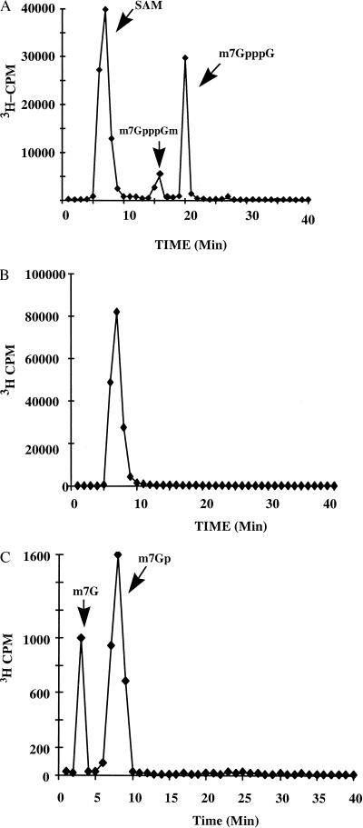
Certain viruses have guanylyltransferase and methyltransferase activities encoded by a single viral protein (25). In our previous report, we showed that both genomic double-stranded RNA and mRNA synthesized by virus cores in vitro have 5′-terminal cap (m7GpppGm) structures (18). Recently, we documented a guanylyltransferase activity associated with BTV core protein VP4 (26). Because guanylation of mRNA is usually followed by methylation, it was possible that VP4 may also possess a methyltransferase activity. To examine this possibility, purified, soluble VP4 was used in an in vitro assay to investigate whether it catalyzed the transfer of a donor methyl group Ado[methyl-3H]Met to an unmethylated cap analogue (GpppG). Reaction products were fractionated by ion-exchange chromatography, and the presence of the label as determined by the radioactivity associated with each fraction. A 30-min reaction led to most of the label coeluting with m7GpppG (Fig. 1A), whereas reactions in which VP4 had been heated to 68°C for 15 min prior to addition had radioactivity associated exclusively with the Ado[methyl-3H]Met marker (Fig. 1B). No other radioactive peaks were identified in the products, consistent with a cap methyltransferase activity associated with purified VP4. To confirm that the product synthesized was indeed m7GpppG, an aliquot of m7GpppG fraction was digested with TAP, and the dephosphorylated products were subsequently analyzed by HPLC using similar conditions. Two peaks were identified, one comigrating with the position of the m7G marker and the other with m7GMP (Fig. 1C).
Figure 1.
Analysis of methylated cap structures synthesized by recombinant VP4. Reaction mixtures (100 μl) contain 50 mM Tris⋅HCl (pH7.9), 9 mM MgCl2, 6 mM DTT, 200 μM GpppG, 2 μMAdo[methyl-3H]Met (87 Ci/mmol), and 15 μg of purified recombinant VP4. Parallel reactions were performed with VP4 protein that had been heated to 68°C for 15 min prior to addition. After incubation for two hr at 30°C and proteinase K treatment, the reaction mix was extracted with phenol/chloroform and precipitated with ethanol. Samples were analyzed by HPLC on a Beckman BioSys 510 system using a SAX 5 ion-exchange column. The products recovered from HPLC were dried and counted in the scintillation counter, and the radioactivity associated with each fraction is plotted. The positions of unincorporated AdoMet (SAM) and m7GpppG are indicated. (A) Reaction products from mixtures containing VP4 as enzyme. (B) Reaction products from mixtures containing VP4 that had been heated to 68°C for 15 min prior to addition to the reaction mix. (C) Reanalysis of the peak fraction from A after TAP treatment. The reduction in scale reflects the fact that only ≈10% of the peak from A was analyzed.
VP4 Has Both Methyltransferase 1 and 2 Activities.
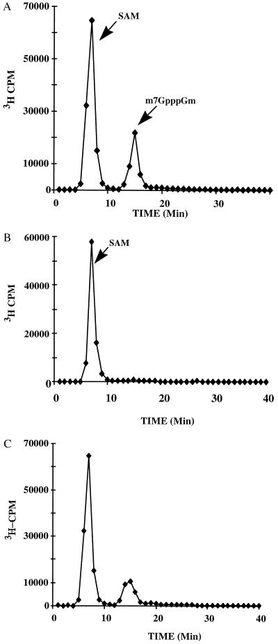
When VP4 is expressed with the BTV core structural proteins VP7 and VP3 it is encapsidated in CLPs (23). Thus, CLPs containing or lacking VP4 were purified and assayed for transferase activity. When analyzed by HPLC, the products of the reaction mixtures containing CLPs + VP4 produced three peaks (Fig. 2A), whereas the products of the CLPs lacking VP4 contained only the methyl donor [3H-CH3]-AdoMet peak (Fig. 2B). By comparison with the marker nucleotides, it was deduced that the second peak in Fig. 2A corresponded to m7GpppGm (methylation of the 2′ position of the guanosine ribose) with a smaller peak at the position of m7GpppG (Fig. 2A). These data suggest that VP4 has both 2′-O-methylation and 5′ cap methylation activities.
Figure 2.
Analysis of methyltransferase activity of VP4 within CLPs. Reactions were performed as described for Fig. 1 except that CLPs containing VP4 were used as enzyme instead of VP4. Parallel reactions were performed with CLPs alone as control. Products were analyzed by HPLC and plotted as described. The positions of unincorporated AdoMet, m7GpppGm, and m7GpppG are indicated. (A) CLPs + VP4. (B) CLPs alone.
To confirm the ability of CLPs + VP4 to catalyze the guanosine 2′-O-methyltransferase activity, an unlabeled methylated cap analogue (m7GpppG) was provided as an alternative substrate in a reaction similar to the one described above. The products of this reaction gave only two labeled peaks corresponding to m7GpppGm and unincorporated [3H-CH3]-AdoMet (Fig. 3A). CLPs without VP4 produced no m7GpppGm (Fig. 3B). To determine whether VP4 alone was sufficient for the reaction, purified VP4 protein was incubated with m7GpppG and [3H-CH3]-AdoMet, and the reaction products were similarly analyzed. As was the case for the CLP + VP4 reaction, two peaks were identified (Fig. 3C), although the level of m7GpppGm produced was much lower compared with the reactions in which VP4 was present within a CLP. It was concluded that VP4 is responsible for both the 7-methyltransferase and the 2-O-methyltransferase activities (methyltransferase reactions 1 and 2) associated with BTV cores (18).
Figure 3.
Identification of methyltransferase type 2 activity of VP4 within CLPs. Thirty micrograms of either CLP + VP4 or CLP alone or 15 μg of purified VP4 was incubated with 200 μM unlabeled methylated cap structure (m7GpppG) and 2 μm Ado[methyl-3H]Met (87 Ci/mmol) in the presence of 50 mM Tris⋅HCl (pH 7.9), 9 mM MgCl2, and 6 mM DTT. After incubation for 2 hr at 30°C, samples were digested with 10 μg of nuclease P1 for 60 min at 37°C. The reaction products were phenol/chloroform-extracted and ethanol-precipitated before analysis by HPLC as described. The positions of unincorporated AdoMet and m7GpppGm are indicated. (A) CLPs + VP4. (B) CLPs alone. (C) Purified VP4. Note that the relative molar concentrations of VP4 in reactions A and C vary greatly and indicate that the relative efficiency of the pure protein is poor.
GTP and AdoMet Form Capped Nucleotides in the Presence of VP4.
To define further the capping process mediated by VP4, purified protein was incubated with GTP, GDP, or GMP in the presence of Ado[methyl-3H]Met, and the products were analyzed by HPLC. When GDP or GMP was used, only one peak, corresponding to the unincorporated Ado[methyl-3H]Met, was detected (not shown). In contrast, the products from reaction mixtures containing GTP and AdoMet gave two major peaks, one corresponding to AdoMet and the other to m7GpppG (Fig. 4A). A minor peak was observed ahead of the m7GpppG products that coeluted with the marker for m7GpppGm, although confirmation of its identity was not possible because of the small amounts of material recovered.
Figure 4.
Acceptor nucleotide for VP4 methyltransferase activity. (A) Recombinant VP4 was incubated with cold GTP in the presence of 2 μM Ado[methyl-3H]Met (87 Ci/mmol), 50 mM Tris⋅HCl (pH 7.9), 9 mM MgCl2, and 6 mM DTT. After incubation for 2 hr at 30°C and treatment with proteinase K, the reaction products were extracted with phenol/chloroform and precipitated with ethanol. Samples were analyzed by HPLC. The positions of unincorporated AdoMet and m7GpppG are indicated. (B) Purified CLPs encapsidating VP4 (30 μg) were incubated with 6 μM ([α-32P]GTP (400 Ci/mmol) and 2 μM AdoMet in 50 mM Tris⋅HCl (pH 7.9), 9 mM MgCl2, and 6 mM DTT. Samples were analyzed as described. Position of m7GpppGm is indicated.
The ability of CLPs + VP4 to use GTP as the acceptor was assessed by incubation with [α-32P]GTP and AdoMet. The reaction products gave a major radioactive peak corresponding to m7GpppGm (Fig. 4B). The results of this and previous experiments suggest that the ribose methylation is significantly enhanced when VP4 is present within CLPs. Because of the low levels of radioactivity, the minor peaks shown in Fig. 4B were not characterized further, although the peaks eluting in fractions 19–21 and fractions 25–27 most likely represent the products m7GpppG and GpppG, respectively.
VP4 in the Presence of AdoMet Modifies de Novo BTV mRNA to Form a Fully Methylated Cap Structure.
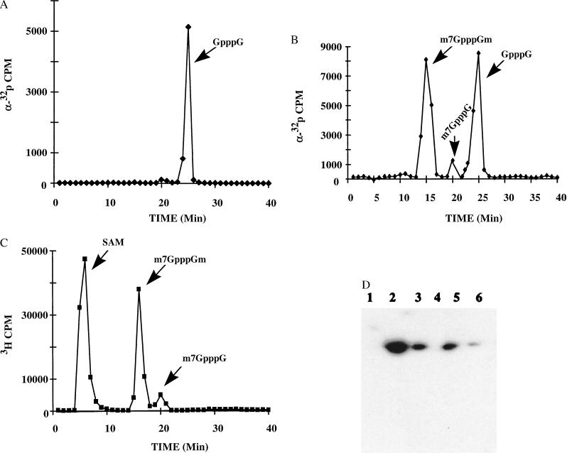
To investigate whether purified recombinant VP4 can methylate BTV mRNA, a transcript of BTV segment 5 was prepared from cDNA by using T7 polymerase purified from unincorporated nucleotides, and used as a cap structure and methylation acceptor in the presence of GTP and VP4. The derived RNA products were recovered, and the cap structures were released from the 5′-end by digestion with nuclease P1 to recover GpppG from unmethylated capped mRNA, m7GpppG from cap 0 mRNAs, and m7GpppGm from cap 1 or cap 2 mRNAs. When the T7-produced transcripts were incubated with purified VP4 and [α-32P]GTP in the absence of AdoMet, 32P-labeled products were recovered at the position of the GpppG marker (Fig. 5A), indicating that mRNA transcripts are substrates for guanylylation by VP4. The transfer of radioactivity to segment 5 transcripts was also confirmed by polyacrylamide gel electrophoresis (Fig. 5D). The lower level of RNA labeling observed with CLP + VP4 as compared with purified VP4 (Fig. 5D), despite previous indications that encapsidated VP4 is more efficient, doubtless reflects better access by the mRNA to the unencapsidated enzyme. When the nuclease P1-resistant products of the reaction mixtures, which also contained AdoMet, were analyzed, two major 32P-labeled peaks were recovered at the positions of m7GpppGm and GpppG, with an additional minor peak of m7GpppG (Fig. 5B). To confirm these data, reactions were repeated with unlabeled GTP and Ado[methyl-3H]Met. A major peak containing tritium label was recovered at the position of m7GpppGm with a minor peak at m7GpppG (Fig. 5C). These data suggest that full cap 1 formation activity can be induced by provision of an authentic substrate (mRNA) to purified VP4.
Figure 5.
Modification of 5′ end of BTV mRNA by VP4. Purified recombinant VP4 (15 μg) was incubated with in vitro-transcribed segment 5 RNA in the presence of 6 μM [α-32P]GTP (400 Ci/mmol) in 50 mM Tris-⋅HCl (pH 7.9), 9 mM MgCl2, and 6 mM DTT either in the presence or in the absence of 2 μM AdoMet. After incubation at 30°C for 2 hr, RNA in the reaction was purified by precipitation and desalted by passage through a G-25 spin column. Samples were analyzed on 4% acrylamide gels containing 8 M urea, or aliquots were digested with 10 μg of nuclease P1 for 1 hr at 37°C. The phenol/chloroform-extracted and ethanol-precipitated products were analyzed by HPLC. The positions of GpppG, m7GpppG, and m7GpppGm are shown. (A) VP4 in the absence of AdoMet. (B) VP4 in the presence of AdoMet. (C) VP4 in the presence of unlabeled GTP and Ado[methyl-3H]Met. (D) Autoradiograph of 4% acrylamide gel containing 8 M urea after electrophoresis. Tracks 1 and 4, CLPs only; tracks 2 and 5, purified VP4; tracks 3 and 6, CLP + VP4. In tracks 1–3, the reactions contained only GTP and RNA whereas in tracks 4–6, AdoMet was also present.
DISCUSSION
Cellular methyltransferase proteins typically appear to encode only a single activity (13), whereas a number of viral methyltransferases, such as that encoded by vaccinia virus, have an additional enzymatic activity such as guanylyltransferase (25, 27). The nsP1 proteins of Semliki Forest virus and Sindbis virus (both positive-strand RNA viruses) also encode both methyltransferase and guanylyltransferase activities (28, 29). In vaccinia virus RNA triphosphatase, RNA guanylyltransferase and RNA guanine-7-methyltransferase are components of a capping enzyme complex containing two subunits of 95 kDa and 31 kDa (30). However, RNA 2-O-methyltransferase activity is mediated by an additional protein, VP39 (31). In contrast to these viruses, BTV maximizes its coding capacity by having the minor core protein, VP4, catalyze all of the capping and methylation steps necessary, i.e., RNA triphosphatase, RNA guanylyltransferase, RNA guanine-7-methyltransferase, and RNA 2-O-methyltransferase activity.
When GpppG was incubated with purified VP4 and Ado[methyl-3H]Met, most of the radioactivity was transferred to GpppG, yielding m7GpppG, thereby demonstrating that VP4 is responsible for methyltransferase activity. When m7GpppG was used as substrate, the completed cap structure (i.e., m7GpppGm) was also detectable in the reaction. In contrast, a fully modified 5′-terminal structure was the major labeled product when GpppG was incubated with VP4 encapsidated in CLPs. The data presented, together with our failure to detect any GpppGm in the reaction products, indicate that the methyltransferase reactions involved in type 1 cap formation are sequential. The methyl transfer to 7-guanine appears to occur first, providing a m7GpppG… substrate for the second methyltransferase reaction and formation of m7GpppGm. The second methylation activity appeared to be limited when VP4 was used alone, indicating that VP4 may adopt a conformation that is more favorable for the complete reaction when encapsidated within a CLP, perhaps as a result of association with VP3 inside the subcore layer.
The fact that VP4 in solution has the capability to produce the complete cap structure was confirmed by subsequent experiments using authentic BTV transcripts. As expected, when purified VP4 protein was incubated with [α-32P]GTP, RNA, and AdoMet, the full cap structure, i.e., m7GpppGm, was produced at the 5′ terminus of the RNA, although intermediate products GpppG and m7GpppG were also detected. The production of the m7GpppGm cap 1 structure is evidently the final modification step. These data generally agree with those of Martin and Moss (16), who examined cap formation by vaccinia viral cores. Vaccinia virus cores, however, did not produce blocked 5′ termini as a result of the inhibitory effect of inorganic pyrophosphate on the guanylyltransferase reaction. BTV VP4 protein, however, in the presence of [α-32P]GTP, produced RNA with the blocked terminal structure G(5′)pppG. This is consistent with the previous observation that VP4 protein has inorganic pyrophosphatase activity (26).
Generally, capping is considered to be a posttranscriptional modification of mRNA. But the data from these studies suggest that, in addition to the capping of existing RNA molecules, VP4 can also condense GTP to form a capped dinucleotide, thereby confirming its guanylyltransferase activity. This provides an alternative mechanism for capping mRNA (which the formation of GpppG from GTP may precede) or for coupling to the initiation of mRNA synthesis. Such a scenario would be consistent with BTV biology, in which transcription and RNA modification occur in a transcriptionally active core from which only mature capped mRNAs emerge. In addition, VP4 provides an exciting model protein in which all of the capping functions are combined and from which there is much to learn through further biochemical and structural analysis.
Acknowledgments
The authors are grateful to Stephanie Price for assistance in the preparation of the manuscript. The financial support of Biotechnology and Biological Sciences Research Council (BBSRC) and the Ministry of Agriculture, Fisheries and Food is acknowledged.
ABBREVIATIONS
- BTV
bluetongue virus
- CLPs
core-like particles
- AdoMet
S-adenosyl-l-methionine
- TAP
tobacco acid pyrophosphatase
References
- 1.Banerjee A K. Microbiol Rev. 1980;44:175–205. doi: 10.1128/mr.44.2.175-205.1980. [DOI] [PMC free article] [PubMed] [Google Scholar]
- 2.Filipowicz W. FEBS Lett. 1978;96:1–11. doi: 10.1016/0014-5793(78)81049-7. [DOI] [PubMed] [Google Scholar]
- 3.Shatkin A J. Cell. 1976;9:645–653. doi: 10.1016/0092-8674(76)90128-8. [DOI] [PubMed] [Google Scholar]
- 4.Furuichi Y, LaFiandra A, Shatkin A J. Nature (London) 1977;266:235–239. doi: 10.1038/266235a0. [DOI] [PubMed] [Google Scholar]
- 5.Ahola T, Kaariainen L. Proc Natl Acad Sci USA. 1995;92:507–511. doi: 10.1073/pnas.92.2.507. [DOI] [PMC free article] [PubMed] [Google Scholar]
- 6.Ensinger M J, Martin S A, Paoletti E, Moss B. Proc, Natl Acad Sci USA. 1975;72:2525–2529. doi: 10.1073/pnas.72.7.2525. [DOI] [PMC free article] [PubMed] [Google Scholar]
- 7.Furuichi Y, Muthukrishnan S, Tomasz J, Shatkin A J. Prog Nucleic Acid Res Mol Biol. 1976;19:3–20. doi: 10.1016/s0079-6603(08)60905-8. [DOI] [PubMed] [Google Scholar]
- 8.Furuichi Y, Shatkin A J. Virology. 1977;77:566–578. doi: 10.1016/0042-6822(77)90482-2. [DOI] [PubMed] [Google Scholar]
- 9.Liao H J, Stollar V. Virology. 1997;228:19–28. doi: 10.1006/viro.1996.8365. [DOI] [PubMed] [Google Scholar]
- 10.Mao Z X, Joklik W K. Virology. 1991;185:377–386. doi: 10.1016/0042-6822(91)90785-a. [DOI] [PubMed] [Google Scholar]
- 11.Shuman S. Virology. 1997;227:1–6. doi: 10.1006/viro.1996.8305. [DOI] [PubMed] [Google Scholar]
- 12.Shuman S, Hurwitz J. Proc Natl Acad Sci USA. 1981;78:187–191. doi: 10.1073/pnas.78.1.187. [DOI] [PMC free article] [PubMed] [Google Scholar]
- 13.Reddy R, Singh R, Shimba S. Pharmacol Ther. 1992;54:249–267. doi: 10.1016/0163-7258(92)90002-h. [DOI] [PubMed] [Google Scholar]
- 14.Venkatesan S, Gershowitz A, Moss B. J Biol Chem. 1980;255:2829–2834. [PubMed] [Google Scholar]
- 15.Venkatesan S, Moss B. Proc Natl Acad Sci USA. 1982;79:340–344. doi: 10.1073/pnas.79.2.340. [DOI] [PMC free article] [PubMed] [Google Scholar]
- 16.Martin S A, Moss B. J Biol Chem. 1975;250:9330–9335. [PubMed] [Google Scholar]
- 17.Mizumoto K, Kaziro Y. Prog Nucleic Acid Res Mol Biol. 1987;34:1–28. doi: 10.1016/s0079-6603(08)60491-2. [DOI] [PubMed] [Google Scholar]
- 18.Mertens P P C, Burroughs J N, Wade-Evans A M, Leblois H, Oldfield S, Basak A. Proceedings of the Second International Symposium on Bluetongue, African Horsesickness, and Related Orbiviruses. Boca Raton, FL: CRC; 1992. pp. 404–415. [Google Scholar]
- 19.Stauber N, Martinez Costas J, Sutton G, Monastyrskaya K, Roy P. J Virol. 1997;71:7220–7226. doi: 10.1128/jvi.71.10.7220-7226.1997. [DOI] [PMC free article] [PubMed] [Google Scholar]
- 20.Urakawa T, Ritter D G, Roy P. Nucleic Acids Res. 1989;17:7395–7401. doi: 10.1093/nar/17.18.7395. [DOI] [PMC free article] [PubMed] [Google Scholar]
- 21.Le Blois H, French T, Mertens P P, Burroughs J N, Roy P. Virology. 1992;189:757–761. doi: 10.1016/0042-6822(92)90600-t. [DOI] [PubMed] [Google Scholar]
- 22.Ramadevi N, Roy P. J Gen Virol. 1998;280:859–866. doi: 10.1099/0022-1317-79-10-2475. [DOI] [PubMed] [Google Scholar]
- 23.French T J, Marshall J J, Roy P. J Virol. 1990;64:5695–5700. doi: 10.1128/jvi.64.12.5695-5700.1990. [DOI] [PMC free article] [PubMed] [Google Scholar]
- 24.Ramadevi N, Rodriguez J, Roy P. J Virol. 1998;72:2983–2990. doi: 10.1128/jvi.72.4.2983-2990.1998. [DOI] [PMC free article] [PubMed] [Google Scholar]
- 25.Martin S A, Moss B. J Biol Chem. 1976;251:7313–7321. [PubMed] [Google Scholar]
- 26.Costas J M, Sutton G, Ramadevi N, Roy P. J Mol Biol. 1998;79:2475–2480. [Google Scholar]
- 27.Martin S A, Paoletti E, Moss B. J Biol Chem. 1975;250:9322–9329. [PubMed] [Google Scholar]
- 28.Laakkonen P, Hyvonen M, Peranen J, Kaariainen L. J Virol. 1994;68:7418–7425. doi: 10.1128/jvi.68.11.7418-7425.1994. [DOI] [PMC free article] [PubMed] [Google Scholar]
- 29.Mi S, Durbin R, Huang H V, Rice C M, Stollar V. Virology. 1989;170:385–391. doi: 10.1016/0042-6822(89)90429-7. [DOI] [PubMed] [Google Scholar]
- 30.Venkatesan S, Gershowitz A, Moss B. J Biol Chem. 1980;255:903–908. [PubMed] [Google Scholar]
- 31.Barbosa E, Moss B. J Biol Chem. 1978;253:7698–7702. [PubMed] [Google Scholar]


