

Abstract
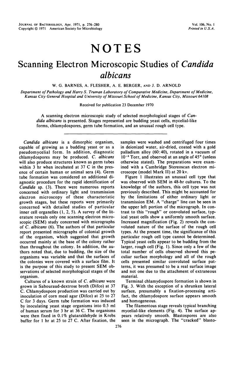
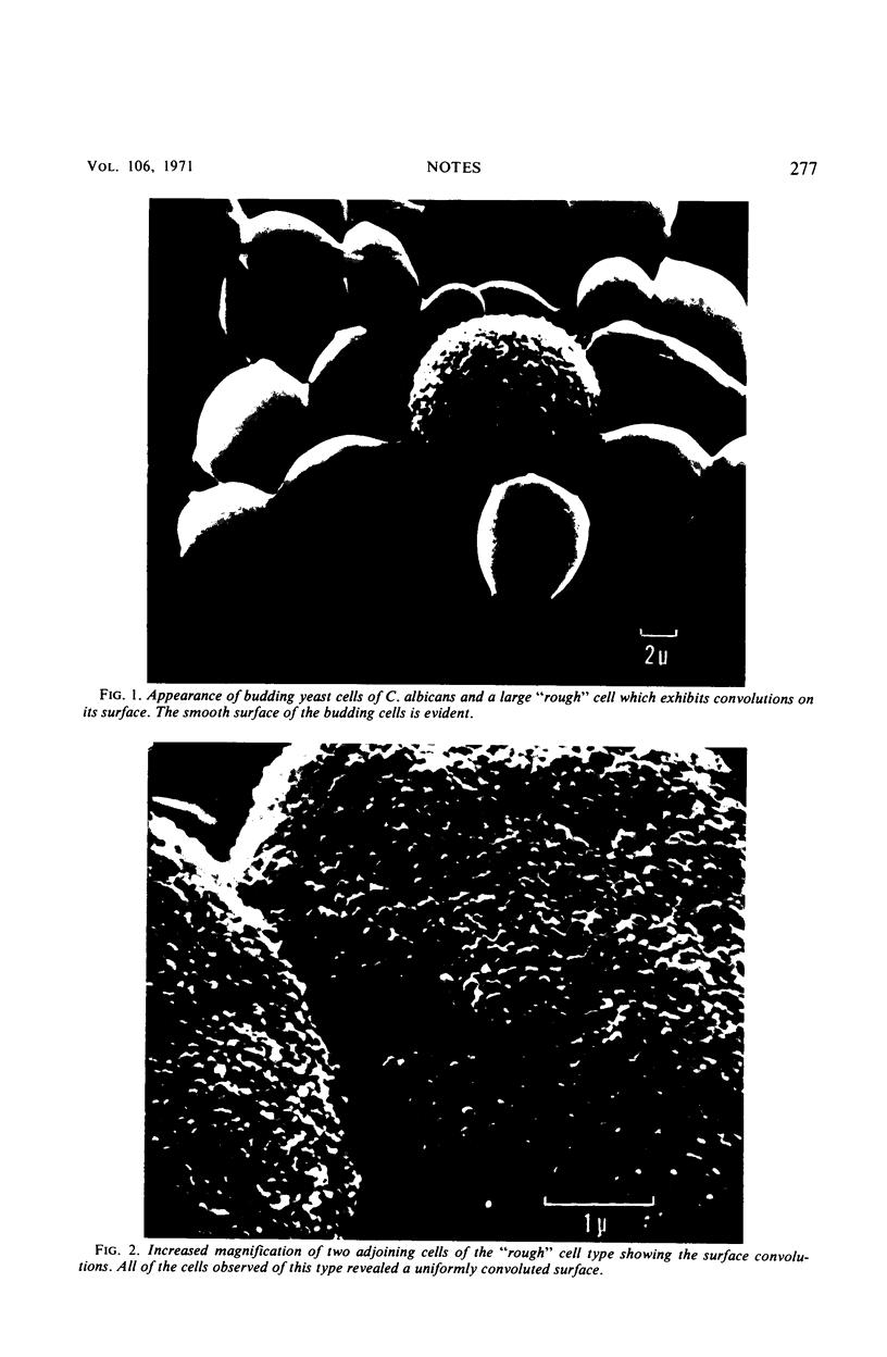
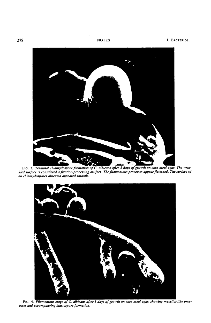
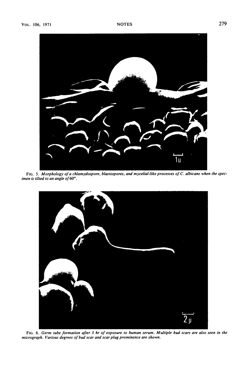
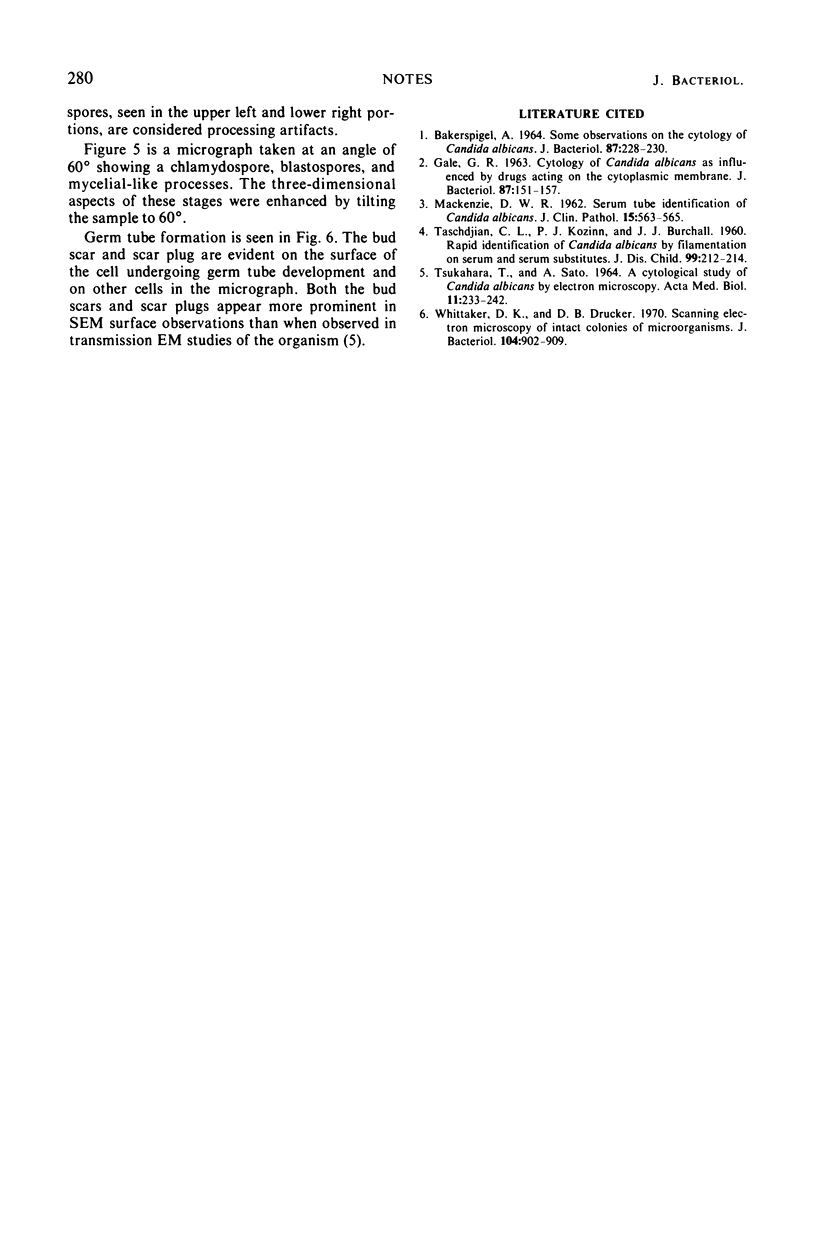
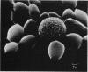
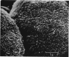
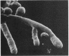
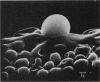
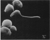
A scanning electron microscopic study of selected morphological stages of Candida albicans is presented. Stages represented are budding yeast cells, mycelial-like forms, chlamydospores, germ tube formation, and an unusual rough cell type.
Full text
PDF
Images in this article
Selected References
These references are in PubMed. This may not be the complete list of references from this article.
- BAKERSPIGEL A. SOME OBSERVATIONS ON THE CYTOLOGY OF CANDIDA ALBICANS. J Bacteriol. 1964 Jan;87:228–230. doi: 10.1128/jb.87.1.228-230.1964. [DOI] [PMC free article] [PubMed] [Google Scholar]
- GALE G. R. CYTOLOGY OF CANDIDA ALBICANS AS INFLUENCED BY DRUGS ACTING ON THE CYTOPLASMIC MEMBRANE. J Bacteriol. 1963 Jul;86:151–157. doi: 10.1128/jb.86.1.151-157.1963. [DOI] [PMC free article] [PubMed] [Google Scholar]
- Mackenzie D. W. Serum tube identification of Candida albicans. J Clin Pathol. 1962 Nov;15(6):563–565. doi: 10.1136/jcp.15.6.563. [DOI] [PMC free article] [PubMed] [Google Scholar]
- TASCHDJIAN C. L., BURCHALL J. J., KOZINN P. J. Rapid identification of Candida albicans by filamentation on serum and serum substitutes. AMA J Dis Child. 1960 Feb;99:212–215. doi: 10.1001/archpedi.1960.02070030214011. [DOI] [PubMed] [Google Scholar]
- TSUKAHARA T., SATO A. A CYTOLOGICAL STUDY OF CANDIDA ALBICANS BY ELECTRON MICROSCOPY. Acta Med Biol (Niigata) 1964 Mar;11:233–242. [PubMed] [Google Scholar]
- Whittaker D. K., Drucker D. B. Scanning electron microscopy of intact colonies of microorganisms. J Bacteriol. 1970 Nov;104(2):902–909. doi: 10.1128/jb.104.2.902-909.1970. [DOI] [PMC free article] [PubMed] [Google Scholar]