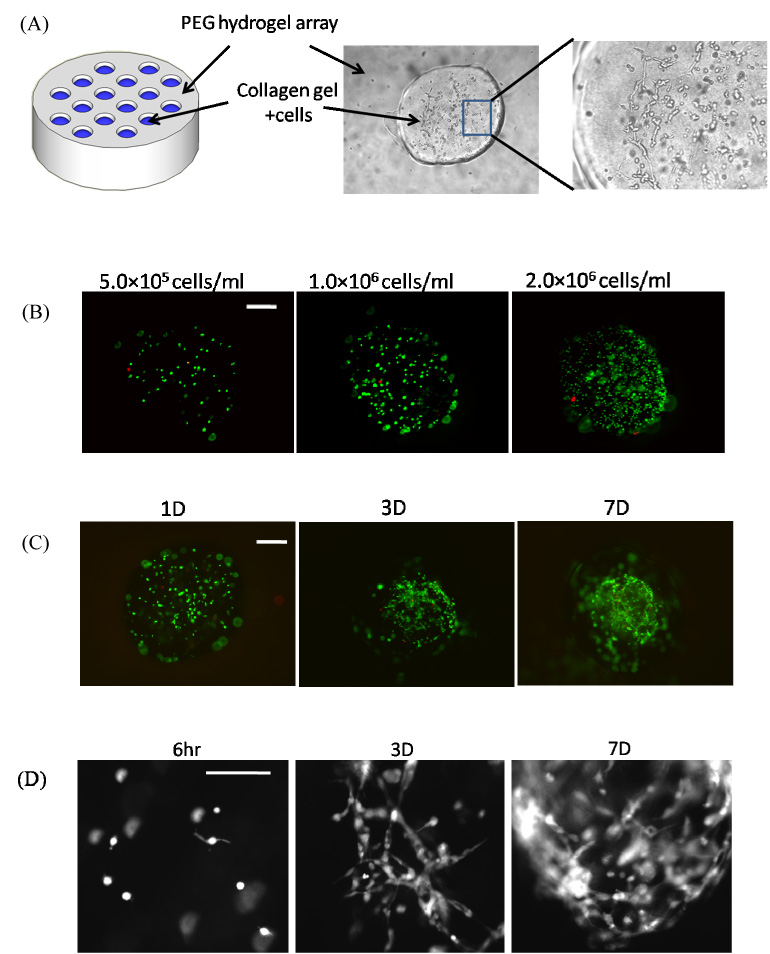
Figure 6.

A) Schematic representation of PEG hydrogel array filled with collagen hydrogels in spots. B) Seeding of NIH/3T3 fibroblasts into collagen hydrogel spots with various cell seeding densities. C) Live/dead images of NIH/3T3 fibroblasts cultured in collagen hydrogel spots for various times. D) Higher magnification (100X) images of NIH/3T3 fibroblasts spreading over time in collagen hydrogel array spots.
