Abstract
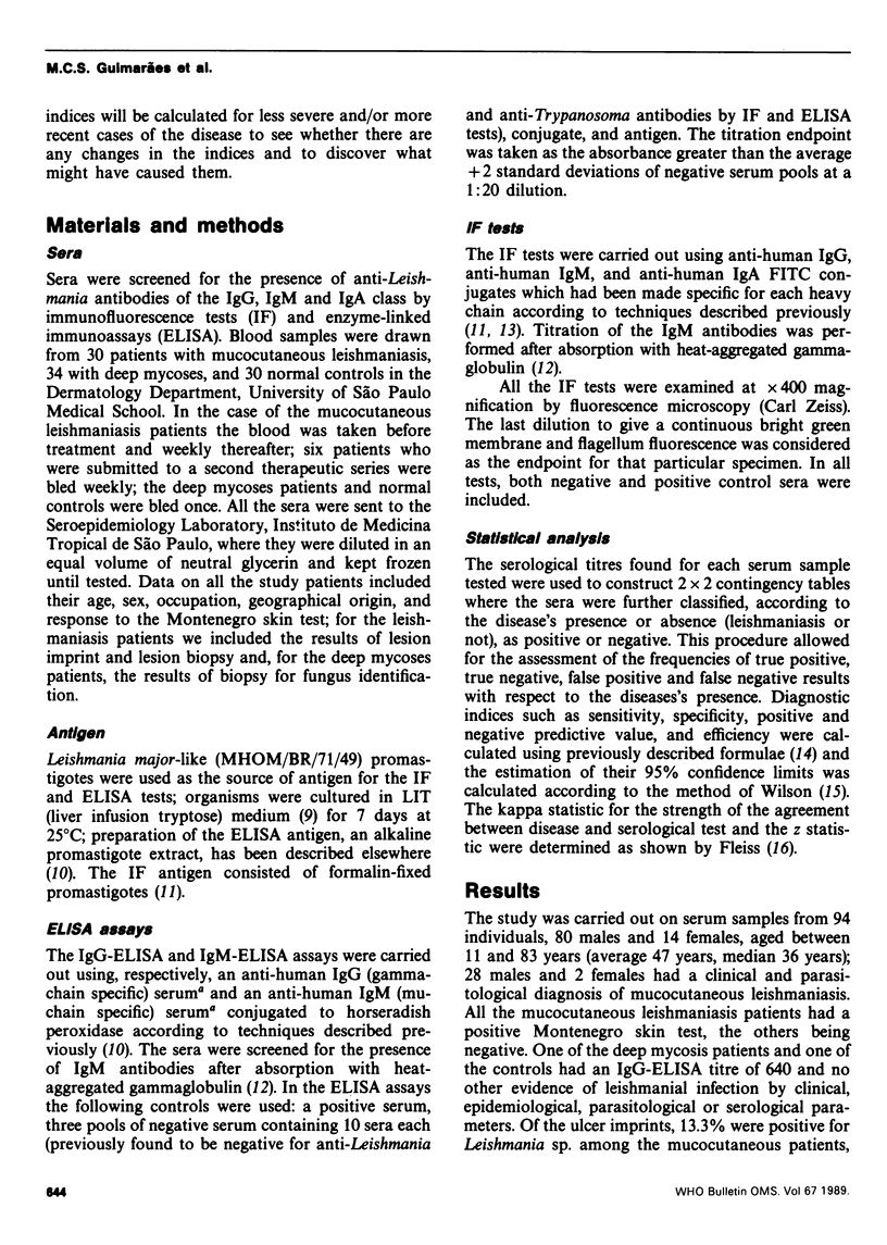
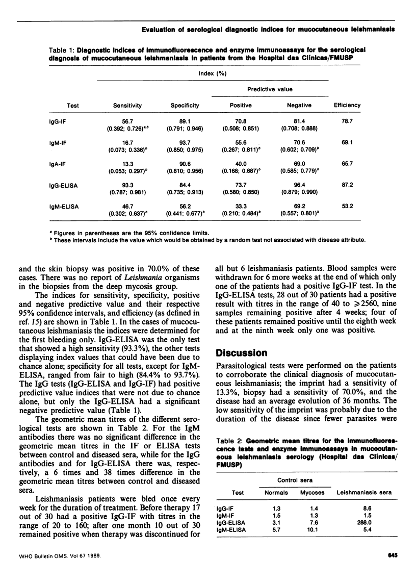
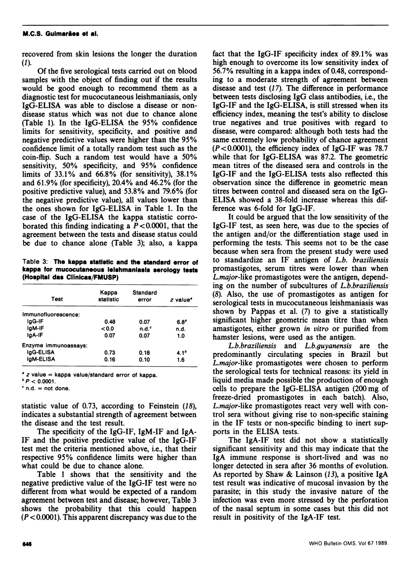
The sensitivity, specificity, positive predictive value, negative predictive value, and efficiency of immunofluorescence (IF) and enzyme-linked immunoassays (ELISA) for IgG, IgM and IgA antibodies were assessed on sera from mucocutaneous leishmaniasis patients and controls. The sensitivity of the IgG-ELISA test was 93.3% with 95% confidence interval higher than what could be due to a random test not associated with the disease. The specificity of all tests, except the IgM-ELISA, gave indices that could not have been due to chance. The IgG-ELISA and IgG-IF had the highest positive predictive value and the kappa statistic showed that the strength of agreement between the disease and the test was strongest for IgG-ELISA. The IgG-ELISA had a negative predictive value with 95% confidence limits that were not due to chance alone. Efficiency was highest for IgG-ELISA and IgG-IF. These results were obtained using sera from patients with severe or long-standing disease and from controls in whom the disease was ruled out by a negative Montenegro skin test. In field surveys where the differences between cases and controls are less easy to define the diagnostic indices of these tests may vary with the disease prevalence.
Full text
PDF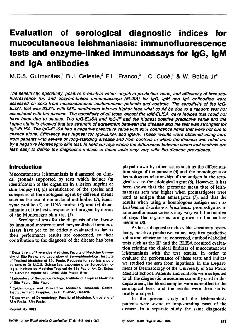
Selected References
These references are in PubMed. This may not be the complete list of references from this article.
- Anthony R. L., Williams K. M., Sacci J. B., Rubin D. C. Subcellular and taxonomic specificity of monoclonal antibodies to New World Leishmania. Am J Trop Med Hyg. 1985 Nov;34(6):1085–1094. doi: 10.4269/ajtmh.1985.34.1085. [DOI] [PubMed] [Google Scholar]
- Camargo M. E., Leser P. G., Rocca A. Rheumatoid factors as a cause for false positive IgM anti-toxoplasma fluorescent tests. A technique for specific results. Rev Inst Med Trop Sao Paulo. 1972 Sep-Oct;14(5):310–313. [PubMed] [Google Scholar]
- Celeste B. J., Guimarães M. C. Growth curves of Leishmania braziliensis braziliensis promastigotes and surface antigen expression before and after adaptation to Schneider's Drosophila medium as assessed by anti-Leishmania human sera. Rev Inst Med Trop Sao Paulo. 1988 Mar-Apr;30(2):63–67. doi: 10.1590/s0036-46651988000200001. [DOI] [PubMed] [Google Scholar]
- Cuba Cuba C. A., Marsden P. D., Barreto A. C., Rocha R., Sampaio R. R., Patzlaff L. Diagnóstico parasitológico e immunológico de leishmaniasis tegumentaria americana. Bol Oficina Sanit Panam. 1980 Sep;89(3):195–208. [PubMed] [Google Scholar]
- Miles M. A. Biochemical identification of the leishmanias. Bull Pan Am Health Organ. 1985;19(4):343–353. [PubMed] [Google Scholar]
- Pappas M. G., McGreevy P. B., Hajkowski R., Hendricks L. D., Oster C. N., Hockmeyer W. T. Evaluation of promastigote and amastigote antigens in the indirect fluorescent antibody test for American cutaneous leishmaniasis. Am J Trop Med Hyg. 1983 Nov;32(6):1260–1267. doi: 10.4269/ajtmh.1983.32.1260. [DOI] [PubMed] [Google Scholar]
- Shaw J. J., Lainson R. Leishmaniasis in Brazil. XIV. Leishmanial and trypanosomal IgA antibody in patients with leishmaniasis and Chagas's disease. Trans R Soc Trop Med Hyg. 1981;75(2):254–257. doi: 10.1016/0035-9203(81)90329-1. [DOI] [PubMed] [Google Scholar]


