Abstract
Objectives
To determine normal spleen dimensions in a healthy collegiate athletic population.
Methods
631 Division I collegiate athletes from one university participated in the study. During pre‐participation examinations, demographic data collected were collected from volunteer athletes including sex, race, measurement of height and weight, and age. Subjects also completed a medical history form to determine any history of mononucleosis infection, platelet disorder, sickle cell disease (or trait), thalassaemia, or recent viral symptoms. Subjects then underwent a limited abdominal ultrasound examination, where splenic length and width were recorded.
Results
Mean (SD) splenic length was 10.65 (1.55) cm and width, 5.16 (1.21) cm. Men had larger spleens than women (p<0.001). White subjects had larger spleens than African‐American subjects (p<0.001). A previous history of infectious mononucleosis or the presence of recent cold symptoms had no significant affect on spleen size. In more than 7% of athletes, baseline spleen size met current criteria for splenomegaly.
Conclusions
There is a wide range of normal spleen size among collegiate athletes. Average spleen size was larger in men and white athletes than in women and black athletes. A single ultrasound examination for determination of splenomegaly is of limited value in this population.
Keywords: spleen, ultrasound, athletics
Splenomegaly is a clinically important finding, particularly for physicians required to make decisions on an athlete's ability to resume athletic activity safely. For the most part, the ability to detect the presence of an enlarged spleen by physical examination alone has both poor sensitivity and poor specificity.1 The identification of splenomegaly by clinical examination is particularly difficult when dealing with mild splenomegaly. As a result, objective diagnostic measures have been proposed as a potentially useful step in making return to play decisions in athletes with suspected splenomegaly.2
While many imaging techniques can be used to determine spleen size, ultrasonography is particularly useful because of ease of use and lack of radiation exposure. Diagnostic imaging to assess spleen size is routinely accomplished by ultrasonographic measurement along its long axis. However, there is variation among radiological texts in defining the upper limits of normal for longitudinal diameter, with values ranging from 12 to 14 cm in adults.3,4,5
Normal spleen size has been found to vary significantly depending on age and sex.6 In paediatric populations, there is a significant correlation between spleen size and height, weight, and body surface area.7,8,9 While there are previous published studies documenting normal splenic dimensions in both paediatric and adult populations, the study populations were often heterogeneous and the individual sample sizes small.6,7,8,9,10,11,12
Furthermore, there are no studies specifically designed to evaluate special populations such as athletes. Given the wide range of body types encountered among athletes and the potential importance of defining splenomegaly in these individuals, normative values for spleen size would be clinically useful.
Methods
The study protocol was approved by the university's institutional review board. Informed consent was obtained from each participant before data collection was begun.
Volunteer athletes from a Division I university were recruited to participate in the study. Individuals were excluded if they were less than 18 years of age or had previously undergone a splenectomy. Demographic data were collected on each participant at the time of their pre‐participation physical examination. This information included sex, measurement of height and weight, and race. Subjects also completed a medical history form to ascertain any history of mononucleosis infection, platelet disorder, sickle cell disease (or trait), thalassaemia, or symptoms suggesting recent viral illness. Subjects then underwent their annual pre‐sports physical examination, followed by limited abdominal ultrasound to obtain splenic measurements.
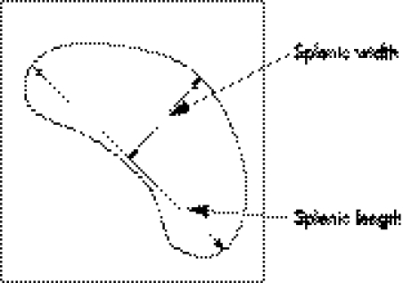
Ultrasonography was undertaken by an experienced and licensed technician at the university medical centre. The examination was done using an ATL HDI 3500 or 5000 ultrasound machine and a curved 5.2 MHz transducer (Philips Medical Systems, Bothel, Washington). The spleen was visualised with the participant in the right lateral decubitus position. Measurements were then taken in the sagittal (longitudinal) and transverse planes (measurement of width), with the maximum dimension being recorded in each plane (fig 1). Images were also saved onto a compact disc for review by a radiologist experienced in reading abdominal ultrasound scans. The radiologist confirmed the splenic measurements and noted any significant additional findings.
Figure 1 Diagram showing the method for measuring splenic length and with by ultrasound.
A subgroup of participants underwent two splenic ultrasound scans done a week apart by two different ultrasonographers to determine inter‐ and intrarater reliability.
Data analysis
Body mass index (BMI) was calculated (weight (kg)/height (m2)). Pearson moment correlation coefficients were calculated between height, weight, BMI, and spleen size. Three separate univariate analyses of variance (UNIANOVA) were carried out to compare spleen size by race and sex, with height, weight, and BMI as covariates. One‐way analysis of variance (ANOVA) was used to compare spleen size in individuals with and without a previous history of infectious mononucleosis or a report of recent or current “cold symptoms”. Intraclass correlation coefficients (ICCs) were calculated to assess inter‐ and intrarater reliability for ultrasound measurements, using the SPSS statistical software package, version 10.14 (SPSS, Chicago, Illinois, USA).
Results
Data were collected on 631 subjects (table 1). Mean (SD) spleen length was 10.65 (1.55) cm (range 5.59 to 17.06), and mean width, 5.16 (1.21) cm (range 2.83 to 12.81) (fig 2). There was a moderate correlation of both height and weight with splenic length, with correlation coefficients (r) of 0.48 and 0.47, respectively. Height and weight were highly correlated (r = 0.80). Men had significantly larger spleens than women (p<0.001), while white participants had larger spleens than African Americans (p<0.001) (table 2).
Table 1 Anthropometric and spleen size data.
| n | Age (years) | Height (m) | Weight (kg) | BMI (kg/m2) | Spleen width (cm) | Spleen length (cm) | |
|---|---|---|---|---|---|---|---|
| All | 631 | 19.42 (1.47) | 1.76 (0.12) | 76.27 (20.30) | 24.22 (4.03) | 5.16 (1.21) | 10.65 (1.55) |
| Female | 290 | 19.04 (1.16) | 1.46 (0.09) | 62.38 (11.00) | 22.31 (2.68) | 4.74 (0.91) | 9.91 (1.27) |
| Male | 341 | 19.74 (1.62) | 1.84 (0.07) | 88.12 (18.88) | 25.85 (4.26) | 5.54 (1.28) | 11.29 (1.49) |
| African American | 97 | 19.56 (1.38) | 1.82 (0.11) | 88.69 (21.02) | 26.40 (4.29) | 5.03 (1.20) | 9.80 (1.32) |
| White | 524 | 19.41 (1.49) | 1.75 (0.12) | 74.12 (19.48) | 23.84 (3.88) | 5.20 (1.21) | 10.82 (1.55) |
Values are mean (SD).
BMI, body mass index.
Figure 2 Scatterplot of spleen dimensions for men and women. (A) Spleen width. (B) Spleen length.
Table 2 Results of univariate analysis of variance comparing spleen size by race and sex with height, weight, and body mass index as covariates.
| Height | Weight | BMI | |
|---|---|---|---|
| Sex | |||
| Spleen length | p<0.001 | p<0.001 | p<0.001 |
| Spleen width | p = 0.022 | p = 0.002 | p<0.001 |
| Race | |||
| Spleen length | p<0.001 | p<0.001 | p<0.001 |
| Spleen width | p<0.001 | p<0.001 | p<0.001 |
BMI, body mass index.
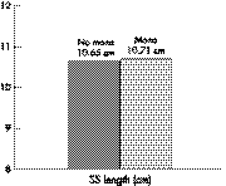
These significant differences were evident when controlling for height, weight, and BMI independently. There was no significant difference in spleen size in athletes with a previous history of infectious mononucleosis (fig 3) or those who reported recent or current viral symptoms (n = 30) compared with those who reported a negative history (p = 0.89 and p = 0.37, for spleen length, and p = 0.78 and p = 0.22 for spleen width, respectively). Three individuals reported a history of sickle cell trait, and two reported idiopathic thrombocytopenia. No athletes reported a history of thalassaemia.
Figure 3 Average spleen length (SS length) for athletes with (n = 79) and without (n = 552) a history of infectious mononucleosis (mono) (p = 0.89).
Intrarater reliability for technician No 1 was ICC(2,1) = 0.898 for sagittal measurements (length) and ICC(2,1) = 0.59 for transverse (width). For technician No 2 these values were ICC(2,1) = 0.895 and ICC(2,1) = 0.87, respectively. Inter‐rater reliability was ICC(2,1) = 0.90 for sagittal measurements and ICC(2,1) = 0.67 for transverse.
Discussion
The morphology of visceral organs varies from person to person. During the maturation process from infancy through adolescence, growth of visceral organs, including the spleen, shows a high correlation with gains in height, weight, and body surface area.7,8 Splenic length measured by ultrasonography provides an objective and reliable way to assess spleen size.9,10 Although previous studies have measured splenic size in normal individuals, the numbers of subjects of this particular age group were small and from varied populations. Loftus et al reported normal ultrasound spleen length in 783 subjects in a Chinese population, but of these only 35 were within the age range of 15 to 30 years.6 Capaccioli et al found a mean splenic length of 10.5 cm in a population of 180 Italian adults, without stratifying for age.11 Other studies documenting normal spleen growth and dimensions in paediatric age groups likewise fail to include populations representative of college age subjects.7,8,9 Still other studies have been oriented to developing normal standards for splenic weight or volume.12,13,14,15,16 These studies have similar limitations, with small numbers of subjects within each specific age group. Additionally, the calculations used to measure splenic volume are often cumbersome or rely on computed tomography (CT) for measurement. This process is unlikely to add significant clinical value as linear measurements taken by ultrasound show a high correlation with CT volume assessments.17
To our knowledge, this is the first study to define normative values for spleen size in a large cohort of college age athletes, representing a diverse population with regard to body morphology. It is also the first study to describe variation in normal spleen size by race. Our overall average for splenic length of 10.65 cm is consistent with previous normal values reported for the general adult population.11 We have further noted a large range of normal splenic length with more than 7% of athletes having a spleen length of ⩾13 cm—a value that has been used as a loose cut off point to define splenomegaly.
Measurement of splenic length by ultrasound is reliable within and between technicians. Measurement of splenic width, however, is less reliable, as evidenced by only moderate intra‐ and inter‐rater reliability. This finding supports the historical assessment of splenomegaly based on spleen length. Because the measurement of splenic width is less reliable, defining splenomegaly on the basis of splenic volume may be more uncertain.
Moderate correlations between height and weight and splenic length were observed. These are far less than those seen in paediatric and adolescent populations. This observation probably results from the cessation of rapid body growth that occurs with attainment of mature body morphology. Thus it is difficult to predict spleen size reliably on the basis of these variables alone.
Sex and race differences in normal splenic length and width were found to be significant. As there were moderate correlations between spleen size and both height and weight, we would expect a larger average spleen size in men on the basis of their larger body size. The fact that these significant differences persisted when controlling for height and weight independently may suggest that spleen size varies more as a product of these two variables, or that there are additional factors involved. Certainly this seems to be the case in African American individuals. As a group, African Americans in our study population had smaller spleens despite being taller and heavier than the white athletes. Although this has been alluded to in radiological texts,3 no rationale for the finding has been proposed. One could surmise that certain haemoglobinopathies, such as sickle cell anaemia, which are more prevalent among African Americans could result in smaller spleen size because of splenic infarction. However, only three African American athletes in our sample reported a history of sickle cell trait and none reported sickle cell disease or other haemoglobinopathy. Thus it is difficult to explain this finding, other than to surmise that it may be an evolutionary characteristic.
What is already known on this topic
There is a large range of normal splenic dimensions among individuals
The determination of spleen size based on clinic examination is difficult at best and ultrasonographic measurements are often employed for more accurate assessment
What this study adds
Normative data for spleen size among a morphologically diverse athletic population are given
The study underscores the need for caution in defining splenomegaly on the basis of isolated ultrasonographic measurements
Splenomegaly, which may be caused by any number of viral illnesses, is a common reason for restriction from athletic activity. Perhaps the most notable viral illness associated with an enlarged spleen is infectious mononucleosis. Physicians caring for athletes with infectious mononucleosis are often faced with the decision about whether or not to obtain diagnostic imaging to assess spleen size. Our data suggest that a single ultrasound examination for determining splenomegaly is of limited value as there is such a large variation in normal spleen size. For example, an individual with infectious mononucleosis who has a splenic length of 11 cm on ultrasound would probably be regarded as normal. However, this person may have had a baseline spleen length of 7 cm, in which case a significant increase in spleen size (and relative splenomegaly) could be misinterpreted as normal. Conversely, an athlete may have a baseline (normal) spleen size of >13 cm, and be erroneously diagnosed as having pathological splenomegaly.
Conclusions
This study defines normative values for spleen size for a college athletic population. The variation in normal splenic dimensions in this study group underlies the diversity of body types observed in college athletes. Setting an absolute cut off point for defining splenomegaly may be difficult because of the wide range of normal values encountered. This dataset may prove useful in future research to identify the natural course of splenic enlargement followed by normalisation in athletes with suspected splenomegaly.
Acknowledgements
This study was funded in part by a grant from the Agency for Healthcare Research and Quality (AHRQ).
We wish to thank all the athletic trainers, sports medicine fellows, ultrasound technicians, and the Department of Athletics at the University of Kentucky for their help and support of this project.
Footnotes
Competing interests: none declared
References
- 1.Tamayo S G, Rickman L S, Mathews W C.et al Examiner dependence on physical diagnostic tests for the detection of splenomegaly: a prospective study with multiple observers. J Gen Intern Med 1993869–75. [DOI] [PubMed] [Google Scholar]
- 2.Kindernecht J J. Infectious mononucleosis and the spleen. Curr Sports Med Rep 20021116–120. [DOI] [PubMed] [Google Scholar]
- 3.Meire H, Farrant P. The liver. In: Baxter GM, Allan PLP, Morley P, eds. Clinical diagnostic ultrasound. Oxford: Blackwell Science, 1999379–380.
- 4.Ayers A B. The spleen. In: Grainger RG, Allison DJ, eds. Diagnostic radiology: an Anglo‐American textbook of imaging. 2nd edn. Edinburgh: Churchill Livingstone, 19922403
- 5.Fried A M. Retroperitoneum, pancreas, spleen, and lymph nodes. In: McGahan JP, Goldberg BB, eds. Diagnostic ultrasound: a logical approach. Philadelphia: Lippincott‐Raven, 1998777
- 6.Loftus W K, Metrewili C. Normal splenic size in a Chinese population. J Ultrasound Med 199716345–347. [PubMed] [Google Scholar]
- 7.Konus O. Normal liver, spleen, and kidney dimensions in neonates, infants, and children: evaluation with sonography. Am J Roentenol 19981711693–1698. [DOI] [PubMed] [Google Scholar]
- 8.Megremis S D, Vlachonikolis I G, Tsilimigaki A M. Spleen length in childhood with US: normal values based on age, sex, and somatometric parameters. Radiology 2004231129–134. [DOI] [PubMed] [Google Scholar]
- 9.Rosenberg H K, Markowitz R I, Kolberg H.et al Normal splenic size in infants and children: sonographic measurements. Am J Roentgenol 1991157119–121. [DOI] [PubMed] [Google Scholar]
- 10.Loftus W K, Chow L T, Metreweli C. Sonographic measurement of splenic length: correlation with measurement at autopsy. J Clin Ultrasound 19992771–74. [DOI] [PubMed] [Google Scholar]
- 11.Capaccioli L, Stecco A, Vanzi E.et al Ultrasonographic study on the growth and dimensions of healthy children and adult organs. Int J Anat Embryol 20001051–50. [PubMed] [Google Scholar]
- 12.DeLand F H. Normal spleen size. Radiology 197097589–592. [DOI] [PubMed] [Google Scholar]
- 13.Kaneko J, Sugawara Y, Matsui Y.et al Normal splenic volume in adults by computed tomography. Hepato‐gastroenterology 2002491726–1727. [PubMed] [Google Scholar]
- 14.Pietri H, Boscaini M. Determination of a splenic volumetric index by ultrasonic scanning. J Ultrasound Med 1984319–23. [DOI] [PubMed] [Google Scholar]
- 15.Prassopoulos P, Daskalogiannaki M, Raissaki M. Determination of normal splenic volume on computed tomography in relation to age, gender, and body habitus. Eur Radiol 19977246–248. [DOI] [PubMed] [Google Scholar]
- 16.Rodrigues A J, Rodrigues C J, Germano M A.et al Sonographic assessment of normal spleen volume. Clin Anat 19958252–255. [DOI] [PubMed] [Google Scholar]
- 17.Lamb P M, Kanagasabay R R, Martin A.et al Spleen size: how well do linear ultrasound measurements correlate with three‐dimensional CT volume assessments? Br J Radiol 200275573–577. [DOI] [PubMed] [Google Scholar]


