Abstract
Objective
To evaluate the effect of massage on force production and neuromuscular recruitment.
Methods
Ten healthy male subjects performed isokinetic concentric contractions on the knee extensors at speeds of 60, 120, 180, and 240°/s. These contractions were performed before and after a 30 minute intervention of either rest in the supine position or lower limb massage. Electromyography (EMG) and force data were captured during the contractions.
Results
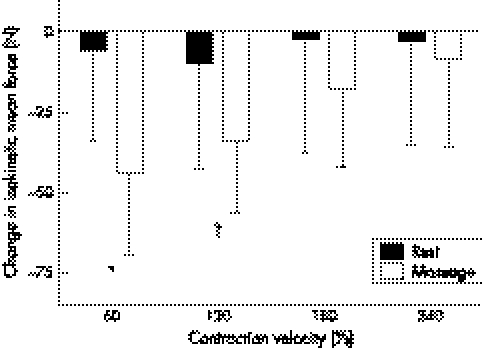
The change in isokinetic mean force due to the intervention showed a significant decrease (p<0.05) at 60°/s and a trend for a decrease (p = 0.08) at 120°/s as a result of massage compared with passive rest. However, there were no corresponding differences in any of the EMG data. A reduction in force production was shown at 60°/s with no corresponding alteration in neuromuscular activity.
Conclusions
The results suggests that motor unit recruitment and muscle fibre conduction velocity are not responsible for the observed reductions in force. Although experimental confirmation is necessary, a possible explanation is that massage induced force loss by influencing “muscle architecture”. However, it is possible that the differences were only found at 60°/s because it was the first contraction after massage. Therefore muscle tension and architecture after massage and the duration of any massage effect need to be examined.
Keywords: massage, electromyography, mean percentile frequency shift, force, muscle architecture
It has recently been shown that massage is mainly used in major sporting events for preparation before competition and recovery from events as opposed to treating specific problems.1 However, the benefit of massage before a bout of high intensity exercise has recently been questioned.2 Robertson et al2 examined the effect of massage on recovery after subjects had completed six high intensity 30 second cycling bouts. This recovery was examined by a 30 second Wingate anaerobic test performed eight minutes after the intervention. The results showed a significantly lower fatigue index after the massage. The authors proposed that this was probably a result of a slightly lower peak power output being generated. This would suggest that massage impaired peak power generation.
This evidence concurs with that of Goodwin,3 who showed a decrement in vertical jump performance after massage treatment. The author proposed that this was due to reduced muscle stiffness and neural activation. This proposal is plausible as, firstly, reduced muscle stiffness has been evidenced by a lengthening of massaged muscle.4,5 This would suggest that massage has a similar effect to stretching, which has recently been shown to result in a loss of force production when incorporated as part of a warm up.6 Secondly, there is evidence to suggest that massage causes a decline in motor unit activation.7,8,9,10,11 However, these studies used H‐reflex before and after massage, and, although they are a valid measure of motor unit activation, they are a different measure from the recording of surface electromyography (EMG) during a voluntary contraction. In addition, none of these studies related a decline in motor unit activation to any alteration in performance characteristics of the massaged muscle. This suggests that more studies need to be performed for the results to be adequately applied to an exercise and sporting context.
To our knowledge no previous studies have examined the effect of massage on neuromuscular recruitment during concentric force production. Accordingly, we decided to examine the effects of massage on four concentric contraction speeds (60, 120, 180, and 240°/s) and capture EMG data simultaneously. As previous studies have shown a decrement in power after massage treatment, we decided to also incorporate a standing vertical jump test with countermovement.
Methods
Subjects
Ten healthy male subjects who were physically active on a regular basis volunteered for the study. Their mean (SD) age and mass were 21.5 (0.5) years (range 20–24) and 74.4 (11.3) kg (range 64–91) respectively. All subjects were given written information about the study, after which they provided written consent before participation. The local ethics of research committee approved the study.
Experimental design
Subjects visited the laboratory on three separate occasions one week apart and at the same time of day. Two of these visits consisted of either a massage intervention or 30 minutes of rest in random order. The first visit was used for familiarisation to ensure that all subjects knew the protocol and could satisfactorily perform maximal contractions at the different speeds. Two days before the familiarisation visit, normal dietary intake (food and fluid) was recorded; this procedure was replicated before the two subsequent visits. Subjects were told to refrain from any heavy exercise during the 24 hours preceding each test session. On arrival at the laboratory, they were questioned about their compliance with the dietary intake and exercise controls.
Standing jump test
On reporting to the laboratory, subjects were asked to perform a five minute warm up on the cycle ergometer at 70 rpm with 100 W load. They then performed three vertical jump tests from a standing position with arms fixed with countermovement included. These jump tests were repeated immediately after the rest and massage intervention.
Muscle function tests
To normalise EMG recordings during isokinetic contractions, it was first necessary to test maximal isometric force output. The strength of the subjects' right knee extensors was measured on an isokinetic dynamometer (Kin‐Com Chattanooga Group Inc, Chattanooga, Tennessee, USA). Subjects sat on the dynamometer, and their hips, thighs, and upper bodies were firmly strapped to the seat. In this position their hip angle was at 100° angle of flexion. The right lower leg was then attached to the arm of the dynamometer at a level slightly above the lateral malleolus of the ankle joint, and the axis of rotation of the dynamometer arm was aligned with the lateral femoral condyle. The dynamometer arm was then set at a start and stop angle of 65° and 60° respectively from full leg extension, which means that during an isometric setting, the lever arm automatically alternated between these two angles for each contraction. Each subject performed four submaximal familiarisation contractions before performing two maximal voluntary contractions (MVCs); the latter at 60° was used for normalisation of EMG data. All subjects were encouraged verbally to exert maximal effort during both MVCs.
After both the MVCs (pre‐intervention) and after 30 minutes of massage or rest (post‐intervention), the subjects performed isokinetic knee extensions at 60, 120, 180, and 240°/s. They performed one warm up at each speed and then completed three maximal effort contractions at each of the speeds (always starting with 60°/s and increasing to 240°/s). Subjects were instructed to exert effort as hard and as fast as possible for all contractions. A 10 second rest was given between each of the contractions and a one minute rest between the different velocities. The lever arm was pushed back by the investigator so that only the concentric phase of the contractions was measured.
Massage
After the subjects had performed the pre‐intervention muscle function tests, they received either 30 minutes of passive rest (in the supine position) or massage in random crossover fashion for both visits. The massage was applied for 30 minutes in 7 minute 30 second segments to the back of each leg, with the subject lying prone on a standard treatment couch. The subject then assumed a supine position, and massage was again applied for the same duration to the anterior aspect of both legs. Table 1 shows the massage protocol followed during each 7 minute 30 second period. Most strokes were grade 1 or 2, but three grade 3 effleurage strokes, using a clenched fist, were applied in a centripetal direction to the left and right iliotibial band midway through the supine massage. All massage was administered by the same charted physiotherapist using a conventional bland mineral oil (40 ml contact medium was used per massage area).
Table 1 Massage protocol.
| Massage technique | Description | Grade |
|---|---|---|
| Stroking | Whole hand, one and two handed, centripetal and multidirectional | Four strokes grade 1 (very light contact to give sedative effect), 2 strokes grade 2 (slightly firmer to produce minimal effect on superficial vessels) |
| Effleurage | Whole hand, two handed, and reinforced centripetal and centrifugal | Grades 1 (sufficient depth to influence onward flow of superficial vessels), 2 (affecting deeper vessels), and 3 (reinforced) |
| Petrissage | Picking up, whole hand, two handed V shaped, centripetal and centrifugal | Grade 1 (sufficient to influence superficial vessels and on underlying structures compress superficial soft tissue) and 2 (sufficient to compress deep tissue on underlying structures and affect deeper tissue drainage) |
| Wringing | Whole hand, two handed, centripetal, centrifugal, multidirectional | Grade 1 (same as Petrissage grade) |
| Rolling | Muscle rolling, centripetal | Grade 2 (muscle rolling, lifting muscle tissue, and affecting deeper structures) |
This protocol represents the procedures followed during each of the four 7 minute 30 second massage periods. All petrissage was interspersed with effleurage grade 2 in a centripetal direction. Grading as described by Watt.15
EMG
Before maximal isometric strength testing on the Kin‐Com isokinetic dynamometer, EMG dual electrodes (PNS Dual Element Electrode; Vermed, Vermont, USA) were attached to the subject's lower limb midway between the superior surface of the patella and the anterior superior iliac crest of the “belly” of the rectus femoris after preparation of the skin as described previously.13 The electrodes were linked to the BioPac EMG apparatus (Biopac Systems, Santa Barbara, California, USA) and host computer. The EMG data were automatically anti‐aliased by the hardware (Biopac Systems). Each activity was sampled at a 2000 Hz capture rate. This gave route mean square (RMS) of the EMG signal, giving a measure of the power of the signal, which was used for subsequent analyses. Recordings were taken during the second maximal isometric trial and for all the isokinetic contractions, yielding a raw signal. MVC EMG data were recorded before the first set of isokinetic contractions for both conditions to ensure similar normalisation of EMG in the two trials. The raw data were divided into four epochs, which captured all the electrical activity recorded in each contraction. The first epoch included all data collected during the second MVC trial, and the remaining three epochs included data collected for the three maximal isokinetic contractions at each speed.
The spectrum of the frequency for each epoch of data collected during the cycle ride was assessed using the raw EMG data by using a fast Fourier transformation algorithm. The analyses of frequency spectrum were restricted to frequencies in the 5–500 Hz range, because the EMG signal content is mostly noise when it is outside of this bandwidth. The frequency spectrum from each epoch of data was compared with that derived from the MVC, and the amount of spectral compression was estimated. This technique was performed as described by Lowery et al,12 which is a modification of the work of Lo Conte and Merletti13 and Merletti and Lo Conte.14 The spectrum of the raw signal of each epoch was obtained, and the normalised cumulative power at each frequency was calculated for each epoch. The shift in percentile frequency was then examined—that is, at 0%…50%…100% of the total cumulative. The percentile shift was then estimated by calculating the mean shift in all percentile frequencies throughout the mid‐frequency range—that is, 5–500 Hz. This method has been suggested as a more accurate estimate of spectral compression than median frequency analysis, which uses a single value (50th) of percentile frequency.15 This change in mean percentile frequency shift (MPFS) data was used for subsequent analyses.
Statistical analysis
A two way analysis of variance for repeated measures was used to evaluate statistical significance of all the variables measured. Significance was accepted at p⩽0.05. All data are expressed as mean (SD).
Results
Force
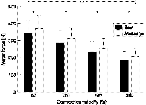
There was a significant (p<0.05) difference in the decline in isokinetic mean force from before to after the intervention for the massage condition for the 60°/s contraction speed only, as well as a trend (p = 0.08) for a decline in isokinetic mean force for the contraction at 120°/s (fig 1). No significant differences were observed for the subsequent 180 and 240°/s contractions, and no significant decline in force was observed over the passive rest intervention (fig 1). However, there was a significantly (p<0.05) greater absolute mean force before the massage intervention and a highly significant (p<0.01) decline in force from 60°/s through to 240°/s, without any interaction effect in all assessments (fig 2). After the intervention, there was no difference in absolute force production between the massage and passive rest trials, but a similar highly significant (p<0.01) drop off in force was shown for both trials as the contraction velocity increased.
Figure 1 Mean (SD) change in isokinetic mean force after the intervention for rest and massage conditions. The decrement in decrease in force for the massage condition was significant (*p<0.05) at 60°/s and showed a trend (†p = 0.08) at 120°/s.
Figure 2 Mean (SD) isokinetic force before the intervention for rest and massage conditions. A highly significant (**p<0.01) reduction in force was shown for both groups as the contraction velocity increased, and a significantly (*p<0.05) higher amount of force was produced before the massage than the rest intervention.
EMG
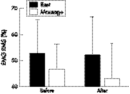
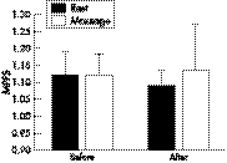
There were no significant differences observed in either EMG RMS (fig 3) or MPFS (fig 4) for contraction velocities of 60, 120, 180, and 240°/s and between the massage and rest conditions, despite a reduction in force decrement for the 60°/s contraction during the massage condition.
Figure 3 Mean (SD) electromyographic (EMG) amplitude (route mean square (RMS)) values normalised as a % of maximal voluntary contraction before and after the massage and rest conditions at a contraction speed of 60°/s. There were no significant differences between any of the values.
Figure 4 Mean (SD) electromyographic frequency (mean percentile frequency shift (MPFS)) values normalised against maximal voluntary contraction before and after the massage and rest conditions at a contraction speed of 60°/s. There were no significant differences between any of the values.
Vertical jump
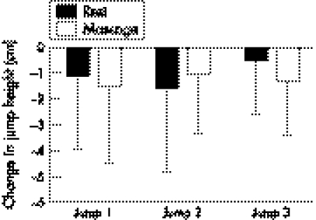
Despite a slight non‐significant reduction in jump height after the intervention, there were no significant differences between the massage and rest conditions (fig 5).
Figure 5 Mean (SD) change in vertical jump height after passive rest and massage interventions.
Subjective response
All subjects reported that their legs felt “light” after receiving the massage treatment, and therefore perceived greater effort generating force during the isokinetic contractions.
Discussion
A greater decrement in force production was observed after the massage treatment at the 60°/s contraction, with a near significant decrement at 120°/s and no decrement in the remaining contractions (180 and 240°/s) compared with the passive rest intervention. There was no corresponding alteration in motor unit recruitment and firing rate for these slow contractions, which is shown by the unchanged RMS and MPFS data.
We hypothesised that any decline in force would coincide with a similar rate of decline in both motor unit recruitment and firing rate.16 This would be a direct result of smaller and fewer motor units being recruited, displaying a reduced RMS.17,18,19 A reduced MPFS could also be hypothesised as a result of reduced drive from the central nervous system and/or an accumulation of metabolites lowering the pH of the muscle resulting in slowed conduction velocity,20,21 which slows down the firing rate. Therefore unchanged RMS and MPFS data suggest that mechanisms other than neuromuscular recruitment must be responsible for this decrement in force. Alternatively, it could be suggested that there should have been an increase in neural recruitment to compensate for the impaired force‐generating capacity after massage treatment, particularly as it is clear from fig 3 that a large amount of recruitment reserve was available for all of the contractions. However, when additional motor units are recruited—for example during submaximal fatigue—this would depend on afferent receptors signalling the central nervous system to increase the force.22 As the protocol used in this study used concentric only isokinetic maximal non‐fatiguing contractions, there would be minimal involvement of afferent receptors to modulate motor unit recruitment.
Evidence suggests that both acute and chronic massage will result in lengthening of the muscle.4,5 Consequently, the length‐tension relation may have been affected in this study, resulting in reduced force output.23,24,25,26 Furthermore, it has been suggested that, when skeletal muscle is lengthened, the number of prospective actin/myosin cross bridges declines,27 resulting in a loss of force without a corresponding reduction in neural activation.27 This suggests that a possible cause of this greater reduction in force after massage is an alteration in muscle architecture rather than any alteration in motor unit recruitment or firing rate.
Interestingly, in our study, the standing vertical jump data were the same for the two conditions. This may be because the contraction velocity in a vertical jump is closer to 240°/s than 60°/s, and therefore the impairment resulting from massage is less, which is also shown in the isokinetic contractions in this study. Contrary to our findings, Goodwin3 did show a decrement in vertical jump height in the same jump test. However, that study combined massage with stretch, which may have resulted in greater lengthening of the muscles than massage alone.
Alternatively, the significant reduction in force after the massage treatment may be the result of the greater force production shown before the massage treatment. However, if this was the only explanation, then we would also expect to see a significant reduction in force for all the contraction velocities rather than just 60°/s. However, the higher force before massage is an interesting observation; it is unclear why it occurred given the pre‐test controls for exercise, diet, and time of day, but a possible explanation is that it is an anticipatory response to receiving a massage.
Furthermore, it is possible that the force decrement was only observed at 60°/s because it was the first contraction after massage, and the effect of the massage on all subsequent contractions to be performed diminished as a result of time or prior contraction. A similar response has also been shown at low contraction velocities after stretching.28 However, Hinds et al29 performed contractions before and after massage at one contraction speed of 240°/s and showed no decrement in force production. The combined data from these studies suggest that massage affects force production only at low contraction velocities. A logical explanation for this originates from the force‐velocity relation described by Hill,30 which clearly shows that greater velocities produce less force, as confirmed in this study. According to Spurway,31 skeletal muscle will shorten fastest under the lightest loads. Therefore, at the low contraction velocities, there is slower shortening, resulting in a greater force‐generating capacity, and therefore the chances of observing an effect from massage will be greater at the lower velocities. This would result in a greater impairment of force production after massage, as the muscle has been lengthened and therefore has a reduced ability to shorten sufficiently to produce the necessary force output.
In conclusion, this study shows that lower limb massage appears to produce a reduction in force during concentric isokinetic contractions of the knee extensors at 60°/s, with no changes in force at higher contraction velocities. We propose that this reduction does not result from altered neuromuscular recruitment, but from a change in muscle architecture affecting the length‐tension relation. Further work is needed to examine both muscle tension and architecture after massage, and consideration needs to be given to the duration of any massage effect.
What is already known on this topic
Massage is widely used by sportspersons in preparation for competition
The perceived performance benefits and physiological mechanisms of this treatment are not fully understood
What this study adds
Immediately after massage treatment, force production in a muscle is reduced at low contraction velocities
This is not caused by an alteration in neuromuscular recruitment
Abbreviations
EMG - electromyography
MPFS - mean percentile frequency shift
MVC - maximal voluntary contraction
RMS - route mean square
Footnotes
Competing interests: none declared
References
- 1.Galloway S D, Watt J M. Massage provision by physiotherapists at major athletics events between 1987 and 1998. Br J Sports Med 200438235–236. [DOI] [PMC free article] [PubMed] [Google Scholar]
- 2.Robertson A, Watt J M, Galloway S D. Effects of leg massage on recovery from high intensity cycling exercise. Br J Sports Med 200438173–176. [DOI] [PMC free article] [PubMed] [Google Scholar]
- 3.Goodwin J E. A comparison of massage and sub‐maximal exercise as warm‐up protocols combined with a stretch for vertical jump performance. J Sports Sci 20022048–49. [Google Scholar]
- 4.Hernandez‐Reif M, Field T, Krasnegor J.et al Lower back pain is reduced and range of motion increased after massage therapy. Int J Neurosci 2001106131–145. [DOI] [PubMed] [Google Scholar]
- 5.Wiktorsson‐Moller M, Oberg B, Ekstrand J.et al Effects of warming up, massage, and stretching on range of motion and muscle strength in the lower extremity. Am J Sports Med 198311249–252. [DOI] [PubMed] [Google Scholar]
- 6.Behm D G, Button D C, Butt J C. Factors affecting force loss with prolonged stretching. Can J Appl Physiol 200126261–272. [PubMed] [Google Scholar]
- 7.Goldberg J, Sullivan S J, Seaborne D E. The effect of two intensities of massage on H‐reflex amplitude. Phys Ther 199272449–457. [DOI] [PubMed] [Google Scholar]
- 8.Goldberg J, Seaborne D E, Sullivan S J.et al The effect of therapeutic massage on H‐reflex amplitude in persons with a spinal cord injury. Phys Ther 199474728–737. [DOI] [PubMed] [Google Scholar]
- 9.Dishman J D, Bulbulian R. Comparison of effects of spinal manipulation and massage on motoneuron excitability. Electromyogr Clin Neurophysiol 20014197–106. [PubMed] [Google Scholar]
- 10.Sullivan S J, Williams L R, Seaborne D E.et al Effects of massage on alpha motoneuron excitability. Phys Ther 199171555–560. [DOI] [PubMed] [Google Scholar]
- 11.Morelli M, Seaborne D E, Sullivan S J. H‐reflex modulation during manual muscle massage of human triceps surae. Arch Phys Med Rehabil 199172915–919. [DOI] [PubMed] [Google Scholar]
- 12.Lowery M, O'Malley M, Vaughan C.et al A physiologically based stimulation of the electromyographic signal. Proceedings of the 12th International Society of Electrophysiology and Kinesiology, 1998
- 13.Lo Conte L R, Merletti R. Estimating EMG spectral compression: comparison of four indices. 18th Ann Int Cong IEEE Eng Med Biol Sci 199652–5. [Google Scholar]
- 14.Merletti R, Lo Conte L R. Advances in processing of surface myoelectric signals: part 1. Med Biol Eng Comput 199533362–372. [DOI] [PubMed] [Google Scholar]
- 15.Watt J.Massage for sport. Marlborough: Crowood Press, 1999
- 16.De Luca C J, Erim Z. Common drive of motor units in regulation of muscle force. Trends Neurosci 199417299–305. [DOI] [PubMed] [Google Scholar]
- 17.DeVries H A. Method for evaluation of muscle fatigue and endurance from electromyographic fatigue curves. Am J Phys Med 196847125–135. [PubMed] [Google Scholar]
- 18.deVries H A, Moritani T, Nagata A.et al The relation between critical power and neuromuscular fatigue as estimated from electromyographic data. Ergonomics 198225783–791. [DOI] [PubMed] [Google Scholar]
- 19.Moritani T, Nagata A, Muro M. Electromyographic manifestations of muscular fatigue. Med Sci Sports Exerc 198214198–202. [PubMed] [Google Scholar]
- 20.Lindstrom L, Magnusson R, Petersen I. Muscular fatigue and action potential conduction velocity changes studied with frequency analysis of EMG signals. Electromyography 197010341–356. [PubMed] [Google Scholar]
- 21.Stulen F B, DeLuca C J. Frequency parameters of the myoelectric signal as a measure of muscle conduction velocity. IEEE Trans Biomed Eng 198128515–523. [DOI] [PubMed] [Google Scholar]
- 22.Leonard C T, Kane J, Perdaems J.et al Neural modulation of muscle contractile properties during fatigue: afferent feedback dependence. Electroencephalogr Clin Neurophysiol 199493209–217. [DOI] [PubMed] [Google Scholar]
- 23.Marginson V, Eston R. The relationship between torque and joint angle during knee extension in boys and men. J Sports Sci 200119875–880. [DOI] [PubMed] [Google Scholar]
- 24.Rassier D E, MacIntosh B R, Herzog W. Length dependence of active force production in skeletal muscle. J Appl Physiol 1999861445–1457. [DOI] [PubMed] [Google Scholar]
- 25.Edman K A, Reggiani C. Redistribution of sarcomere length during isometric contraction of frog muscle fibres and its relation to tension creep. J Physiol 1984351169–198. [DOI] [PMC free article] [PubMed] [Google Scholar]
- 26.Gordon A M, Huxley A F, Julian F J. The variation in isometric tension with sarcomere length in vertebrate muscle fibres. J Physiol 1966184170–192. [DOI] [PMC free article] [PubMed] [Google Scholar]
- 27.Aratow M, Ballard R E, Crenshaw A G.et al Intramuscular pressure and electromyography as indexes of force during isokinetic exercise. J Appl Physiol 1993742634–2640. [DOI] [PubMed] [Google Scholar]
- 28.Nelson A G, Guillory I K, Cornwell C.et al Inhibition of maximal voluntary isokinetic torque production following stretching is velocity‐specific. J Strength Cond Res 200115241–246. [PubMed] [Google Scholar]
- 29.Hinds T, McEwan I, Perkes J.et al Effects of massage on limb and skin blood flow after quadriceps exercise. Med Sci Sports Exerc 2004361308–1313. [DOI] [PubMed] [Google Scholar]
- 30.Hill A V. The heat of shortening and the dynamic constants of muscle. Proc R Soc B 1938122–138.
- 31.Spurway N C.Muscle. In: Basic and applied sciences for sports medicine. Guildford: Butterworth Heineman, 19991–47.


