Abstract
Currently, the only accepted assay with which to detect active Clostridium botulinum neurotoxin is an in vivo mouse bioassay. The mouse bioassay is sensitive and robust and does not require specialized equipment. However, the mouse bioassay is slow and not practical in many settings, and it results in the death of animals. Here, we describe an in vitro cleavage assay for SNAP-25 (synaptosome-associated proteins of 25 kDa) for measuring the toxin activity with the same sensitivity as that of the mouse bioassay. Moreover, this assay is far more rapid, can be automated and adapted to many laboratory settings, and has the potential to be used for toxin typing. The assay has two main steps. The first step consists of immunoseparation and concentration of the toxin, using immunomagnetic beads with monoclonal antibodies directed against the 100-kDa heavy chain subunit, and the second step consists of a cleavage assay targeting the SNAP-25 peptide of the toxin, labeled with fluorescent dyes and detected as a fluorescence resonance energy transfer assay. Our results suggest that the sensitivity of this assay is 10 pg/ml, which is similar to the sensitivity of the mouse bioassay, and this test can detect the activity of the toxin in carrot juice and beef. These results suggest that the assay has a potential use as an alternative to the mouse bioassay for analysis of C. botulinum type A neurotoxin.
Clostridium botulinum is an anaerobic, gram-positive, spore-forming rod-shaped organism that produces the most biologically potent poisons currently known. There are seven serotypes of botulinum toxin, designated BoNT-A to BoNT-G. Types A, B, and E are most commonly associated with illness in humans. Exposure to these toxins generally occurs through the consumption of contaminated low-acid canned foods (4). Recently, there was an incident of contamination of carrot juice that caused outbreaks of botulism in Georgia and Florida (3). Also, Wein and Liu (11) discuss the outcome of a case of deliberate contamination of milk and cold drinks with BoNT. That article reiterates the threat that botulinum toxin could be released deliberately by bioterrorists, causing hundreds of thousands of deaths and billions of dollars in economic losses.
There is a great need to develop better methods to detect active botulinum neurotoxin. The “gold standard” test to detect active botulinum toxin is an in vivo mouse bioassay (6). This procedure, developed in 1927, is still being used by the scientific community. This assay involves the intraperitoneal injection of suspected contaminated food into a mouse and can take 4 to 6 days to obtain results. Because injection of the food product itself or the damage to vital organs during inoculation can cause the mouse's death, the lethal activity must be shown to be neutralized with an antibody against one of the botulinum toxin serotypes (6). This iterative procedure, requiring many animals, is expensive to perform and impractical for testing a large number of samples, and it raises ethical concerns with regard to the use of experimental animals.
Several attempts to replace the mouse bioassay have been made; for example, immunoassays, which speed detection, are useful for testing large numbers of samples (7, 10). However, such immunoassays cannot distinguish between the active form of the toxin, which poses a threat to life, and the inactive form. Also, samples that have tested positive by these immunoassays still require confirmation by the mouse bioassay (6). BoNT activity assays, based on the toxins' ability to cleave specific soluble N-ethylmaleimide-sensitive factor (NSF) attachment protein (SNAP) receptor (SNARE) proteins, have been developed (9). Such cleavage activity has been detected by mass spectrometry (1) and by using fluorescence sensors to detect fluorescence resonance energy transfer (FRET)-based reactions (5). However, such assays are not currently used for food analyses due to interference by peptidases and protease inhibitors in food matrices. The main objective of this study was to develop a rapid in vitro activity assay for the detection of BoNT-A, with a sensitivity level comparable to that of the mouse bioassay. To achieve this objective, we utilized the BoNT-A cleavage activity of SNAP-25, which was detected by FRET assay, and we applied immunomagnetic bead technology to concentrate the toxin from food matrices and to separate it from food peptidases and protease inhibitors.
MATERIALS AND METHODS
Chemicals and reagents.
BoNT-A was obtained from Metabiologics (Madison, WI). The BoNT-A dual-labeled fluorogenic substrate is N-terminally linked to fluorescein-isothiocyanate (FITC), and the carboxyl-terminal Lys is labeled with a quencher, 4-(4-dimethylaminophenyl)diazenylbenzoic acid (DABCYL): (FITC)HN-Thr-(d)Arg-Ile-Asp-Gln-Ala-Asn-Gln-Arg-Ala-Thr-Lys(DABCYL)-Nle-CONH2. The 12-amino-acid synthetic peptide corresponds to the natural sequence (amino acids 190 to 201) of SNAP-25 (2) and was more than 90% pure as determined by reverse-phase high-performance liquid chromatography by List Biological Laboratories (Campbell, CA). The recombinant BoNT-A light chain (LcA) was also obtained from List. Antibodies targeting the BoNT-A heavy and light chains were kindly provided by Larry Stanker from the U.S. Department of Agriculture, Agricultural Research Service, Western Regional Research Center, Food-borne Contaminants Research Unit, Albany, CA. All other reagents were obtained from Sigma-Aldrich (St. Louis, MO).
ELISA.
Preparation of enzyme-linked immunosorbent assay (ELISA) was carried out as described previously (12) and as briefly explained. Microtiter plates (NUNC, Rochester NY) were coated with purified BoNT-A (100 μl/well) at concentrations of 400, 200, 100, 50, 25, 12.5, and 6.25 ng/ml in 0.1 M bicarbonate buffer (pH 9.6). Plates were sealed and incubated overnight at 4°C. Plates were then washed three times with Tris-buffered saline (TBS), 50 mM Tris, 150 mM NaCl, titrated to pH 7.6 with HCl. Plates were blocked with 3% skim milk in TBS, 0.05% Tween-20 at 37°C for 2 h (200 μl/well) and washed. Monoclonal antibodies, diluted in TBS and 0.05% Tween-20, were added to the wells (100 μl/well), incubated for 1 h at 37°C, and washed. Anti-BoNT-A, goat anti-mouse immunoglobulin G (IgG), peroxidase-labeled antibody (Sigma) was diluted in blocking buffer and added to the plates. After plates were incubated at 37°C for 60 min, they were washed six times, and 3,3,5,5-tetramethylbenzidine substrate (Sigma) was added (100 μl/well). After 10 min, the reaction was stopped with 0.3 N HCl (100 μl/well), and the plates were read at 450 nm, using a SpectraMax M2 microplate reader (Molecular Devices, Sunnyvale, CA).
Coating beads with IgG.
Dynabeads M-280 (100 μl), tosylactivated or coated with protein G (Invitrogen, Carlsbad, CA), were washed twice with 600 μl of 0.1 M sodium borate buffer (pH 9.5) (catalog no. B-0394; Sigma) and diluted in the same buffer to 2 × 10⁹ beads/ml. Purified anti-BoNT-A antibody (30 μg) was added to 1 × 10⁸ beads (50 μl). The antibody and beads were incubated for 24 h at 37°C on a slow shaker to facilitate covalent binding. The coated beads were washed twice for 5 min at 4°C with 1 ml of phosphate-buffered saline (PBS) (pH 7.4), containing 0.1% bovine serum albumin (BSA), washed once for 4 h at 37°C with 0.2 M Tris-HCl (pH 8.5) containing 0.1% BSA, and washed once more for 5 min at 4°C with PBS (pH 7.4) containing 0.1% BSA. The beads were resuspended in 50 μl of the Tris-BSA buffer.
Assays of BoNT-A or LcA protease activities.
Recombinant toxin light chain stock solution (250 nM) was diluted in 20 mM HEPES and 0.2% Tween-20 (pH 8.2). BoNT-A was diluted to 250 nM in 1 mg/ml BSA and 20 mM HEPES buffer (pH 8.0). Liquid food samples to be spiked with BoNT-A were diluted in sample buffer (20 mM HEPES [pH 8.0], 5 mM dithiothreitol [DTT], 0.3 mM ZnCl2), and 8 μM of the dual-labeled fluorescein substrate was added to the food samples with various concentrations of BoNT-A. Liquid food samples to be spiked with LcA were diluted in 20 mM HEPES and 0.2% Tween-20 (pH 8.2), and 8 μM of the dual-labeled fluorogenic substrate was added to the food samples with various concentrations of LcA. The samples were transferred to a black 96-well plate (Costar-Corning Inc., NY). After incubation at 37°C for various time points, fluorescence emission at 523 nm was measured, with an excitation filter of 490 nm and a cutoff filter of 495 nm, using a SpectraMax M2 microplate reader (Molecular Devices, Sunnyvale, CA).
Preparation of beef sample.
Preserved beef paste (20 g; Gerber) was added to 20 ml of PBS buffer, vortexed, and centrifuged at 1,900 × g for 5 min at 4°C. The supernatant below the layer of fat was transferred to a new tube and spun at 21,000 × g for 5 min. The supernatant was removed and spiked with BoNT-A.
Sample binding.
The immunomagnetic beads (50 μl) were incubated with a tilting motion at 4°C with 40 ml of spiked carrot juice. After samples were incubated for 16 h, the tube was placed on a magnet for 2 min to collect the beads. The beads were washed twice with PBS (pH 7.4) containing 0.1% BSA and resuspended with 1 ml of reduction buffer (20 mM HEPES [pH 8.0], 5 mM DTT, 0.3 mM ZnCl2, and 1 mg/ml BSA). After 8 μM of substrate was added to 1 ml of sample, the tube was incubated at 37°C for 4 h, and then 250 μl of this mixture was transferred to a black 96-well plate. Fluorescence emission at 523 nm was measured, with an excitation filter of 490 nm and a cutoff filter of 495 nm, using a SpectraMax M2 microplate reader (Molecular Devices, Sunnyvale, CA).
Statistical analysis.
Statistical analysis was performed using SigmaStat 3.5 for Windows (Systat Software, San Jose, CA). Multiple comparisons of the spiked food items were made, using several methods. One-way analysis of variance (ANOVA) was used to compare unspiked food with food items that contained increasing concentrations of BoNT-A or LcA. Two-way ANOVA was used to compare various food concentrations with or without the toxin and spiked food items over various time points. The experiments were repeated at least three times, and results with P values of <0.05 were considered statistically significant.
RESULTS
In vitro peptide cleavage assay to detect BoNT-A activity in foods.
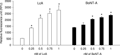
To evaluate the ability of the assay to detect BoNT-A in carrot juice, we spiked carrot juice with increasing concentrations of BoNT-A (Fig. 1, right) or LcA (Fig. 1, left) together with the 12-amino-acid synthetic internally quenched peptide and analyzed it fluorometrically. Our results show that after 30 min of incubation, the fluorescence emission increased in a dose-dependent manner. Activity levels for BoNT-A are slightly lower than those for LcA. This may be because the reduction buffer we used for BoNT-A might not have been entirely efficient, leaving some light chains still attached to the heavy chains. Thus, not enabling full cleavage of the dual-labeled fluorescein substrate resulted in a lower signal. Longer incubation (24 h) did not significantly change the relative fluorescence units. However, there was a significant difference between carrot juice spiked with BoNT-A or LcA and unspiked carrot juice. The limit of detection was 12.4 ng/ml of LcA and 37.6 ng/ml of BoNT-A.
FIG. 1.
Direct detection of BoNT-A in carrot juice. Increasing concentrations of BoNT-A (right) or LcA (left) were incubated for 30 min with the internally quenched peptide. Error bars represent standard errors, and an asterisk indicates significant differences (P < 0.05) between carrot juice containing toxin and juice without toxin.
Increasing the specificity and sensitivity of the assay.
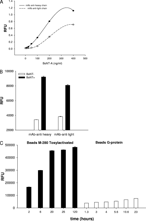
To increase the specificity and sensitivity of the assay, we evaluated monoclonal antibodies (MAb) that contained high affinity to the toxin, did not bind to the toxin activity site, and did not inhibit the cleavage activity and reduce the signal of the assay. We first evaluated two types of MAb for coating the immunomagnetic beads: a MAb directed against the 100-kDa heavy chain subunit (MAb-anti heavy) and a MAb directed against the 50-kDa light chain (MAb-anti light). To determine which MAb obtains a higher binding capacity to the toxin, we utilized an ELISA. As shown in Fig. 2A, MAb-anti heavy produces a higher signal than MAb-anti light. This shows that MAb-anti heavy has a higher affinity for the toxin. Also, when we employed both MAb on tosylactivated beads (Fig. 2B), the MAb anti-heavy obtained a higher signal-to-noise ratio. This demonstrates that MAb-anti heavy has a higher binding capacity to the toxin than that of MAb-anti light. We next evaluated two types of magnetic beads: tosylactivated and G protein beads. As shown in Fig. 2C tosylactivated immunomagnetic beads capture at least five times more toxin than protein G immunomagnetic beads. Thus, to obtain a highly sensitive assay, we utilized tosylactivated beads coated with a MAb against the heavy chain subunit.
FIG. 2.
Assay optimization. Comparison between the MAb-anti light and MAb-anti heavy chain using ELISA (A) or FRET assay (B) showed the MAb-anti heavy has a stronger binding affinity. (C) Comparison of immunomagnetic beads showed that the tosylactivated beads (left) produced a higher signal than the protein G beads (right). RFU, relative fluorescence units.
BoNT-A could be detected in beef by using a bead cleavage assay.
To evaluate the ability of the assay to detect BoNT-A in a matrix containing large amounts of protein, beef was spiked with 37.45 ng/ml (1 nM) of BoNT-A.
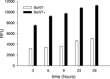
Our results showed that in spiked beef, the peptide cleavage was higher than in beef without the toxin (Fig. 3). During extended incubation, the signal continued to increase, even in beef without the toxin, due to nonspecific cleavage of the peptide by the beef proteases; however, beef containing BoNT-A still showed significantly higher signal than the control.
FIG. 3.
Detection of BoNT-A in beef. Beef was spiked with BoNT-A (150 ng/ml) and incubated for 16 h with tosylactivated beads coated with MAb-anti heavy chain. The beads were washed and incubated with the fluorogenic peptide for 4 h. Significant differences (P < 0.05) are observed between beads incubated with beef containing toxin and beads from beef not containing toxin. Error bars represent standard errors. RFU, relative fluorescence units.
Limit of detection with bead cleavage assay.
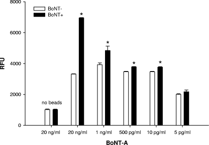
To determine the limit of detection for the presence of biologically active BoNT-A in a food item that has been associated with the recent outbreak, carrot juice samples were spiked with various concentrations of BoNT-A, ranging from 20 ng/ml to 5 pg/ml. As shown in Fig. 4, the FRET fluorescence decreased in a dose-dependent manner, and the limit of detection was 10 pg/ml, which is comparable to the sensitivity of the mouse bioassay (8).
FIG. 4.
Limits of detection shown with the bead cleavage assay of 40 ml of carrot juice. Carrot juice was spiked with decreasing concentrations of BoNT-A and incubated for 16 h with tosylactivated beads coated with MAb-anti heavy chain. The beads were washed and incubated with the fluorogenic peptide for 4 h. Error bars represent standard errors, and an asterisk (*) indicates significant differences (P < 0.05) between beads incubated with carrot juice containing toxin and beads from carrot juice not containing toxin. RFU, relative fluorescence units.
DISCUSSION
In the present study we developed a rapid, in vitro assay with which to detect active BoNT-A in food, with a level of sensitivity comparable to that of the mouse bioassay. Our assay utilizes immunomagnetic beads to remove interference by food matrix endopeptidases and concentrates the toxin from the food to obtain higher sensitivity. Currently, the mouse bioassay is utilized due to its high sensitivity and specificity. However, one disadvantage of the mouse bioassay is that it takes 4 to 6 days to obtain results. This is because the assay requires three steps: toxin screening, toxin titer, and toxin neutralization by monovalent binding of an antibody. Our assay can obtain results in less than 24 h. The use of a MAb specific to BoNT-A assists in the detection, which allows the quantification and subtyping of BoNT to be performed in one step.
The mouse bioassay determines the subtype of the BoNT by using antibodies specific to each subtype. The contaminated food must be shown to be neutralized by one of the subtypes. Our assay also utilizes antibodies that are specific to each subtype; in addition, we use a synthetic peptide sequence that the toxin recognizes and cleaves at a specific site. Thus, our method of subtyping is more accurate and has less room for error than the method used by the mouse bioassay.
We also compared two different monoclonal antibodies. We utilized monoclonal antibodies because we previously demonstrated that when we employed commercially available polyclonal antibodies that were directed against the entire toxin, including the toxin activity site, these antibodies partially inhibited the enzymatic activity of the toxin and reduced the assay sensitivity.
To optimize the assay, we compared two different types of immunomagnetic beads: tosylactivated beads and beads coated with staphylococcal protein G. The difference between the two immunomagnetic beads is that protein G binds to the Fc domain of the IgG, allowing the Fab fragment to be accessible, whereas tosylactivated beads will bind to some IgG in the wrong orientation, hiding the Fab fragment. We had assumed that the beads coated with protein G would produce better results, because all of the MAb would be in the optimal orientation for efficient toxin binding. However, the tosylactivated beads performed better (Fig. 2C); this may be because the number of IgGs binding to tosylactivated beads was higher than the number of IgGs binding to beads coated with protein G. Thus, the higher number of IgGs on the tosylactivated beads compensated for some of the IgGs that may have been in the incorrect orientation.
Using the immunomagnetic beads offers several advantages; unlike the mouse bioassay, which can only utilize up to 0.5 ml of food matrix, the immunomagnetic beads concentrate the toxin from a large volume, thus increasing the sensitivity of the assay. The toxin, captured by the antibody, is less toxic, allowing for separation of the hazardous phase, which is performed under containment conditions, from the nonhazardous phase, which is used for detection of fluorescence emission. Also, this approach could be readily adapted to the detection of all BoNT serotypes in one reaction by using more than one synthetic peptide with a unique cleavage site labeled with different fluorogenic dyes, to distinguish between the different toxin serotypes. The main advantages of the peptide cleavage assay are that it eliminates animal testing, it offers faster diagnostic testing, and its sensitivity levels are similar to those of the mouse bioassay. Because this peptide cleavage assay does not require the use of animals, the number of laboratories capable of performing BoNT serotype diagnostic assays will increase.
Footnotes
Published ahead of print on 30 May 2008.
REFERENCES
- 1.Barr, J. R., H. Moura, A. E. Boyer, A. R. Woolfitt, S. R. Kalb, A. Pavlopoulos, L. G. McWilliams, J. G. Schmidt, R. A. Martinez, and D. L. Ashley. 2005. Botulinum neurotoxin detection and differentiation by mass spectrometry. Emerg. Infect. Dis. 11:1578-1583. [DOI] [PMC free article] [PubMed] [Google Scholar]
- 2.Boldt, G. E., J. P. Kennedy, M. S. Hixon, L. A. McAllister, J. T. Barbieri, S. Tzipori, and K. D. Janda. 2006. Synthesis, characterization and development of a high-throughput methodology for the discovery of botulinum neurotoxin A inhibitors. J. Comb. Chem. 8:513-521. [DOI] [PMC free article] [PubMed] [Google Scholar]
- 3.Centers for Disease Control and Prevention. 2006. Botulism associated with commercial carrot juice-Georgia and Florida. MMWR Morb. Mortal. Wkly. Rep. 55:1098-1099. [PubMed] [Google Scholar]
- 4.Del Torre, M., M. L. Stecchini, A. Braconnier, and M. W. Peck. 2004. Prevalence of Clostridium species and behaviour of Clostridium botulinum in gnocchi, a REPFED of Italian origin. Int. J. Food Microbiol. 96:115-131. [DOI] [PubMed] [Google Scholar]
- 5.Dong, M., W. H. Tepp, E. A. Johnson, and E. R. Chapman. 2004. Using fluorescent sensors to detect botulinum neurotoxin activity in vitro and in living cells. Proc. Natl. Acad. Sci. USA 101:14701-14706. [DOI] [PMC free article] [PubMed] [Google Scholar]
- 6.Ferreira, J. L., S. Maslanka, E. Johnson, and M. Goodnough. 2003. Detection of botulinal neurotoxins A, B, E, and F by amplified enzyme-linked immunosorbent assay. J. AOAC Int. 86:314-331. [PubMed] [Google Scholar]
- 7.Ferreira, J. L., S. J. Eliasberg, P. Edmonds, and M. A. Harrison. 2004. Comparison of the mouse bioassay and enzyme-linked immunosorbent assay procedures for the detection of type A botulinal toxin in food. J. Food Prot. 67:203-206. [DOI] [PubMed] [Google Scholar]
- 8.Notermans, S., and J. Nagel. 1989. Assays for botulinum and tetanus toxins, p. 319-331. In L. L. Simpson (ed.), Botulinum neurotoxin and tetanus toxin. Academic Press, San Diego, CA.
- 9.Schmidt, J. J., and R. G. Stafford. 2003. Fluorogenic substrates for the protease activities of botulinum neurotoxins, serotypes A, B, and F. Appl. Environ. Microbiol. 69:297-303. (Erratum, 69:3025.) [DOI] [PMC free article] [PubMed] [Google Scholar]
- 10.Sharma, S. K., J. L. Ferreira, B. S. Eblen, and R. C. Whiting. 2006. Detection of type A, B, E, and F Clostridium botulinum neurotoxins in foods by using an amplified enzyme-linked immunosorbent assay with digoxigenin-labeled antibodies. Appl. Environ. Microbiol. 72:1231-1238. [DOI] [PMC free article] [PubMed] [Google Scholar]
- 11.Wein, L. M., and Y. Liu. 2005. Analyzing a bioterror attack on the food supply: the case of botulinum toxin in milk. Proc. Natl. Acad. Sci. USA 102:9984-9989. [DOI] [PMC free article] [PubMed] [Google Scholar]
- 12.Yang, G. H., K. S. Kim, H. W. Kim, S. T. Jeong, G. H. Huh, J. C. Kim, and H. H. Jung. 2004. Isolation and characterization of a neutralizing antibody specific to internalization domain of Clostridium botulinum neurotoxin type B. Toxicon 44:19-25. [DOI] [PubMed] [Google Scholar]


