Abstract
We demonstrate that apolipoprotein E -deficient (ApoE−/−) mice are highly susceptible to tuberculosis and that their susceptibility depends on the severity of hypercholesterolemia. Wild-type (WT) mice and ApoE−/− mice fed a low-cholesterol (LC) or high-cholesterol (HC) diet were infected with ∼50 CFU Mycobacterium tuberculosis Erdman by aerosol. ApoE−/− LC mice were modestly more susceptible to tuberculosis than WT LC mice. In contrast, ApoE−/− HC mice were extremely susceptible, as evidenced by 100% mortality after 4 weeks with tuberculosis. The lung pathology of ApoE−/− HC mice was remarkable for giant abscess-like lesions, massive infiltration by granulocytes, elevated inflammatory cytokine production, and a mean bacterial load ∼2 log units higher than that of WT HC mice. Compared to WT HC mice, the gamma interferon response of splenocytes restimulated ex vivo with M. tuberculosis culture filtrate protein was delayed in ApoE−/− HC mice, and they failed to control M. tuberculosis growth in the lung. OT-II cells adoptively transferred into uninfected ApoE−/− HC mice had a weak proliferative response to their antigen, indicating impaired priming of the adaptive immune response. Our studies show that ApoE−/− deficiency is associated with delayed expression of adaptive immunity to tuberculosis caused by defective priming of the adaptive immune response and that elevated serum cholesterol is responsible for this effect.
Dyslipidemia is a growing health problem in many industrialized countries and developing nations (33). High cholesterol is one of the most common forms of dyslipidemia, with 17% of adults in the United States alone being hypercholesterolemic (15). The impact of hypercholesterolemia on inflammation and atherosclerosis has received considerable attention, but much less is known about its potential effects on protective immunity. However, limited work with hypercholesterolemic apolipoprotein E-deficient (ApoE−/−) and low-density lipoprotein receptor deficient (LDL-R−/−) mice has demonstrated that the effects of hypercholesterolemia on the immune system extend beyond promoting inflammation.
The LDL-R on hepatocytes removes cholesterol from the blood by binding to LDL particles via ApoE; thus, LDL-R- and ApoE-deficient mice are spontaneously hypercholesterolemic. ApoE−/− mice were reported to have impaired defense against Candida albicans, Listeria monocytogenes, Klebsiella pneumoniae, and lymphocytic choriomeningitis virus (LCMV) infection (1, 5, 11, 25). Decreased resistance of LDL-R−/− mice to LCMV has also been described (11). Immunologic disorders, including Th2 skewing of antibody responses and impaired dendritic cell (DC) trafficking, have been described in ApoE−/− mice (2, 24, 35). The proposed mechanisms for the increased susceptibility of hypercholesterolemic mice to infectious diseases are diverse and range from increased availability of lipids as a nutrient source for microbes to detoxification of lipopolysaccharide by ApoE, reduced phagocytic capacity, and impaired cytotoxic-T-lymphocyte activation. An improved understanding of how hyperlipidemia affects immunity will allow a more complete assessment of the health risks of hyperlipidemia and the populations most at risk for infectious diseases.
The World Health Organization predicts there will be 1 billion new Mycobacterium tuberculosis infections, 150 million new active tuberculosis (TB) cases, and 37 million TB-related deaths by 2020 (34). About one-third of the world's population is M. tuberculosis infected, and of this population, ∼10% will develop active TB disease. Increased risk for TB disease is based on both inherited and acquired factors. Given the large number of persons with latent TB infection, even a modest impairment of protective immunity by hyperlipidemia could have a substantial impact on the public health burden of TB.
Here, we have investigated the effect of hypercholesterolemia on TB immunity by challenging wild-type (WT) and ApoE−/− mice with a low aerosol dose of M. tuberculosis Erdman. To test whether the ApoE−/− phenotype in the TB model was a function of hypercholesterolemia and not ApoE deficiency per se, mice were fed a low-cholesterol (LC) or high-cholesterol (HC) “Western” diet. ApoE−/− LC mice were slightly more susceptible to TB, as evidenced by increased lung inflammation and bacterial lung burden. In contrast, ApoE−/− HC mice demonstrated extreme TB susceptibility characterized by massive lung inflammation, heavy bacterial burden, and early mortality. Their failure to mount a timely protective Th1 immune response to TB was due in part to impaired priming of the adaptive immune response.
MATERIALS AND METHODS
Animals.
C57BL/6 and B6.129P2-Apoetm1UncN11 (ApoE−/−) mice were purchased from Taconic Farms or bred at the University of Massachusetts Medical School (UMMS) with breeders purchased from The Jackson Laboratory. ApoE−/− mice were originally derived by Piedrahita et al. (23) and backcrossed on a C57BL/6 background for more than 10 generations. C57BL/6 OT-II mice were a kind gift from Kenneth Rock, UMMS. The mice were housed within the Animal Medicine facility at UMMS, and the Institutional Animal Care and Use Committee approved these experiments.
Reagents.
The mice were fed either an LC (0.12% cholesterol) or an HC (1.25% cholesterol, 0.5% sodium cholate; Research Diets) diet 2 weeks before and during each experiment. Ovalbumin (OVA)-coated iron beads were a kind gift from Lian Jun Shen, UMMS. Chicken OVA was covalently bound to iron oxide beads according to the manufacturer's protocol (BioMag Amine; Polyscience Inc.). Culture filtrate protein (CFP) was obtained by growing M. tuberculosis Erdman in Sauton's broth for 1 month and then concentrating the culture supernatant and dialyzing it against phosphate-buffered saline (PBS) with a cutoff of 6 kDa.
Serum cholesterol.
Sera were incubated at 60°C for 30 min before being shipped to IDEXX Preclinical Research Services for total-cholesterol measurement.
M. tuberculosis infection.
A frozen stock of M. tuberculosis Erdman, ∼108 CFU/ml in PBS-0.05% Tween 80, was thawed, the concentration was adjusted with PBS-0.05% Tween 80 to deliver ∼50 CFU per mouse, and it was sonicated for 5 min in a cup horn sonifier (Branson Ultrasonics Corporation) and mice were infected in a Glass-Col Inhalation Exposure System (Glas-Col, LLC). In every experiment, two mice were sacrificed 24 h postinfection (p.i.) to confirm the delivered dose.
Bacterial load.
Tissues were homogenized in PBS-0.05% Tween 80, serially diluted 10-fold over 4 to 5 log units, and plated in duplicate on Middlebrook 7H11 agar (Difco, Becton Dickinson) supplemented with oleic acid-albumin-dextrose-catalase, and cultured at 37°C, and the colonies were counted 3 weeks later.
Histopathology.
Lungs were inflated and fixed with 10% buffered formalin for ≥24 h and then processed for staining. Tissue sections were stained with hematoxylin and eosin (H&E) or for inducible nitric oxide synthase (iNOS) production with a 1:100 dilution of anti-iNOS/NOS II rabbit polyclonal immunoglobulin G (Millipore), and bound antibody was detected with a peroxidase-based ABC staining kit according to the manufacturer's protocol (Vector Laboratories). As a negative control, sections were stained in the absence of the primary antibody. All staining was done by the Diabetes and Endocrinology Research Center histopathology core facility at the UMMS.
Fluorescence-activated cell sorter analysis.
Lungs were perfused through the heart with PBS, minced, digested for 30 min at 37°C in collagenase IV (1 mg/ml) and DNase (25 μg/ml) (both from Sigma-Aldrich), and passed through a 40-μm cell strainer, and the remaining red blood cells were lysed with Gey's solution. Viable cells were counted using a hemocytometer and trypan blue dye staining. Lung leukocytes or splenocytes (1 × 10⁶ to 2 × 10⁶) were treated with Fc-blocking monoclonal antibody (clone 2.4G20; BD Bioscience Pharmingen) and then stained with allophycocyanin-cyanine 7-conjugated anti-CD3 (145-2C11), peridinin chlorophyll α-conjugated anti-CD4 (RM4-5), phycoerythrin (PE)-conjugated anti-CD8 (53.6-7), and PE-cyanine 7-conjugated anti-Gr-1 (RB6-8C5) (all from BD Bioscience Pharmingen) and allophycocyanin-conjugated anti-F4/80 (BM8; eBioscience). Flow cytometry was performed on an LSRII flow cytometer (BD Bioscience Pharmingen), 50,000 leukocyte-gated events were collected, and data analysis was done with FlowJo PC (TreeStar, Inc.). Isotype control antibodies were purchased from BD Bioscience Pharmingen and eBioscience.
Lung cytokine production.
Lungs were homogenized in PBS-0.05% Tween 80; an equal volume of cell lysis buffer was added (0.5% Trion X-100, 150 mM NaCl, 15 mM Tris, 1 mM CaCl2, and 1 mM MgCl2, pH 7.4); the lungs were vortexed, incubated for 20 min at 4°C, vortexed, and centrifuged for 10 min at 12,000 × g; and the supernatant was sterile filtered. Lung lysates were assayed for interleukin 1α (IL-1α), IL-1β, IL-2, IL-4, IL-10, IL-17, tumor necrosis factor (TNF), macrophage inflammatory protein 1α, and transforming growth factor beta by multiplex enzyme-linked immunosorbent assay (ELISA) (Searchlight; Pierce Biotechnology). Gamma interferon (IFN-γ) in lung lysates was measured with an ELISA kit according to the manufacturer's protocol (R&D Systems).
Ex vivo T-cell restimulation.
Splenocytes were cultured for 48 h at 37°C and 5% CO2 in medium with 4 μg/ml concanavalin A (Sigma-Aldrich) or 2 μg/ml M. tuberculosis Erdman CFP in 24-well plates with 2 × 10⁶ cells per well in 2 ml. After 48 h, the contents of each well were sterile filtered and assayed for IFN-γ by ELISA.
In vivo antigen presentation.
Leukocytes from the spleen and lymph nodes (LN) of OT-II mice were labeled with 2 μM carboxyfluorescein diacetate succinimidyl ester (CFSE), their viability was confirmed by trypan blue dye exclusion, CFSE labeling was confirmed by fluorescence microscopy, and ∼15 million cells were injected intravenously into recipient mice. Twenty-four hours later, the recipient mice were injected subcutaneously (s.c.) near the left inguinal LN with 100 μl of OVA-coated iron beads (0.1 μg/μl) or with uncoated iron beads as a negative control; 72 h later, leukocytes were isolated from the left inguinal LN. The cells were stained with peridinin chlorophyll α-conjugated anti-CD4 (clone RM4-5) and PE-conjugated anti-CD45.1 (Ly-5.1; clone A20), and 200,000 leukocyte-gated events were collected on a LSRII flow cytometer and analyzed as described above.
Statistical analysis.
An F test was performed to confirm that variances between two groups were not statistically significant. Student's t test for samples with equal variance or unequal variance was performed as appropriate for comparisons between two groups. Comparison between more than two groups was done by analysis of variance, and a Tukey-Kramer multiple-comparison posttest was performed when analysis of variance results were significant. All tests were performed with GraphPad Instat (version 3.05 for Windows 95; GraphPad Software). P values of <0.05 were considered significant.
RESULTS
Hypercholesterolemia increases the TB susceptibility of ApoE−/− mice.
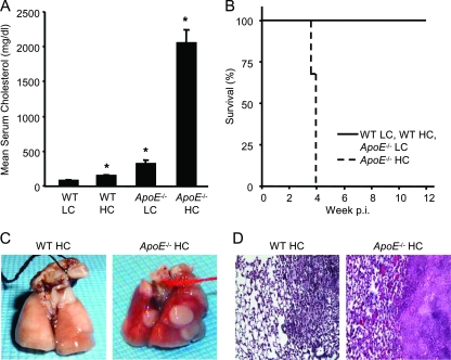
Mice were fed LC or HC diets in an effort to correlate TB resistance with different levels of serum cholesterol. The mean serum cholesterol for WT LC, WT HC, ApoE−/− LC, and ApoE−/− HC mice are shown in Fig. 1A. Serum total cholesterol of >200 mg/dl indicated hypercholesterolemia. The mice were maintained on these diets for 2 weeks before and during an experiment. Serum cholesterol measurements taken before infection and at the conclusion of experiments did not significantly differ within treatment groups (n = 4 or 5) (data not shown). WT and ApoE−/− mice on LC or HC diets were infected with ∼50 CFU of M. tuberculosis Erdman, and survival was monitored for 3 months (Fig. 1B). All five ApoE−/− HC mice in this experiment died within 4 weeks after M. tuberculosis infection. By 3 weeks p.i., they appeared hunched, lethargic, poorly groomed, and visibly smaller than their WT LC, WT HC, and ApoE−/− LC counterparts. On gross inspection, their lungs had large abscess-like masses with extensive inflammation and tissue necrosis visible by histology (Fig. 1C and D). All of the WT LC, WT HC, and ApoE−/− LC mice survived until the conclusion of the experiment 3 months p.i. and showed no overt signs of illness.
FIG. 1.
ApoE−/− HC mice are extremely susceptible to TB. (A) Serum cholesterol of WT and ApoE−/− mice after 2 weeks on an LC or HC diet. The values are means plus standard deviations. *, P < 0.05 WT HC, ApoE−/− LC, and ApoE−/− HC mice versus WT LC mice (n = 5). (B) Survival times of WT and ApoE−/− mice fed an LC or HC diet and then infected by aerosol with ∼50 CFU M. tuberculosis Erdman (n = 5). (C) Representative lungs from WT HC and ApoE−/− HC mice 30 days p.i. by aerosol with M. tuberculosis. (D) Representative lung tissue sections from WT HC and ApoE−/− HC mice 30 days after aerosol infection with ∼50 CFU M. tuberculosis stained with H&E (magnification, ×200). The results shown are representative of at least two independent experiments.
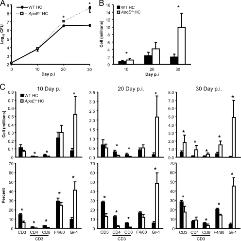
WT and ApoE−/− HC mice had comparable lung bacterial burdens up to 20 days p.i., suggesting that a defect of innate immunity or an enhanced intrinsic rate of M. tuberculosis replication was not responsible for the TB susceptibility of ApoE−/− HC mice. By 30 day p.i., the bacterial lung burden reached a plateau of 6.5 log units CFU in WT HC mice while it was still increasing exponentially in ApoE−/− HC mice, reaching 8.5 log units at the time of death (Fig. 2A). The bacterial lung burden of ApoE−/− LC mice was ∼0.5 log CFU greater than that of WT LC mice 30 day p.i. (P < 0.05; n = 4) (data not shown). Since control of M. tuberculosis growth in the lungs requires an effective adaptive immune response, these results suggested that adaptive immunity was severely compromised in ApoE−/− HC mice.
FIG. 2.
Despite a massive inflammatory response, ApoE−/− HC mice fail to control M. tuberculosis growth. (A) M. tuberculosis lung burdens of WT HC and ApoE−/− HC mice 10 days, 20 days, and 30 days after aerosol infection. The data are presented as mean log10 CFU ± standard deviations (SD). *, P < 0.05 (n = 4). (B) Lung leukocyte counts for the right caudal and left lung lobes of WT HC and ApoE−/− HC mice 10 days, 20 days, and 30 days after aerosol infection with M. tuberculosis. The values are the mean cell counts expressed in millions plus SD. *, P < 0.05 (n = 5). (C) The total numbers (top graphs) and percentages (bottom graphs) of T cells (CD3+ CD4+/CD8+), macrophages (F4/80+), or granulocytes (Gr-1+ CD3− F4/80−) isolated from the right caudal and left lung lobes of WT HC and ApoE−/− HC mice 10 days, 20 days, and 30 days after aerosol infection with M. tuberculosis Erdman. The total number of each cell population was determined by multiplying the percentage of cells positive for the above-mentioned surface markers by the total number of lung leukocytes isolated from each mouse. The values are the mean numbers of cells expressed in millions plus SD. *, P < 0.05 (n = 5). The results shown are representative of one experiment per time point, except for the CFU data for day 30 p.i., which is representative of two independent experiments.
Increased lung inflammation in ApoE−/− mice with TB.
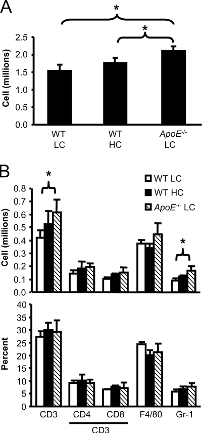
The pattern and kinetics of lung leukocyte recruitment were investigated in M. tuberculosis-infected ApoE−/− HC and WT HC mice. Lung leukocytes were isolated from the left lung lobe and the right caudal lobe by enzymatic digestion, and T-cell (CD3+ CD4+/CD8+), macrophage/monocyte (F4/80+), and granulocyte (GR-1+ CD3− F4/80−) populations were measured by flow cytometry. Signs of increased lung inflammation were detectable in ApoE−/− HC mice as early as 10 days p.i. and involved a massive influx of granulocytes (Fig. 2B and C). Roughly 45% of the lung leukocytes of ApoE−/− HC mice at this time point were granulocytes versus <10% for WT HC mice. Most of the granulocytes appeared to be neutrophils, with few basophils or eosinophils visible in H&E-stained lung tissue sections. Despite the massive early influx of neutrophils into the lungs of ApoE−/− HC mice, extensive lung tissue necrosis and inflammation were not evident by histology until after 20 days p.i. While ApoE−/− HC lungs had more T cells and macrophages than those of WT HC mice by 30 days p.i., they comprised a smaller proportion of the total lung leukocyte population than in WT HC mice. Uninfected ApoE−/− HC mice also had a higher proportion of resident lung granulocytes (24% versus 14%) but a lower proportion of T cells (3% versus 7%) than WT HC mice (P < 0.05; n = 5). The exaggerated innate immune response and relatively low T-cell numbers despite the presence of a high antigen burden served as additional indicators that adaptive immunity was impaired in ApoE−/− HC mice. Differences between WT LC, WT HC, and ApoE−/− LC mice were not as dramatic as those observed with ApoE−/− HC mice; therefore, these mice were examined 3 months p.i. The degree of lung inflammation after M. tuberculosis infection coincided with increasing serum cholesterol, as ApoE−/− LC mice had a modest but statistically significant increase in lung leukocytes compared with WT LC and WT HC mice after 3 months of TB disease (Fig. 3A). There was no difference in the proportions of T-cell, macrophage/monocyte, and granulocyte populations between WT LC, WT HC, and ApoE−/− LC mice 3 months p.i. (Fig. 3B). However, ApoE−/− LC mice had significantly more T cells and granulocytes than WT LC and WT HC mice (Fig. 3B).
FIG. 3.
Lung inflammation increases with increasing serum cholesterol. (A) Lung leukocyte counts for the right caudal and left lung lobes of WT LC, WT HC, and ApoE−/− LC mice 12 weeks after aerosol infection with M. tuberculosis. The values are the mean numbers of cells expressed in millions plus standard deviations (SD). *, P < 0.05 (n = 5). (B) The total numbers (top graphs) and percentages (bottom graphs) of T cells (CD3+ CD4+/CD8+), macrophages (F4/80+), or granulocytes (Gr-1+ CD3− F4/80−) isolated from the right caudal and left lung lobes of WT LC, WT HC, and ApoE−/− LC mice 12 weeks after aerosol infection with M. tuberculosis. The total number of each cell population was determined by multiplying the percentage of cells positive for the above-mentioned surface markers by the total number of lung leukocytes isolated from each mouse. The values are the mean numbers of cells expressed in millions plus SD. *, P < 0.05 (n = 4). The results shown are representative of one experiment.
Lung cytokine production of WT and ApoE−/− mice with TB.
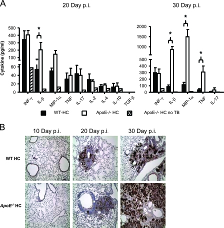
A Th1 cell-mediated immune response is required for protection against TB, and it is well established that IFN-γ is essential for resistance to TB in mice and humans (6, 10, 18). We assayed lung lysates from WT and ApoE−/− mice on LC or HC chow for Th1, Th2, and proinflammatory cytokines. The mean IFN-γ levels in the lungs of WT LC, WT HC, and ApoE−/− LC mice were 268 pg/ml, 253 pg/ml, and 166 mg/ml, respectively, 3 months p.i. Although ApoE−/− LC mice had significantly lower IFN-γ production than WT LC and HC mice (P < 0.05; n = 4), it is unclear whether this small difference accounts for the fivefold-higher bacterial lung burden of ApoE−/− LC mice than WT LC mice. Interestingly, there was no difference in lung IFN-γ production between WT HC and ApoE−/− HC mice 20 days and 30 days p.i. (Fig. 4A). This raised the possibility that the lung macrophages of ApoE−/− HC mice might have had a reduced capacity to respond to IFN-γ, thereby reducing their ability to kill internalized bacilli. Stimulation of macrophages to produce iNOS is one of the most important antimycobacterial functions of IFN-γ (9, 27, 12). To assess IFN-γ responsiveness, immunohistochemical staining for iNOS was performed on lung tissue sections from WT HC and ApoE−/− HC mice 10 days, 20 days, and 30 days p.i. (Fig. 4B). Abundant iNOS production was detected in the TB lung lesions of ApoE−/− HC mice, indicating that their macrophages retained the capacity to respond to IFN-γ.
FIG. 4.
Production of inflammatory cytokines in ApoE−/− HC lungs increases with the M. tuberculosis burden, while iNOS production is comparable to that in WT HC lungs. (A) Cytokine production in lung homogenates from WT HC and ApoE−/− HC mice 20 days or 30 days after aerosol infection with M. tuberculosis. The values are the mean cytokine concentrations expressed as pg/ml plus standard deviatons. *, P < 0.05 (n = 4). Lung homogenate from an uninfected ApoE−/− HC mouse was used as a background control. (B) Lung tissue sections from WT HC and ApoE−/− HC mice stained for iNOS production by immunohistochemistry 10 days, 20 days, and 30 days after aerosol infection with M. tuberculosis. Cells expressing iNOS are identified by brown staining; magnification, ×40. The lung sections shown are representative of three mice per mouse strain per time point. The results shown are representative of one experiment.
Levels of Th1, Th2, and proinflammatory cytokines in lung homogenates were similar between WT LC, WT HC, and ApoE−/− LC mice 3 months p.i. (data not shown). Th1 and Th2 cytokine production levels were also comparable in WT HC and ApoE−/− HC lungs, and neither group had detectable production of transforming growth factor beta 20 days p.i. Production of the inflammatory cytokines IL-1β, macrophage inflammatory protein 1α, and TNF increased dramatically in the lungs of ApoE−/− HC mice between 20 days and 30 days p.i. (Fig. 4A). ApoE−/− HC mice lost ∼7% of their preinfection body weight, consistent with the elevated production of TNF, while WT HC mouse preinfection body weight increased ∼14% by 30 days p.i. (data not shown). Interestingly, IL-17 production levels were similar in WT HC and ApoE−/− HC mice. IL-17 is associated with neutrophilic inflammation (16) but appears not to play a significant role in the inflammation seen in the lungs of M. tuberculosis Erdman-infected ApoE−/− HC mice. The overproduction of inflammatory cytokines in ApoE−/− HC lungs was consistent with the histology and lung leukocyte data and presumably reflected the persistent stimulation of innate immunity in the absence of an effective adaptive immune response.
Response to ex vivo antigen stimulation by WT and ApoE−/− mice with TB.
Any delay in the adaptive immune response to low-dose aerosol M. tuberculosis challenge in mice results in a higher bacterial lung burden before bacterial growth is restricted. To monitor the kinetics of the adaptive immune response to M. tuberculosis in WT HC and ApoE−/− HC mice, splenocytes were stimulated ex vivo for 48 h with M. tuberculosis CFP, and IFN-γ release was measured by ELISA. Splenocytes incubated with medium or concanavalin A were used as negative and positive controls, respectively. A strong IFN-γ response by T cells from WT HC mice to ex vivo CFP stimulation was seen as early as 20 days p.i., while the response by T cells from ApoE−/− HC mice was weak at this time point (Fig. 5A). By 30 days p.i., the CFP-stimulated IFN-γ response of splenocytes from ApoE−/− HC mice was comparable to that of WT HC mice. The spleen bacterial burdens were comparable between WT HC and ApoE−/− HC mice 30 days p.i., 4.7 log units and 4.9 log units, respectively (n = 4), indicating that differences in IFN-γ responses were not caused by unequal antigen loads. The number of splenocytes in WT HC mice increased during M. tuberculosis infection but remained unchanged in ApoE−/− HC mice (Fig. 5B). ApoE−/− HC mice had more granulocytes in the spleen than WT HC mice despite having fewer total splenocytes (Fig. 5C). At 20 days and 30 days p.i., WT HC mice had almost fivefold more T cells than ApoE−/− HC mice. Taken together, these results indicate that by 30 days p.i., IFN-γ production on a per-T-cell basis was actually higher in the spleens of ApoE−/− HC mice than in WT HC mice. Uninfected WT HC and ApoE−/− HC mice had similar numbers of splenocytes, but ApoE−/− HC mice had slightly more granulocytes (data not shown). T cells from WT LC and ApoE−/− LC mice 3 months p.i. produced equivalent amounts of IFN-γ after ex vivo stimulation with CFP (data not shown). WT LC and ApoE−/− LC mice had comparable numbers of splenocytes with no differences in the proportion or number of T cells, macrophages/monocytes, or granulocytes (data not shown).
FIG. 5.
Delayed M. tuberculosis antigen-specific T-cell response in ApoE−/− HC mice. (A) Amounts of IFN-γ released by splenocytes 10 days, 20 days, and 30 days after aerosol infection with M. tuberculosis from WT HC and ApoE−/− HC mice that were cultured for 48 h with 2 μg/ml M. tuberculosis Erdman CFP. Individual measurements are shown, and the mean concentration is indicated with a solid line. Day 10 was below the lower limit of detection of 31 pg/ml. *, P < 0.05 (n = 5). (B) Splenocyte counts from WT HC and ApoE−/− HC mice 10 days, 20 days, and 30 days after aerosol infection with M. tuberculosis. The values are the mean splenocyte counts plus standard deviations (SD). *, P < 0.05 (n = 5). (C) The total numbers (top graphs) and percentages (bottom graphs) of T cells (CD3+ CD4+/CD8+), macrophages (F4/80+), or granulocytes (Gr-1+ CD3− F4/80−) isolated from the spleens of WT HC and ApoE−/− HC mice 10 days, 20 days, and 30 days after aerosol infection with M. tuberculosis. The total number of each cell population was determined by multiplying the percentage of cells positive for the above-mentioned surface markers by the total number of splenocytes isolated from each mouse. The values are the mean numbers of cells expressed in millions plus SD. *, P < 0.05 (n = 4). The results shown for panels A and B are representative of two experiments, and the results for panel C are representative of one experiment.
In vivo antigen presentation by WT and ApoE−/− mice.
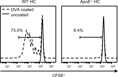
The M. tuberculosis-specific T-cell response of ApoE−/− HC mice was delayed compared to that of WT HC mice, suggesting that inefficient priming of the adaptive immune response might be responsible for their increased TB susceptibility. To test this possibility, an adoptive-transfer experiment was done using leukocytes from OT-II T-cell receptor (TCR) transgenic mice. These mice express a TCR that recognizes OVA peptide (amino acids 323 to 339) in the context of a major histocompatibility complex class II I-Ab molecule (3). Their cells were distinguishable from WT and ApoE−/− leukocytes by the allelic marker CD45.1. CFSE-labeled OT-II leukocytes were injected intravenously into WT HC and ApoE−/− HC mice, and the next day, OVA-coated or uncoated iron beads were injected s.c. near the left inguinal LN. After 3 days, leukocytes were harvested from the left inguinal LN, and the proliferative response of OT-II T cells was measured by CFSE dilution. OT-II T cells from WT HC mice injected with OVA-coated iron beads underwent about five rounds of cell division, while significantly less proliferation of OT-II T cells was seen in identically treated ApoE−/− HC mice (Fig. 6). No proliferation of OT-II T cells was seen in WT HC or ApoE−/− HC mice injected with uncoated iron beads. The frequency of CD4+ CD45.1+ OT-II T cells in the inguinal LN of ApoE−/− HC mice receiving OVA-coated iron beads was comparable to that in WT HC and ApoE−/− HC mice that received uncoated iron beads (data not shown). This indicated equivalent seeding and survival of OT-II T cells in WT HC and ApoE−/− HC inguinal LN. These results support the conclusion that priming of the adaptive immune response is significantly impaired in ApoE−/− HC mice prior to M. tuberculosis infection.
FIG. 6.
In vivo antigen presentation is impaired in ApoE−/− HC mice. Shown are representative histograms of CFSE fluorescence of OT-II CD45.1+ CD4+ cells recovered from the left inguinal LN of WT HC and ApoE−/− HC mice. OT-II cells were stimulated in vivo for 3 days by s.c. injection of OVA-coated or uncoated iron beads near the left inguinal LN. The results are representative of three mice per group that were stimulated with OVA-coated beads; 66.6% and 13.4% of OT-II CD45.1+ CD4+ cells underwent ≥1 cell division in WT HC and ApoE−/− HC mice, respectively. *, P < 0.05 (n = 3). The results shown are representative of one experiment.
DISCUSSION
Limited work with mouse models has demonstrated that hypercholesterolemia can have detrimental affects on the host defense. The underlying mechanisms responsible for this remain poorly understood and are likely to differ depending on the pathogen studied. The susceptibility of ApoE−/− mice to L. monocytogenes and K. pneumoniae has been attributed to impaired innate immunity (25, 5), while defective cytotoxic-T-lymphocyte function was linked to LCMV susceptibility in ApoE−/− and LDL-R−/− mice (11). We investigated the impact of elevated cholesterol on host defense using an M. tuberculosis aerosol infection model. Unexpectedly, ApoE−/− HC mice exhibited extreme susceptibility to M. tuberculosis with a survival time comparable to that of IFN-γ-deficient mice, which are the most TB-susceptible knockout mouse strain known (14, 19). Even moderately elevated cholesterol, as seen in ApoE−/− LC mice, influenced TB immunity. The TB susceptibility of ApoE−/− mice increased with serum cholesterol, indicating that their susceptibility was dependent on cholesterol and not ApoE deficiency per se. ApoE−/− HC mice mount a reasonably robust antigen-specific Th1 immune response to M. tuberculosis, but its expression is delayed during the critical period of logarithmic bacterial growth, allowing a massive increase in the bacterial burden. Although lung inflammation was increased in hypercholesterolemic mice, dramatically so in the case of ApoE−/− HC mice, the data suggest that the most significant impact of hypercholesterolemia on TB defense is impaired priming of the adaptive immune response.
A systemic proinflammatory state has been described in ApoE−/− mice (4, 7). Consistent with those observations, the number of resident granulocytes and IL-1β production were slightly elevated in the lungs of uninfected ApoE−/− mice. The rapid influx of granulocytes into the lungs of ApoE−/− HC mice after M. tuberculosis infection also suggests they are primed for inflammation. However, the role granulocytes play in controlling M. tuberculosis growth remains unclear, since neutrophil depletion has been reported to cause no change in the M. tuberculosis lung burden in C57BL/6 mice and only a marginal increase in BALB/c mice (28, 20). Despite mounting a vigorous early inflammatory response involving heavy granulocyte infiltration of the lung, ApoE−/− HC mice had a bacterial lung burden similar to that of WT HC mice up to 20 days after infection. Around the same time, ApoE−/− HC mice started developing atypical massive abscess-like lesions that were heavily infiltrated by neutrophils. In contrast, established TB lesions in C57BL/6 mice are predominately composed of macrophages and T cells that are occasionally organized into granuloma-like structures. Heavy neutrophil infiltration and necrosis have also been described in TCR-α/β−/− mouse TB lung lesions (14); however, unlike ApoE−/− HC mice, iNOS expression was greatly reduced in these mice. The inability of ApoE−/− HC mice to control M. tuberculosis growth may have exacerbated their inflammatory response to M. tuberculosis, leading to the massive lung tissue destruction observed by 30 days p.i.
Following aerosol infection of WT mice, M. tuberculosis grows exponentially in the lungs for about 20 days, after which the bacterial load is held at a plateau by adaptive immunity. Control of TB requires an effective Th1 adaptive immune response, and particularly IFN-γ, to enhance the antimycobacterial functions of macrophages. Mice with impaired adaptive immunity, e.g., SCID mice, OVA-specific TCR transgenic mice, or IFN-γ-deficient mice, succumb rapidly to TB (6, 14, 17, 19). C57BL/6, the background strain of the ApoE−/− mice used in our study, is a TB-resistant mouse strain with an expected survival time of >200 days after low-dose aerosol infection (14). Adaptive immunity to TB was severely impaired in ApoE−/− HC mice, since they were unable to restrict M. tuberculosis growth and died ∼28 days p.i. Hypercholesterolemia did not prevent an antigen-specific T-cell response to M. tuberculosis from developing, but its expression was significantly delayed in ApoE−/− HC mice. Comparable amounts of Th1- and Th2-related cytokines were found in the lungs of WT and ApoE−/− mice with TB, indicating that hypercholesterolemia did not predispose the mice to a less protective Th2 response. The ability of macrophages to respond to appropriate activation by IFN-γ did not appear to be compromised by hypercholesterolemia, since abundant iNOS production was detected in ApoE−/− mice during the course of M. tuberculosis infection.
Severe hypercholesterolemia impaired priming of the adaptive immune response, since OT-II T cells adoptively transferred into uninfected ApoE−/− HC mice proliferated weakly after stimulation with OVA. Reduced proliferation of LCMV-specific transgenic CD4+ T cells to their cognate peptide was also recently reported in ApoE−/− mice fed a high-fat/cholesterol diet (29). DCs play a critical role in the priming of the adaptive immune response, and their impairment by high cholesterol would be consistent with the delayed immune response to M. tuberculosis and subsequent uncontrolled bacterial growth observed in ApoE−/− HC mice. This idea is also supported by a recent report (31) that transient in vivo depletion of CD11c+ DCs in mice delays their antigen-specific immune response to M. tuberculosis and increases their bacterial burden, although not as severely as seen in our experiments. Angeli et al. (2) reported that oxidized LDL (ox-LDL) interferes with the normal trafficking of DCs from the skin to draining LN in ApoE−/− mice fed a high-fat diet. Inefficient migration of lung DCs to the draining LN of ApoE−/− HC mice after M. tuberculosis infection could also explain their delayed adaptive immune response to TB. Recently, ox-LDL was reported to increase the susceptibility of ApoE−/− mice fed a high-fat/cholesterol diet to Leishmania major by priming CD8α− myeloid DCs to induce a nonprotective Th2 immune response (29). However, ApoE−/− HC mice with TB mounted a robust, albeit delayed, Th1 immune response, and IL-4 production in their lungs was not elevated compared to WT HC mice. Although ox-LDL may prime CD8α− myeloid DCs to induce a Th2 immune response, it does not appear to be responsible for the increased TB susceptibility of ApoE−/− HC mice. Our data indicate that hypercholesterolemia negatively impacts the TB host defense by interfering with the priming of the adaptive response, which could reflect impairment of DC antigen uptake or processing, migration to LN, or antigen presentation and costimulatory ligand expression. The molecular basis for this effect of elevated cholesterol remains to be discovered. A number of biochemical pathways for cholesterol-mediated pathology at the cellular level have been proposed (reviewed in reference 30) and will direct future investigation in our model. While the strong antigen-specific T-cell response in the spleen and heavy recruitment of T cells to the lungs 30 days p.i. argues against gross impairment of T cells in ApoE−/− HC mice, the impaired priming of the adaptive immune response could be masking more subtle T-cell deficits. Future studies will examine this possibility in greater detail.
The hypercholesterolemic environment in ApoE−/− mice might directly support more robust M. tuberculosis growth by increasing nutrient availability. Van der Geize et al. (32) recently reported that M. tuberculosis has the capacity to take up and use cholesterol as a source of energy. Further, many of the M. tuberculosis genes related to cholesterol uptake and catabolism have been reported to be upregulated 2 to 4 weeks p.i (26). While ApoE−/− HC mice had a greater bacterial lung burden than WT HC mice beginning 20 days p.i., their delayed M. tuberculosis antigen-specific immune response indicates that TB susceptibility is not likely to be exclusively due to a putative nutrient affect. Whether hypercholesterolemia promoted rapid M. tuberculosis growth and compounded the consequences of the delayed Th1 immune response by ApoE−/− HC mice is unknown. Further investigation will be required to determine what impact, if any, hypercholesterolemia has on the growth rate of M. tuberculosis in vivo.
The increased TB susceptibility of hypercholesterolemic mice suggests that there may be heretofore-unappreciated health risks associated with elevated cholesterol in people. An especially vulnerable group may be people with diabetes mellitus. Diabetes is known to increase TB susceptibility (13, 21), and roughly half of the diabetics in the Unted States have high cholesterol (8). However, Perez-Guzman et al. (22) reported that TB patients receiving a high-cholesterol diet responded to antimycobacterial therapy with a higher sputum sterilization rate than patients on a normal diet. Patients on the high-cholesterol diet had borderline high cholesterol, ∼ 210 mg/dl. In our study ApoE knockout mice had substantially higher serum total cholesterol whether they were on an LC or an HC diet; cholesterol was elevated prior to infection, and they did not receive antimycobacterial therapy. Further, our data indicate that hyperlipidemia has its greatest impact on the initiation of the adaptive immune response; whether cholesterol influences preexisting adaptive immune responses is unknown. Based upon our observations and the reports by Van der Geize et al. (32) and Sassetti et al. (26) that M. tuberculosis has the ability to use cholesterol as an energy source, we suggest exercising caution when considering cholesterol treatment of TB patients.
Acknowledgments
This publication was made possible by grant number 5 P30 DK32520 from the National Institute of Diabetes and Digestive and Kidney Diseases to Aldo Rossini and HL081149 to H.K.
We thank Birgit Stein, Mathumathi Thiruvengadam, and Jonathan Eskander for technical assistance with data collection and animal care. Special thanks are due to Kim Wigglesworth for her help with the adoptive transfer experiments.
The authors have no conflicting financial interests.
Editor: J. L. Flynn
Footnotes
Published ahead of print on 27 May 2008.
REFERENCES
- 1.Alieke, G. V., N. de Bont, M. G. Netea, P. N. Demacker, J. W. van der Meer, A. F. Stalenhoef, and B. J. Kullberg. 2004. Apolipoprotein-E-deficient mice exhibit an increased susceptibility to disseminated candidiasis. Med. Mycol. 42341-348. [DOI] [PubMed] [Google Scholar]
- 2.Angeli, V., J. Llodra, J. X. Rong, K. Satoh, S. Ishii, T. Shimizu, E. A. Fisher, and G. J. Randolph. 2004. Dyslipidemia associated with atherosclerotic disease systemically alters dendritic cell mobilization. Immunity 21561-574. [DOI] [PubMed] [Google Scholar]
- 3.Barnden, M. J., J. Allison, W. R. Heath, and F. R. Carbone. 1998. Defective TCR expression in transgenic mice constructed using cDNA-based alpha- and beta-chain genes under the control of heterologous regulatory elements. Immunol. Cell Biol. 7634-40. [DOI] [PubMed] [Google Scholar]
- 4.Bjorkbacka, H., V. V. Kunjathoor, K. J. Moore, S. Koehn, C. M. Ordija, M. A. Lee, T. Means, K. Halmen, A. D. Luster, D. T. Golenbock, and M. W. Freeman. 2004. Reduced atherosclerosis in MyD88-null mice links elevated serum cholesterol levels to activation of innate immunity signaling pathways. Nat. Med. 10416-421. [DOI] [PubMed] [Google Scholar]
- 5.de Bont, N., M. G. Netea, P. N. Demacker, B. J. Kullberg, J. W. van der Meer, and A. F. Stalenhoef. 2000. Apolipoprotein E-deficient mice have an impaired immune response to Klebsiella pneumoniae. Eur. J. Clin. Investig. 30818-822. [DOI] [PubMed] [Google Scholar]
- 6.Flynn, J. L., and J. Chan. 2001. Immunology of tuberculosis. Annu. Rev. Immunol. 1993-129. [DOI] [PubMed] [Google Scholar]
- 7.Grainger, D. J., J. Reckless, and E. McKilligin. 2004. Apolipoprotein E modulates clearance of apoptotic bodies in vitro and in vivo, resulting in a systemic proinflammatory state in apolipoprotein E-deficient mice. J. Immunol. 1736366-6375. [DOI] [PubMed] [Google Scholar]
- 8.Imperatore, G., B. L. Cadwell, L. Geiss, J. B. Saadinne, D. E. Williams, E. S. Ford, T. J. Thompson, K. M. Narayan, and E. W. Gregg. 2004. Thirty-year trends in cardiovascular risk factor levels among US adults with diabetes: National Health and Nutrition Examination Surveys, 1971-2000. Am. J. Epidemiol. 160531-539. [DOI] [PubMed] [Google Scholar]
- 9.Jung, Y. J., R. LaCourse, L. Ryan, and R. J. North. 2002. Virulent but not avirulent Mycobacterium tuberculosis can evade the growth inhibitory action of a T helper 1-dependent, nitric oxide synthase 2-independent defense in mice. J. Exp. Med. 196991-998. [DOI] [PMC free article] [PubMed] [Google Scholar]
- 10.Lammas, D. A., E. De Heer, J. D. Edgar, V. Novelli, A. Ben-Smith, R. Baretto, P. Drysdale, J. Binch, C. MacLennan, D. S. Kumararatne, S. Panchalingam, T. H. Ottenhoff, J. L. Casanova, and J. F. Emile. 2002. Heterogeneity in the granulomatous response to mycobacterial infection in patients with defined genetic mutations in the interleukin 12-dependent interferon-gamma production pathway. Int. J. Exp. Pathol. 831-20. [DOI] [PMC free article] [PubMed] [Google Scholar]
- 11.Ludewig, B., M. Jaggi, T. Dumrese, K. Brduscha-Riem, B. Odermatt, H. Hengartner, and R. M. Zinkernagel. 2001. Hypercholesterolemia exacerbates virus-induced immunopathologic liver disease via suppression of antiviral cytotoxic T cell responses. J. Immunol. 1663369-3376. [DOI] [PubMed] [Google Scholar]
- 12.MacMicking, J. D., R. J. North, R. LaCourse, J. S. Mudgett, S. K. Shah, and C. F. Nathan. 1997. Identification of nitric oxide synthase as a protective locus against tuberculosis. Proc. Natl. Acad. Sci. USA 945243-5248. [DOI] [PMC free article] [PubMed] [Google Scholar]
- 13.Martens, G. W., M. C. Arikan, J. Lee, F., Ren, D. Greiner, and H. Kornfeld. 2007. Tuberculosis susceptibility of diabetic mice. Am. J. Respir. Cell Mol. Biol. 37518-524. [DOI] [PMC free article] [PubMed] [Google Scholar]
- 14.Mogues, T., M. E. Goodrich, L. Ryan, R. LaCourse, and R. J. North. 2001. The relative importance of T cell subsets in immunity and immunopathology of airborne Mycobacterium tuberculosis infection in mice. J. Exp. Med. 193271-280. [DOI] [PMC free article] [PubMed] [Google Scholar]
- 15.National Center for Health Statistics. 2006. Technical notes. Health, United States, 2006 with chartbook on trends in the health of Americans, p. 106-435. U.S. Government Printing Office, Hyattsville, MD.
- 16.Oda, N., P. B. Canelos, D. M. Essayan, B. A. Plunkett, A. C. Myers, and S. K. Huang. 2005. Interleukin-17F induces pulmonary neutrophilia and amplifies antigen-induced allergic response. Am. J. Respir. Crit. Care Med. 17112-18. [DOI] [PubMed] [Google Scholar]
- 17.Orme, I. M. 2001. Immunology and vaccinology of tuberculosis: can lessons from the mouse be applied to the cow? Tuberculosis 81109-113. [DOI] [PubMed] [Google Scholar]
- 18.Ottenhoff, T. H., D. Kumararatne, and J. L. Casanova. 1998. Novel human immunodeficiencies reveal the essential role of type-I cytokines in immunity to intracellular bacteria. Immunol. Today 19491-494. [DOI] [PubMed] [Google Scholar]
- 19.Pearl, J. E., B. Saunders, S. Ehlers, I. M. Orme, and A. M. Cooper. 2001. Inflammation and lymphocyte activation during mycobacterial infection in the interferon-gamma-deficient mouse. Cell Immunol. 21143-50. [DOI] [PubMed] [Google Scholar]
- 20.Pedrosa, J., B. M. Saunders, R. Appelberg, I. M. Orme, M. T. Silva, and A. M. Cooper. 2000. Neutrophils play a protective nonphagocytic role in systemic Mycobacterium tuberculosis infection of mice. Infect. Immun. 68577-583. [DOI] [PMC free article] [PubMed] [Google Scholar]
- 21.Perez, A., H. S. Brown III, and B. I. Restrepo. 2006. Association between tuberculosis and diabetes in the Mexican border and non-border regions of Texas. Am. J. Trop. Med. Hyg. 74604-611. [PMC free article] [PubMed] [Google Scholar]
- 22.Perez-Guzman, C., M. H. Vargas, F. Quinonez, N. Bazavilvazo, and A. Aguilar. 2005. A cholesterol-rich diet accelerates bacteriologic sterilization in pulmonary tuberculosis. Chest 127643-651. [DOI] [PubMed] [Google Scholar]
- 23.Piedrahita, J. A., S. H. Zhang, J. R. Hagaman, P. M. Oliver, and N. Maeda. 1992. Generation of mice carrying a mutant apolipoprotein E gene inactivated by gene targeting in embryonic stem cells. Proc. Natl. Acad. Sci. USA 894471-4475. [DOI] [PMC free article] [PubMed] [Google Scholar]
- 24.Robertson, A. K., X. Zhou, B. Strandvik, and G. K. Hansson. 2004. Severe hypercholesterolaemia leads to strong Th2 responses to an exogenous antigen. Scand. J. Immunol. 59285-293. [DOI] [PubMed] [Google Scholar]
- 25.Roselaar, S. E., and A. Daugherty. 1998. Apolipoprotein E-deficient mice have impaired innate immune responses to Listeria monocytogenes in vivo. J. Lipid Res. 391740-1743. [PubMed] [Google Scholar]
- 26.Sassetti, C. M., D. H. Boyd, and E. J. Rubin. 2003. Genes required for mycobacterial growth defined by high density mutagenesis. Mol. Microbiol. 4877-84. [DOI] [PubMed] [Google Scholar]
- 27.Scanga, C. A., V. P. Mohan, K. Tanaka, D. Alland, J. L. Flynn, and J. Chan. 2001. The inducible nitric oxide synthase locus confers protection against aerogenic challenge of both clinical and laboratory strains of Mycobacterium tuberculosis in mice. Infect. Immun. 697711-7717. [DOI] [PMC free article] [PubMed] [Google Scholar]
- 28.Seiler, P., P. Aichele, B. Raupach, B. Odermatt, U. Steinhoff, and S. H. Kaufmann. 2000. Rapid neutrophil response controls fast-replicating intracellular bacteria but not slow-replicating Mycobacterium tuberculosis. J. Infect. Dis. 181671-680. [DOI] [PubMed] [Google Scholar]
- 29.Shamshiev, A. T., F. Ampenberger, B. Ernst, L. Rohrer, B. J. Marsland, and M. Kopf. 2007. Dyslipidemia inhibits Toll-like receptor-induced activation of CD8α-negative dendritic cells and protective Th1 type immunity. J. Exp. Med. 204441-452. [DOI] [PMC free article] [PubMed] [Google Scholar]
- 30.Tabas, I. 2002. Consequences of cellular cholesterol accumulation: basic concepts and physiological implications. J. Clin. Investig. 10905-911. [DOI] [PMC free article] [PubMed] [Google Scholar]
- 31.Tian, T., J. Woodworth, M. Skold, and S. M. Behar. 2005. In vivo depletion of CD11c+ cells delays the CD4+ T cell response to Mycobacterium tuberculosis and exacerbates the outcome of infection. J. Immunol. 1753268-3272. [DOI] [PubMed] [Google Scholar]
- 32.Van der Geize, R., K. Yam, T. Heuser, M. H. Wilbrink, H. Hara, M. C. Anderton, E. Sim, L. Dijkhuizen, J. E. Davies, W. W. Mohn, and L. D. Eltis. 2007. A gene cluster encoding cholesterol catabolism in a soil actinomycete provides insight into Mycobacterium tuberculosis survival in macrophages. Proc. Natl. Acad. Sci. USA 1041947-1952. [DOI] [PMC free article] [PubMed] [Google Scholar]
- 33.World Health Organization. 2002. Quantifying selected major risks to health. World health report 2002: reducing risks, promoting healthy life, p. 49-97. World Health Organization, Geneva, Switzerland.
- 34.World Health Organization. 2007. Global tuberculosis control: surveillance, planning, financing. WHO report 2007, p. 1-270. World Health Organization, Geneva, Switzerland.
- 35.Zhou, X., G. Paulsson, S. Stemme, and G. K. Hansson. 1998. Hypercholesterolemia is associated with a T helper (Th) 1/Th2 switch of the autoimmune response in atherosclerotic apo E-knockout mice. J. Clin. Investig. 1011717-1725. [DOI] [PMC free article] [PubMed] [Google Scholar]


