Abstract
Strains of Francisella tularensis secrete a siderophore in response to iron limitation. Siderophore production is dependent on fslA, the first gene in an operon that appears to encode biosynthetic and export functions for the siderophore. Transcription of the operon is induced under conditions of iron limitation. The fsl genes lie adjacent to the fur homolog on the chromosome, and there is a canonical Fur box sequence in the promoter region of fslA. We generated a Δfur mutant of the Schu S4 strain of F. tularensis tularensis and determined that siderophore production was now constitutive and no longer regulated by iron levels. Quantitative reverse transcriptase PCR analysis with RNA from Schu S4 and the mutant strain showed that Fur represses transcription of fslA under iron-replete conditions. We determined that fslE (locus FTT0025 in the Schu S4 genome), located downstream of the siderophore biosynthetic genes, is also under Fur regulation and is transcribed as part of the fslABCDEF operon. We generated a defined in-frame deletion of fslE and found that the mutant was defective for growth under iron limitation. Using a plate-based growth assay, we found that the mutant was able to secrete a siderophore but was defective in utilization of the siderophore. FslE belongs to a family of proteins that has no known homologs outside of the Francisella species, and the fslE gene product has been previously localized to the outer membrane of F. tularensis strains. Our data suggest that FslE may function as the siderophore receptor in F. tularensis.
Francisella tularensis, the etiologic agent of the disease tularemia, belongs to a deeply divergent clade of gammaproteobacteria (13). The organism has a small genome and is auxotrophic for numerous amino acids and nutrients that it must therefore acquire from its environment (18). In the mammalian host, the bacterium is intracellular, replicating within macrophages and other cells, such as hepatocytes.
The survival of pathogens within the host is dependent on the ability to acquire requisite nutrients. One such essential nutrient is iron, which is largely sequestered within protein complexes and is consequently limiting in the host environment. To circumvent this problem, pathogenic bacteria express specific mechanisms to acquire iron.
We and others have previously shown that different F. tularensis subspecies express siderophores as one mechanism to acquire iron from the environment (7, 26). The siderophore produced by the live vaccine strain (LVS) (holarctica subspecies) and the virulent Schu S4 strain (tularensis subspecies) of F. tularensis is a polycarboxylate molecule, structurally similar to rhizoferrin, expressed by Rhizopus spp. and Ralstonia pickettii strains (8, 21, 26). Expression of the siderophore is dependent on a putative siderophore synthetase encoded by fslA (also called figA), the first gene in an operon which is conserved across the different F. tularensis subspecies (7, 26).
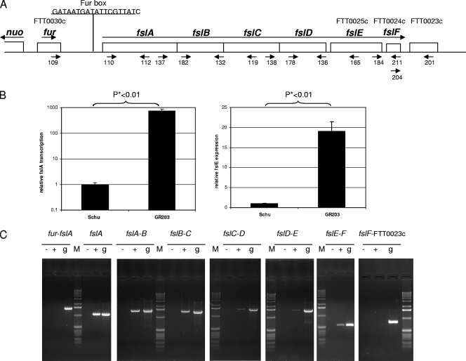
Iron uptake in bacteria is commonly under the control of the Fur (ferric uptake regulator) protein, originally identified because mutation in the fur gene resulted in constitutive expression of multiple iron uptake genes in Salmonella enterica serovar Typhimurium and Escherichia coli (10, 15; reviewed in reference 11). Fur is a Fe2+-dependent DNA binding protein that binds to a specific sequence (the Fur box) present in promoter regions of target genes and functions as a repressor under iron-replete conditions. Under iron limitation, the Fur protein becomes deferrated and consequently loses DNA binding capability, resulting in derepression of transcription. The fsl genes in F. tularensis are located downstream of the fur homolog (locus FTT0030c) in the chromosome, and the fslA gene has a canonical Fur box sequence in its promoter region (see Fig. 2A). This suggests that the fsl genes may be regulated by the Fur repressor in response to iron levels, as seen in other bacteria.
FIG. 2.
The fur-fsl region and transcriptional analysis of fsl genes. (A) Representation of the fur-fsl region of the chromosome. The locations of primers used in RT-PCR are shown. (B) qPCR analysis of fslA and fslE expression in the Δfur mutant. cDNAs prepared from Schu S4 and from GR203 were tested for fslA and fslE expression relative to that of trpB as an internal standard. Reactions were run in triplicate, and results are represented as averages with standard deviations. Note that the y axis for fslA expression is a log scale. (C) RT-PCR to delineate the fsl operon. cDNA prepared from GR203 was used in PCR with primer pairs to detect transcription across genetic loci as follows: 109-112 for fur and fslA, 110-112 for fslA, 137-132 for fslAB, 182-119 for fslBC, 138-136 for fslCD, 178-165 for fslDE, 184-211 for fslEF, and 204-201 for fslF and FTT0023c. “−” represents negative controls, where reverse transcriptase was left out of the cDNA reaction; “+” indicates reactions with cDNA; and “g” represents reactions with gDNA as a template. “M” represents the 1-kb DNA ladder (Invitrogen).
All siderophore-mediated iron uptake systems characterized to date in gram-negative bacteria are dependent on the ubiquitous tonB, exbB, and exbD genes (reviewed in reference 12). The inner membrane proteins encoded by these genes form a complex which is thought to provide the energy requisite for uptake of the siderophore by the cognate siderophore receptor in the outer membrane. This uptake is dependent on interaction of TonB with sequences in the periplasmic amino-terminal domain of the siderophore receptor. While F. tularensis does produce and utilize a siderophore, its genome does not encode identifiable tonB, exbB, or exbD homologs or tonB-dependent receptor homologs for siderophore uptake (18). A receptor for the F. tularensis siderophore is therefore expected to be a novel protein.
We used a Δfur mutant of Schu S4 to demonstrate here that siderophore biosynthesis and expression of the fslA gene in F. tularensis are under the control of the Fur repressor. We determined that fslE, located downstream of the siderophore biosynthetic genes, is also part of the Fur-regulated operon. The fslE gene product has been localized to the outer membrane of the LVS and Schu S4 strains and belongs to a family of sequence-related proteins unique to Francisella species (17, 18). We showed by deletion mutagenesis with the Schu S4 strain that fslE is necessary for siderophore-mediated iron acquisition. FslE may therefore function as the siderophore receptor in F. tularensis.
MATERIALS AND METHODS
Bacterial strains and culture.
Francisella tularensis strains Schu S4 (obtained from the CDC, Fort Collins, CO) and LVS (kindly provided by K. Elkins, CBER, Rockville, MD) were grown at 37°C on cystine heart agar (Difco) plates with 5% defibrinated horse blood or on modified Muller-Hinton agar (Difco) supplemented with ferric pyrophosphate or ferrous sulfate, horse serum, and 0.1% cysteine. Isolates were passaged up to five times before restreaking from stock cultures maintained at −80°C. Chamberlain's defined medium containing 2 μg/ml FeSO4 (CDM) (6) or tryptic soy broth supplemented with 0.1% cysteine (TSB/C) was used for routine liquid culture. CDM prepared without addition of FeSO4 (CDM−Fe) was used as iron-limiting medium for agar plates. For growth under iron limitation, the medium was deferrated with Chelex-100 resin (Bio-Rad) and then supplemented with essential divalent cations (MgSO4 [0.55 mM], ZnCl2 [1.5 μM], and CaCl2 [5 μM]) (che-CDM). Defined levels of iron were added to the che-CDM; iron-limited che-CDM medium contained 0.1 μg/ml FeCl3 (0.37 μM) or FeSO4 (0.36 μM), whereas iron-replete medium contained 2 μg/ml of FeCl3 (7.4 μM) or FeSO4 (7.19 μM). Kanamycin, when required, was used at a 15-μg/ml concentration. All glassware for preparation of che-CDM was washed with 10 mM HCl overnight and rinsed several times in milliQ (Millipore) water. All chemicals were from Sigma Chemical Company unless otherwise stated.
Escherichia coli strain MC1061.1 [Δ(ara-leu)7696 araD139 Δ(lac)X74 galK16 galE15 mcrA mcrB1 repsL(Strr) hsdR2 λ− F− recA] was used for routine cloning and was grown in Luria broth. Ampicillin was used at a concentration of 50 μg/ml (liquid cultures) or at 100 μg/ml (agar plates).
Growth in iron-limiting liquid culture.
F. tularensis cells grown to logarithmic phase in CDM were pelleted, washed three times in che-CDM with no added iron, and then diluted into che-CDM containing defined levels of iron to an optical density at 595 nm (OD595) of 0.01 (∼3 × 10⁷ CFU/ml). For complementation studies, the media were supplemented with 15 μg/ml kanamycin and starting cultures were inoculated to an OD595 of 0.02. Growth was monitored as change in OD595 of the liquid cultures over a period of 48 to 50 h.
Bioassay for siderophore utilization.
Cells from an overnight culture in CDM of Schu S4 or a mutant derivative to be tested for siderophore utilization were washed, and ∼3 × 10⁴ CFU was spread on the CDM−Fe plates. Cells to be used for siderophore production were grown overnight in CDM to logarithmic phase, washed once in che-CDM, and resuspended to an OD595 of 1.0 (∼3 × 10⁹ CFU/ml). These cell suspensions (2.5 μl each) were spotted on the seeded plates and the plates incubated at 37°C for 2 to 4 days. A growth halo of colonies around a spot demonstrated the ability of the indicator strain (seeded on the plate) to grow on the iron-limiting plate utilizing the siderophore secreted by the producer strain (spotted on the plate).
Construction of Δfur and ΔfslE mutants and complementing plasmids.
In-frame deletion mutations in fur and fslE were generated in the Schu S4 strain in a two-step process as previously described for generation of a ΔfslA mutant in the LVS (26). The suicide vectors, described below, are pUC based and carry the kanamycin resistance gene from Tn5 and the Bacillus subtilis gene sacB for counterselection (26).
The suicide plasmid used for generating the Δfur mutation was analogous to the plasmid pGIR457, used to obtain the ΔfslA mutant (26), except that the flanking regions cloned in the plasmid were specific to fur. A 1.958-kb stretch of DNA upstream of and including 24 bp at the 5′ end of the fur gene (encoding the first eight amino acid residues of Fur) was amplified from Schu S4 chromosomal DNA using Turbo Pfu DNA polymerase (Stratagene) and primers 113 and 114. A 2.24-kb 3′ flanking sequence that included the sequence encoding the terminal 10 residues and stop codon of fur was obtained by amplification from Schu S4 DNA using primers 115 and 116. The 5′ and 3′ flanking sequences were cloned as NheI-NotI and NotI-SacI fragments, respectively, to generate plasmid pGIR449, where the NotI restriction site replaced the sequences encoding the central 122 amino acids of fur and retained the reading frame of the amino and carboxy-terminal ends encoded by the gene. In this construct, the cloned 5′ flanking region included part of the divergent nuo operon, with the nuo operon oriented toward the kanamycin resistance gene on the plasmid. Common E. coli promoters are believed to be nonfunctional in F. tularensis, and nuo transcription may be responsible for expression of the kanamycin resistance gene in pGIR449 integrants of F. tularensis.
For generation of the plasmid that was used for deletion of the fslE gene from the chromosome, the suicide plasmid was modified by introducing the F. tularensis groE promoter upstream of the kanamycin resistance gene. The groEp sequence was amplified from the genome using PCR with primers 152 and 153 and cloned as an NheI-BglII fragment to generate plasmid pGIR463. A 1.05-kb region at the 5′ end of fslE and including the sequence encoding the 18 amino-terminal residues was amplified with primers 197 and 198. A 1.13-kb 3′ flanking region including the sequence encoding the terminal eight amino acid residues and the stop codon of fslE was amplified with primers 199 and 200. The 5′ and 3′ flanking sequences were cloned as NheI-BspEI and BspEI-SacI fragments, respectively, in pGIR463 to generate plasmid pGIR467. In this construct, the BspEI site replaced the central 474-amino-acid coding region of fslE and maintained the carboxy-terminal coding region in-frame with the amino-terminal sequence.
The deletion plasmid constructs pGIR449 and pGIR467 were introduced into Schu S4 by electroporation. Schu S4 cells grown to mid-log phase in TSB/C or in CDM were collected and washed in 0.5 M sucrose for electroporation using a Bio-Rad micropulser set at 2.5 kV, 600 Ω resistance, and 10 μF conductance. Kanamycin-resistant colonies arising from the Δfur plasmid transformation were tested for the presence of plasmid integrated in the genome by PCR amplification of genomic DNA using sets of primers where one primer lay within the integrative plasmid and the other was outside of the sequences cloned in the plasmid. Sucrose-resistant colonies arising from integrants were scored for loss of kanamycin resistance, and isolates with the fur or fslE deletion were identified by PCR of genomic DNA. The PCR products from deletion isolates GR203 (Δfur) and GR211 (ΔfslE) were sequenced to verify the deletions.
For complementation of the Δfur strain, the fur gene, along with 178 bp of 5′ flanking DNA containing the putative promoter, was amplified from Schu S4 DNA using primers 161 and 162. This 613-bp fragment was cloned as a NheI-XhoI fragment into the shuttle plasmid pFNLTP6 (19) to generate the fur+ clone pGIR461. The fslE gene, along with 116 bp 5′ of the gene, was amplified from Schu S4 DNA using primers 206 and 207 and cloned into the PCR2.1TOPO vector (Invitrogen). The sequence of the cloned fragment was verified by sequencing, and the insert was then cloned as an NheI-BamHI fragment downstream of the groE promoter in plasmid pFNLTP6gro-GFP (19). The fur+ plasmid pGIR461 and the fslE+ plasmid pGIR469 were introduced into the corresponding mutant strains by electroporation and selection for kanamycin-resistant transformants.
Siderophore detection.
Cultures were grown in iron-replete or iron-limiting CDM. Production of the siderophore in supernatants of cultures was detected by the chromazurol-S (CAS) assay as previously described (24, 26). One hundred microliters of culture supernatants were mixed with 100 μl of the CAS reagent and 2 μl of the shuttle solution. The absorbance at 630 nm was read after 30 min at room temperature in a plate reader. The CAS activity was calculated as follows: (OD630 of blank − OD630 of sample)/OD630 of blank. The CAS activity was normalized to cell density (OD595) to obtain a specific CAS activity. The supernatants were diluted as necessary with milli-Q water to maintain the reaction in the linear range.
RNA isolation and reverse transcription.
F. tularensis cultures were grown overnight in CDM, washed three times in che-CDM, and inoculated into iron-limited or iron-replete che-CDM to an OD595 of 0.01. After 24 h of growth, RNA was isolated from cells using TRIzol (Invitrogen) and further purified on RNeasy columns (Qiagen) as per the manufacturer's protocols. Three micrograms of RNA was reverse transcribed at 42°C for 50 min using Superscript II reverse transcriptase (Invitrogen). Random hexamers were used as primers for the reverse transcriptase reaction. Negative control reactions lacked only reverse transcriptase. Resultant cDNAs were used in standard PCRs and quantitative PCR (qPCR) experiments.
Standard PCR with cDNAs.
Genomic DNA (gDNA) or cDNA samples were used as a template in PCRs with HotStar Taq polymerase (Qiagen) and primers located in the fur-fsl region as denoted in Fig. 1A, 2A, and 4. Primers are listed in Table 1.
FIG. 1.
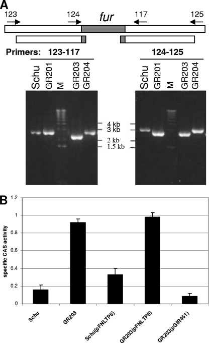
Characterization of Δfur mutant. (A) PCR analysis of Δfur mutant. The fur gene is depicted with flanking sequences and locations of primers used in PCR analysis. Shown beneath is the extent of sequences carried on the deletion plasmid construct. The primer pairs as shown were used to generate PCR products from Schu S4, GR201 (deletion plasmid integrant derivative), GR203 (Δfur), and GR204 (fur+ derivative). “M” indicates lanes with the 1-kb DNA ladder (Invitrogen). (B) Siderophore activity in Δfur mutant and complementation. Schu S4 and GR203 and transformants harboring the control vector pFNLTP6 or the fur+ plasmid pGIR461 were grown overnight in iron-replete CDM, and the specific CAS activities of culture supernatants were determined. Assays were carried out in triplicate, and the averages with standard deviations are represented.
FIG. 4.
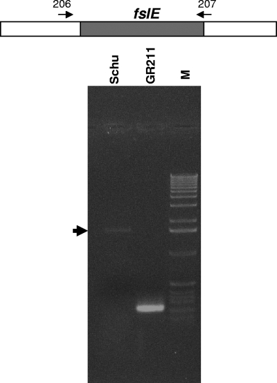
PCR analysis of ΔfslE mutant. The fslE gene is depicted with flanking sequences and locations of primers used in PCR analysis of gDNA from Schu S4 and GR211. Lane M shows the 1-kb DNA ladder (Invitrogen). Schu S4 yields a band of 1.669 kb, including the full-length fslE and indicated by the arrowhead, while the deletion mutant yields a band of 218 bp.
TABLE 1.
Primers used in this work
| Primer | Sequencea |
|---|---|
| 73 | 5′ TCAGCTGGTCTGGATTTTCC 3′ |
| 74 | 5′ GCTAGGGCGTGAGATGATTC 3′ |
| 79 | 5′ ATCACGATGATTGGCAACAA 3′ |
| 80 | 5′ AACTGCTCCCCATTGCTCTA 3′ |
| 109 | 5′ ctactagcatATGTTAAATGCAAATCCTGTCG 3′ |
| 110 | 5′ ctactagcatatgTAAGCTTTTATCGTGAGGGGC 3′ |
| 112 | 5′ GGATTTGTCCTAAAAATTGCTG 3′ |
| 113 | 5′ ctactggctagcGTCCATATAACCAATACTTTGG 3′ |
| 114 | 5′ ctactggcggccgcTAAGTCAAGGTTTTTCGAGTTC 3′ |
| 115 | 5′ ctactggcggccgcTGTTAAATGCAAATCCTGTCG 3′ |
| 116 | 5′ ctactggagctcCTGCTATGATTATAAGCTGAC 3′ |
| 117 | 5′ ctactgggtaccAAGAGTCTCTATATTAAGTGG 3′ |
| 119 | 5′ ctactgggtaccGGAGATATCTATATCAACCTC 3′ |
| 123 | 5′ GCCATCACATAAGCATGCTC 3′ |
| 124 | 5′ AGCAGCACCTAAACCGAAAG 3′ |
| 125 | 5′ ATTCCAGGCATTTACTGGTAG 3′ |
| 132 | 5′ TAGATGATTTAAGGTCAAATAGATAAAGTAG 3′ |
| 136 | 5′ TATCTCTTTTAATACAGAAAAACCAC 3′ |
| 137 | 5′ TTACCAAGCTTTGAAGCAAGCG 3′ |
| 138 | 5′ TAGTTTGGTCGATTATTCAGTAGC 3′ |
| 152 | 5′ ctactggctagcTTGTATGGATTAGTCGAGC 3′ |
| 153 | 5′ ctactgagatctGACGAATGTTCATAACAATCTTAC 3′ |
| 161 | 5′ ctactggctagcATAATTAGACTCTAAGTAC 3′ |
| 162 | 5′ ctactgctcgagTTCTGGATAGTGATTATTGC 3′ |
| 165 | 5′ GACAAAAGCGTTACCCAAAGAG 3′ |
| 178 | 5′ GCTTGTTTGCCTACTTTAGGAGG 3′ |
| 182 | 5′ GATTTGTGCTGTTATAGCTTGC 3′ |
| 184 | 5′ TGGGAGATCATCATCAGGG 3′ |
| 188 | 5′ TGGGCAACAACAACCAATAA 3′ |
| 189 | 5′ TGGTGAAGCGGTTAATGTCA 3′ |
| 197 | 5′ ctactggctagcAACAGATATACTGGTTAATC 3′ |
| 198 | 5′ ctactgtccggaAAAGGGGGTAATTGATAATAC 3′ |
| 199 | 5′ ctactgtccggaACAATAGATATGGCTGTATATC 3′ |
| 200 | 5′ CAAACTGTTTTAAGAGCTCG 3′ |
| 201 | 5′ CAACAAACTGGATATTTGTTTG 3′ |
| 204 | 5′ CGATAATAAATATCAATTATCATTCGG 3′ |
| 206 | 5′ ctactggctagcGTGGTTTTTCTGTATTAAAAGAG 3′ |
| 207 | 5′ ctactgggatccTTAAAGATATACAGCCATATC 3′ |
| 211 | 5′ GTAAGCTTTATTTGATTTCCAG 3′ |
| 219 | 5′ ctactgggatccAAACTCTAATGAACAATCGC 3′ |
| 224 | 5′ ctactgtctagaTTAAAGATATACAGCCATATC 3′ |
Uppercase letters correspond to Francisella genomic sequences; lowercase letters indicate nucleotides added to the primers.
qPCR.
cDNA samples were used as a template for qPCR of fslA and fslE. trpB was used as an internal standard for normalization of transcript levels between different RNA preparations. For each gene, a standard curve was generated with serial dilutions of PCR products from gDNA template. Primers 73 and 74 were used for the real-time reaction for trpB. fslA-specific primers were 79 and 80, and fslE primers were 188 and 189. HotStarTaq polymerase (Qiagen) was used for the amplification reactions, with activation at 94°C for 15 min. The reactions were carried out in a DNA Engine Opticon 2 real-time thermocycler from MJ Research. Reaction mixtures included 0.15% Triton X-100, and Sybr green dye (Bio-Rad) was used for detection of PCR products.
Microarray analysis.
All RNAs were inspected on an Agilent BioAnalyzer to ensure that the peaks corresponding to rRNA were intact and that no degradation had occurred. Approximately 15 μg of total RNA per sample was used for synthesis of cDNA containing amino-allyl-labeled nucleotides (16). The newly synthesized cDNAs were then labeled by a covalent coupling of appropriate cyanine dye (CyDye PostLabeling reactive dye pack; GE Healthcare Bio-Sciences Corp., Piscataway, NJ). The labeling efficiencies of the purified target cDNA preparations were inspected by spectrophotometric analysis, and the total picomoles of dye incorporation (Cy3 or Cy5, accordingly) were calculated for each sample (16). Schu S4 and Δfur mutant GR203 cDNAs labeled with equimolar amounts of different Cy dyes were hybridized to F. tularensis glass slide DNA microarrays procured from the Pathogen Functional Genomics Resource Center at The Institute for Genomic Research. After 24 h of incubation at 42°C, the slides were washed and scanned using a ProScanArray microarray scanner (Perkin-Elmer Life Sciences Inc., Boston, MA). Both Cy5 and Cy3 images from one experiment were analyzed with the ScanArray Express microarray analysis system.
Antibodies to FslE.
The sequence encoding the mature portion of FslE (lacking the 28 amino acids of the putative signal peptide) was amplified by PCR using primers 219 and 224 and cloned in the inducible vector pHis Parallel1 (25). The plasmid was introduced into BL21(DE3) cells by transformation and expression of the gene induced with 0.5 mM isopropyl-β-d-thiogalactopyranoside for 3 h. The expressed protein was almost totally present in inclusion bodies. Cells were lysed with CellLytic IIB (Sigma), and inclusion bodies were purified by centrifugation. One hundred micrograms of protein in complete Freund's adjuvant was used to immunize A/J mice by the subcutaneous and intraperitoneal routes, followed by two boosts. Sera were obtained from the mice and screened by enzyme-linked immunosorbent assay and Western blotting for reactivity to the immunogen.
Western blotting.
Whole-cell lysates normalized to cell density were prepared from F. tularensis cultures. The lysates were separated by sodium dodecyl sulfate-polyacrylamide gel electrophoresis on 10% gels and transferred to polyvinylidene difluoride. The primary anti-FslE serum was used at a 1:2,500 dilution. The blots were developed by chemiluminescence after incubation with secondary goat antimouse-peroxidase conjugate.
RESULTS
Siderophore production is regulated by Fur in F. tularensis.
The siderophore biosynthetic operon fslABCD in F. tularensis is 312 bp downstream of the fur homolog (locus FTT0030c) (see Fig. 2A). The putative fur gene is predicted to encode a protein of 140 amino acid residues with 49% identity and 61% similarity to its closest homolog, the Neisseria gonorrhoeae Fur sequence (GenBank accession no. AAA72351.1) (3). F. tularensis fur appears to be transcribed as a single gene. The nuo operon is 210 bp upstream of fur and is divergent. A canonical Fur box sequence is present in the promoter region of fslA 22 bp upstream of the ATG start codon (see Fig. 2A). We predicted that siderophore-mediated iron uptake in F. tularensis is regulated by the Fur repressor, as seen in other bacterial systems (reviewed in reference 11). We generated an unmarked in-frame Δfur mutation (retaining sequences encoding the 3 amino-terminal and 10 carboxy-terminal residues but not the central 122 amino acid residues) in Schu S4 using a suicide plasmid as detailed in the Methods section.
We carried out PCR analysis of gDNA from strain GR201, harboring the integrated suicide plasmid, using a combination of primers that would help identify the site of integration (Fig. 1A). The plasmid pair 123-117 gave a 2.713-kb band similar to that of Schu S4. The 124-125 combination yielded a band that was 360 bp smaller in size (2.668 kb) than the corresponding Schu S4 product (3.028 kb). These results indicated that the plasmid had integrated at the 3′ flanking sequence of the chromosomal fur gene in GR201. Loss of plasmid sequences by counterselection on sucrose gave rise to isolates that either were mutants (Δfur) or were wild-type (fur+) derivatives. We analyzed the Δfur strain GR203 and a fur+ derivative, GR204, by PCR of chromosomal DNA. The GR204 strain yielded PCR products similar to those of Schu S4 with both sets of primers, while GR203 produced shorter PCR products corresponding to the deletion of the fur gene (Fig. 1A). The PCR product was sequenced to verify the deletion.
To test if Fur was involved in regulation of siderophore production, we used a liquid CAS assay to examine siderophore activity in the supernatant of the strains after growth in iron-replete CDM. Under these conditions, Schu S4 has a minimal level of siderophore activity in the culture supernatant (Fig. 1B) (26). In contrast, the Δfur mutant showed a ∼5×-higher level of siderophore activity, demonstrating that siderophore production in F. tularensis is normally repressed by Fur under iron-replete conditions. We tested the ability of the wild-type fur gene to complement the Δfur mutation in trans. We introduced plasmid pGIR461, carrying the wild-type fur gene under control of its own promoter. As a control, we used the vector pFNLTP6. As expected, GR203 with pFNLTP6 has high siderophore activity compared to Schu S4 harboring the same plasmid (Fig. 1B). In a transformant of GR203 carrying the fur+ plasmid pGIR461, the siderophore activity in the supernatant dropped to a very low level, even lower than that for Schu S4 itself. This is not surprising, since the Fur repressor is expressed from a gene on a multicopy plasmid. These experiments demonstrated that siderophore production is deregulated due to specific mutation of the fur gene in strain GR203.
fslA and fslE transcription is deregulated in Δfur mutant.
To test if the effect of fur on siderophore production was at the transcriptional level, as seen in other bacterial systems, we examined fslA expression in the fur mutant strain GR203 by quantitative real-time PCR with cDNA prepared from cells grown under iron-replete conditions. Expression of fslA was normalized to expression of trpB, an internal control unaffected by iron levels in the medium. As shown in Fig. 2B, relative fslA expression is ∼765-fold higher in the Δfur mutant than in Schu S4, consistent with Fur being a transcriptional repressor of the siderophore biosynthetic gene. A pilot microarray comparing the transcriptional profiles of GR203 and Schu S4 grown in iron-replete media revealed that all of the genes of the fslABCD cluster were deregulated in the fur mutant (data not shown). Additionally, expression of locus FTT0025c, encoding a hypothetical protein and located just downstream of fslD, was also found to be upregulated in the fur mutant. We have designated this gene fslE due to its proximity to the fslABCD operon (Fig. 2A). We carried out quantitative reverse transcriptase PCR (RT-PCR) of cDNA from GR203 and Schu S4 using primers specific to fslE and confirmed that expression of this locus was significantly deregulated (∼19-fold) in the Δfur mutant (Fig. 2B).
fslE is transcribed as part of an operon.
The fslA, -B, -C, and -D genes are closely clustered and have been shown to be transcribed as an operon in the LVS (7, 26). The presumptive start codon of fslE is 85 bp downstream of the fslD stop codon. In addition, a hypothetical gene predicted to encode a 114-amino-acid protein (locus FTT0024c) is located 47 bp downstream of the fslE stop codon (Fig. 2A). We have designated this gene fslF due to its proximity to the other fsl genes. Locus FTT0023c, encoding a putative lipase/acetyltransferase, lies 257 bp downstream of the stop codon of fslF. In order to characterize transcription of the fsl genes in Schu S4 and to define the limits of the transcription unit, we used primers that bridged adjacent loci in RT-PCR using cDNA from GR203 grown under iron-replete conditions. As shown in Fig. 2C, primers spanning fslAB, -BC, -CD, -DE, and -EF all yielded bands of the sizes predicted for cotranscription (1.614 kb, 1.459 kb, 1.585 kb,1.585 kb, and 558 bp, respectively), similar to results with gDNA. Primers 109 and 112, which span the fur and fslA genes, did not amplify a 1.524-kb band from cDNA as seen for gDNA, although primers 110 and 112, which are within fslA, yielded a 1.092-kb product. This analysis established that transcription of the fsl operon initiated independently and downstream of the fur gene. Primers spanning fslF and locus FTT0023c also did not amplify a product of 833 bp from cDNA as seen for gDNA, indicating that FTT0023c is not part of the operon. Our results indicate that the fslE gene is transcribed as part of the fslABCDEF operon in Schu S4.
FslE protein production mirrors transcription.
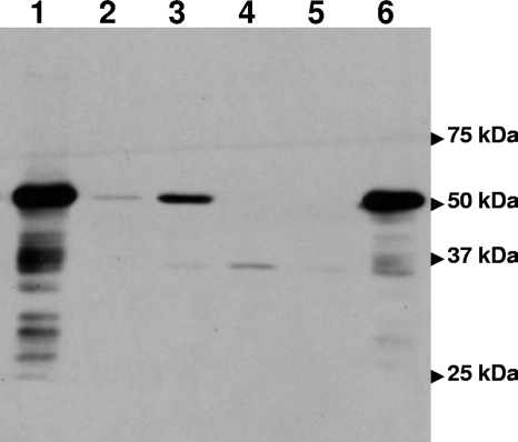
We analyzed production of the FslE protein in Schu S4 using antiserum raised to the recombinant protein. As shown in Fig. 3, lane 2, a band of the expected 50-kDa mass was seen in lysates of Schu S4 grown in iron-replete CDM. The signal in the lysates of fur mutant strain GR203 grown in iron-replete CDM was much higher, which correlated with transcriptional deregulation (Fig. 3, lane 1). We also examined the effect of iron starvation on production of the FslE protein. As shown in Fig. 3, lane 3, FslE was increased in cells grown in iron-limiting medium, although not to the levels seen for the fur mutant. These results indicate that FslE protein production is both Fur and iron regulated.
FIG. 3.
Western blotting with FslE antibodies. Whole-cell lysates normalized for cell density were subjected to sodium dodecyl sulfate-polyacrylamide gel electrophoresis, transferred to a polyvinylidene difluoride membrane, and probed with polyclonal antiserum raised to recombinant FslE. The locations of the prestained standards run on the gel are indicated. The lysates are denoted by lanes: 1, GR203 (Δfur) grown in iron-replete CDM; 2, Schu S4 grown in iron-replete CDM; 3, Schu S4 grown in iron-limiting CDM; 4, GR211 (ΔfslE) grown in iron-limiting CDM; 5, vector (pFNLTP6gro-GFP)-transformed GR211 grown in iron-limiting CDM; 6, fslE+ plasmid (pGIR469)-transformed GR211 grown in iron-limiting CDM.
Generation of ΔfslE mutant.
The fslE gene product, annotated as a hypothetical membrane protein of 509 amino acid residues, has been identified in the outer membrane of LVS and Schu S4 and designated SrfA (17). The proximity to, and its cotranscription with, the siderophore biosynthetic genes suggested that fslE may also play a role in siderophore-mediated iron uptake in F. tularensis. In order to test this possibility, we generated a strain, GR211, with a defined in-frame chromosomal deletion of the gene in the Schu S4 background. The deletion mutation was confirmed by PCR analysis of gDNA using primers flanking the gene, as shown in Fig. 4. The PCR product was also sequenced to confirm that the sequences encoding the amino-terminal 18 residues and carboxy-terminal 8 residues were retained and in-frame.
We tested lysates of GR211 for the presence of the FslE protein by Western blotting of lysates from cells grown in iron-limiting CDM. As shown in Fig. 3, lane 4, the band corresponding to FslE was not produced in these cells, whereas FslE production was induced in Schu S4 under these conditions (Fig. 3, lane 3).
The ΔfslE mutant is defective for growth under iron limitation.
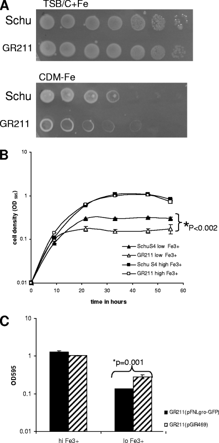
To determine if fslE plays a role in iron acquisition, we tested the fslE mutant strain, GR211, for the ability to grow under iron limitation. We spotted serial dilutions of Schu S4 and of GR211 on an iron-replete agar plate (TSB/C supplemented with iron) and on CDM−Fe agar. As seen in Fig. 5A, both strains grew equally well on the iron-replete plate. On the iron-limiting plate, Schu S4 growth was observed up to the fourth dilution. GR211, however, showed growth only in the margins of the spots at even the very highest concentration of cells (∼3 × 10⁹ CFU/ml). This phenotype was consistent with a poor ability to grow under iron limitation.
FIG. 5.
Growth deficiency of ΔfslE mutant under iron limitation. (A) Growth on iron-replete and iron-deficient agar plates. Cultures of Schu S4 and GR211 in iron-replete CDM were washed in che-CDM−Fe and resuspended to an optical density of 1.0. Tenfold serial dilutions were made in che-CDM−Fe and spotted on an iron-replete or iron-limiting agar plate. Growth was assessed after 3 days on the rich plate and 4 days on the iron-limiting plate. (B) Growth in liquid culture. Washed cells were inoculated into iron-replete (high Fe3+) or iron-limiting (low Fe3+) che-CDM and growth followed over a period of 56 h. Cultures were grown in triplicate, and the means and standard deviations of one representative experiment are shown in the growth plot. (C) Complementation of ΔfslE mutant in trans. GR211 cells transformed with either control vector pFNLTP6gro-GFP or the fslE+ plasmid pGIR469 were washed and inoculated into iron-replete (hi Fe3+) or iron-limiting (lo Fe3+) che-CDM. The growth of cultures in duplicate was monitored over 48 h, and the results at the 48-h time point are shown for one representative experiment.
We also examined growth in liquid medium. The fslE mutant strain, GR211, grew similarly to Schu S4 in standard CDM containing 2 μg/ml of ferrous sulfate (data not shown). We compared growth of Schu S4 and GR211 in che-CDM supplemented with standard high levels (2 μg/ml) of FeCl3 or limiting levels of FeCl3 (0.1 μg/ml). As shown in Fig. 5B, the two strains grew similarly in medium supplemented with high levels of FeCl3. When iron levels were limiting, the Schu S4 strain grew to about a third of its density in iron-replete medium; however, the ΔfslE strain was significantly defective in its ability to grow under this iron limitation (P < 0.002 at all times after 21 h), growing to half the density of the wild type (Fig. 5B). We inferred that the deletion of fslE resulted in a lowered ability to assimilate iron under iron-limiting conditions.
Complementation of ΔfslE by plasmid-borne fslE.
We tested the ability of plasmid pGIR469 carrying the fslE gene under control of the groE promoter to complement the fslE mutation in trans. We analyzed lysates of cells grown in reduced-iron medium for reactivity to FslE antiserum on Western blots. As shown in Fig. 3, lane 5, GR211 transformed with the control plasmid pFNLTP6gro-GFP did not produce the FslE protein. The strain harboring the plasmid pGIR469, however, showed robust FslE levels (Fig. 3, lane 6).
We compared transformants of GR211 carrying either pGIR469 or the control plasmid for their ability to grow in iron-limiting CDM (Fig. 5C). In medium supplemented with 2 μg/ml of ferric chloride as an iron source, the two transformants grew similarly. When levels of ferric iron were limiting, the complemented strain grew significantly better (twofold-higher density) than the control strain. This difference reflected that seen with the parental strain, Schu S4, and the fslE mutant GR211 strain. These results demonstrated that the iron-dependent growth phenotype of GR211 was specifically due to mutation in fslE and could be complemented in trans by the plasmid-borne wild-type gene.
FslE is necessary for siderophore utilization.
In order to determine the nature of the growth defect in ferric iron of the ΔfslE strain, GR211, and if it was related to siderophore-mediated iron uptake, we tested the ability of the strain to both express and utilize the siderophore. We employed a functional plate-based assay which we previously developed for characterizing the LVS siderophore (26).
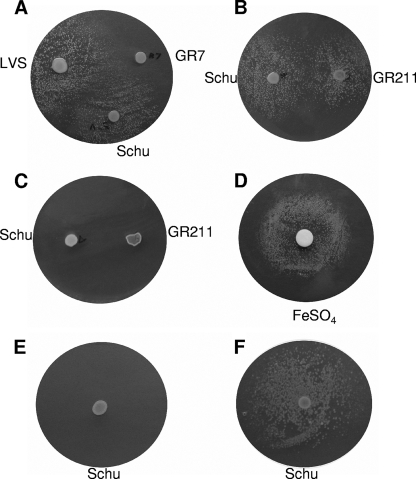
We first established that this assay could be adapted to examine siderophore utilization in Schu S4. We seeded an iron-limiting CDM plate with Schu S4 and spotted on the plate LVS, the siderophore-deficient ΔfslA mutant of LVS (GR7), or Schu S4 (26). After 4 days, a halo of growth was observed around the LVS and Schu S4 spots, indicating that the seeded cells were able to utilize the siderophore secreted by bacteria in both the LVS and Schu S4 spots (Fig. 6A). No halo was observed around GR7, confirming that the growth was siderophore dependent. We then tested GR211 for siderophore production by spotting it on a Schu S4-seeded plate. As seen in Fig. 6B, the seeded Schu S4 cells formed growth halos around GR211 similarly to results with Schu S4, indicating that GR211 was functional in siderophore secretion.
FIG. 6.
Siderophore production and utilization by ΔfslE mutant. Iron-limiting CDM−Fe plates were seeded with Schu S4 (A and B), the ΔfslE strain, GR211 (C and D), or GR211 harboring plasmid vector pFNLTP6groGFP (E) or the fslE+ plasmid, pGIR469 (F). Washed cells of LVS, GR7 (siderophore-deficient derivative of LVS), Schu S4, and GR211 were spotted on the plates as indicated. A filter paper spotted with 10 μl of 20-mg/ml FeSO4 was placed on plate D. Experiments were repeated at least two independent times with similar results; plates from a representative experiment are shown.
In order to test for the ability to utilize a siderophore, we ran parallel assays using GR211 as the tester strain to seed plates. No evidence of any growth was observed around either the Schu S4 or GR211 spots even after 4 days (Fig. 6C). These results indicated that the GR211 cells were unable to utilize a siderophore to grow on the iron-limiting plate. To demonstrate the viability of the starting culture, we spotted ferrous sulfate on a filter paper on a similarly seeded plate and observed robust growth around the filter in just 2 days (Fig. 6D). We then tested transformants of GR211 harboring either a control vector (pFNLTP6gro-GFP) or the fslE+ plasmid pGIR469 for the ability to utilize a siderophore in this plate assay. Vector-transformed Schu S4 bacteria were able to grow utilizing a siderophore, similar to results with untransformed Schu S4 (data not shown). However, vector-transformed GR211 bacteria were unable to form growth halos around a spot of siderophore-secreting Schu S4 (Fig. 6E). The presence of fslE in trans (pGIR469 transformant) was sufficient to restore siderophore-dependent growth (Fig. 6F).
These experiments demonstrated that FslE was not required for siderophore biosynthesis and export but was necessary for siderophore-mediated utilization of iron for growth.
DISCUSSION
Iron acquisition genes, including siderophore biosynthetic and siderophore uptake genes, are commonly under transcriptional control of the iron-responsive repressor Fur in gram-negative and gram-positive bacteria, including E. coli, Bacillus subtilis, Campylobacter jejuni, Pseudomonas aeruginosa, Neisseria spp., and Shewanella (2, 4, 9, 14, 22, 27). Using a defined fur deletion mutant of strain Schu S4, we have shown here that siderophore production in F. tularensis is under Fur regulation. This control is exerted at the level of transcription, as demonstrated for the biosynthetic gene fslA.
We determined that the fslE gene (FTT0025c in the Schu S4 genome), which is downstream of the siderophore biosynthetic gene cluster fslABCD, is also deregulated in the Δfur strain. We determined that fslE is transcribed as part of the fsl operon in the Schu S4 strain. We defined the limits of the operon, showing that the transcript initiates downstream of the fur gene and extends into fslF (FTT0024c) downstream of fslE but does not include the FTT0023c locus.
Genes for the related functions of siderophore biosynthesis and uptake are commonly clustered and coregulated in bacteria. The proximity to fslABCD and the cotranscription suggested that FslE is involved in siderophore-mediated iron uptake. We generated an in-frame deletion in fslE in the Schu S4 background and found that the mutant strain had a diminished ability to grow under iron limitation. Using a plate-based growth assay, we demonstrated that the ΔfslE mutant secreted functional siderophore under iron limitation but was defective in utilizing the siderophore itself. This growth defect was rescued by fslE+, provided in trans. Thus, FslE is essential for siderophore-mediated iron utilization.
It was recently shown that the fslD and fslE homologs in a strain of the closely related novicida subspecies are cotranscribed and that transcription is induced by iron starvation (20). A strain with a mutation in the fslE homolog was competent for siderophore production. Our studies with Schu S4 indicate that parallels may be drawn between the tularensis and novicida subspecies in aspects of siderophore-mediated iron uptake. While they are closely related in sequence, there are also significant sequence differences between the genomes of the subspecies that are reflected in differences in growth characteristics and virulence (23). It is not known at present if the differences may extend to iron acquisition.
The FslE protein belongs to a family of five related open reading frames unique to F. tularensis (18). A signal peptide at the amino terminus of FslE is predicted by the SignalP 3.0 software program (www.cbs.dtu.dk/services/SignalP/), suggesting that it is a secreted protein. In a recent study examining outer membrane proteins of F. tularensis, FslE (referred to as SrfA) was identified in the outer membrane fraction of Schu S4 and of LVS (17). The hidden Markov model-based beta-barrel prediction program PRED-TMBB (1) predicts that residues 178 to 509 of the FslE protein can fold as a 14-stranded beta-barrel in the outer membrane, with the amino-terminal third of the protein (including the putative signal peptide sequences) in the periplasm. The structure is reminiscent of siderophore receptors in gram-negative bacteria, although the typical TonB-dependent transporter is 22 stranded (5). This suggests that FslE may function as the siderophore receptor in F. tularensis in a manner analogous to that for other gram-negative bacteria. How it might do so in the absence of an obvious TonB-ExbB-ExbD system is an interesting question. Alternatively, FslE may mediate uptake of siderophore-bound iron by a completely different mechanism. Future studies will further explore the function of FslE and its role in siderophore-mediated iron uptake.
Acknowledgments
We gratefully acknowledge the F. tularensis oligonucleotide microarrays provided by PFGRC. We thank Barbara J. Mann for critical reading of the manuscript.
This work was supported by grants AI056227 and AI067823 from the NIAID and by an intramural Research and Development grant from the University of Virginia School of Medicine.
Footnotes
Published ahead of print on 6 June 2008.
REFERENCES
- 1.Bagos, P. G., T. D. Liakopoulos, I. C. Spyropoulos, and S. J. Hamodrakas. 2004. PRED-TMBB: a web server for predicting the topology of beta-barrel outer membrane proteins. Nucleic Acids Res. 32W400-W404. [DOI] [PMC free article] [PubMed] [Google Scholar]
- 2.Baichoo, N., T. Wang, R. Ye, and J. D. Helmann. 2002. Global analysis of the Bacillus subtilis Fur regulon and the iron starvation stimulon. Mol. Microbiol. 451613-1629. [DOI] [PubMed] [Google Scholar]
- 3.Berish, S. A., S. Subbarao, C. Y. Chen, D. L. Trees, and S. A. Morse. 1993. Identification and cloning of a fur homolog from Neisseria gonorrhoeae. Infect. Immun. 614599-4606. [DOI] [PMC free article] [PubMed] [Google Scholar]
- 4.Braun, V. 2003. Iron uptake by Escherichia coli. Front. Biosci. 8s1409-s1421. [DOI] [PubMed] [Google Scholar]
- 5.Chakraborty, R., E. Storey, and D. van der Helm. 2007. Molecular mechanism of ferricsiderophore passage through the outer membrane receptor proteins of Escherichia coli. Biometals 20263-274. [DOI] [PubMed] [Google Scholar]
- 6.Chamberlain, R. E. 1965. Evaluation of live tularemia vaccine prepared in a chemically defined medium. Appl. Microbiol. 13232-235. [DOI] [PMC free article] [PubMed] [Google Scholar]
- 7.Deng, K., R. J. Blick, W. Liu, and E. J. Hansen. 2006. Identification of Francisella tularensis genes affected by iron limitation. Infect. Immun. 744224-4236. [DOI] [PMC free article] [PubMed] [Google Scholar]
- 8.Drechsel, H., J. Metzger, S. Freund, G. Jung, J. R. Boelaert, and G. Winkelmann. 1991. Rhizoferrin—a novel siderophore from the fungus Rhizopus-Microsporus var Rhizopodiformis. Biol. Metals 4238-243. [Google Scholar]
- 9.Ducey, T. F., M. B. Carson, J. Orvis, A. P. Stintzi, and D. W. Dyer. 2005. Identification of the iron-responsive genes of Neisseria gonorrhoeae by microarray analysis in defined medium. J. Bacteriol. 1874865-4874. [DOI] [PMC free article] [PubMed] [Google Scholar]
- 10.Ernst, J. F., R. L. Bennett, and L. I. Rothfield. 1978. Constitutive expression of the iron-enterochelin and ferrichrome uptake systems in a mutant strain of Salmonella typhimurium. J. Bacteriol. 135928-934. [DOI] [PMC free article] [PubMed] [Google Scholar]
- 11.Escolar, L., J. Perez-Martin, and V. de Lorenzo. 1999. Opening the iron box: transcriptional metalloregulation by the Fur protein. J. Bacteriol. 1816223-6229. [DOI] [PMC free article] [PubMed] [Google Scholar]
- 12.Faraldo-Gomez, J. D., and M. S. P. Sansom. 2003. Acquisition of siderophores in gram-negative bacteria. Nat. Rev. Mol. Cell Biol. 4105-116. [DOI] [PubMed] [Google Scholar]
- 13.Forsman, M., G. Sandstrom, and A. Sjostedt. 1994. Analysis of 16S ribosomal DNA sequences of Francisella strains and utilization for determination of the phylogeny of the genus and for identification of strains by PCR. Int. J. Syst. Bacteriol. 4438-46. [DOI] [PubMed] [Google Scholar]
- 14.Grifantini, R., E. Frigimelica, I. Delany, E. Bartolini, S. Giovinazzi, S. Balloni, S. Agarwal, G. Galli, C. Genco, and G. Grandi. 2004. Characterization of a novel Neisseria meningitidis Fur and iron-regulated operon required for protection from oxidative stress: utility of DNA microarray in the assignment of the biological role of hypothetical genes. Mol. Microbiol. 54962-979. [DOI] [PubMed] [Google Scholar]
- 15.Hantke, K. 1981. Regulation of ferric iron transport in Escherichia coli K12: isolation of a constitutive mutant. Mol. Gen. Genet. 182288-292. [DOI] [PubMed] [Google Scholar]
- 16.Hegde, P., R. Qi, K. Abernathy, C. Gay, S. Dharap, R. Gaspard, J. E. Hughes, E. Snesrud, N. Lee, and J. Quackenbush. 2000. A concise guide to cDNA microarray analysis. BioTechniques 29548-550, 552-554, 556 passim. [DOI] [PubMed] [Google Scholar]
- 17.Huntley, J. F., P. G. Conley, K. E. Hagman, and M. V. Norgard. 2007. Characterization of Francisella tularensis outer membrane proteins. J. Bacteriol. 189561-574. [DOI] [PMC free article] [PubMed] [Google Scholar]
- 18.Larsson, P., P. C. Oyston, P. Chain, M. C. Chu, M. Duffield, H. H. Fuxelius, E. Garcia, G. Halltorp, D. Johansson, K. E. Isherwood, P. D. Karp, E. Larsson, Y. Liu, S. Michell, J. Prior, R. Prior, S. Malfatti, A. Sjostedt, K. Svensson, N. Thompson, L. Vergez, J. K. Wagg, B. W. Wren, L. E. Lindler, S. G. Andersson, M. Forsman, and R. W. Titball. 2005. The complete genome sequence of Francisella tularensis, the causative agent of tularemia. Nat. Genet. 37153-159. [DOI] [PubMed] [Google Scholar]
- 19.Maier, T. M., A. Havig, M. Casey, F. E. Nano, D. W. Frank, and T. C. Zahrt. 2004. Construction and characterization of a highly efficient Francisella shuttle plasmid. Appl. Environ. Microbiol. 707511-7519. [DOI] [PMC free article] [PubMed] [Google Scholar]
- 20.Milne, T. S., S. L. Michell, H. Diaper, P. Wikstrom, K. Svensson, P. C. Oyston, and R. W. Titball. 2007. A 55 kDa hypothetical membrane protein is an iron-regulated virulence factor of Francisella tularensis subsp. novicida U112. J. Med. Microbiol. 561268-1276. [DOI] [PubMed] [Google Scholar]
- 21.Munzinger, M., K. Taraz, H. Budzikiewicz, H. Drechsel, P. Heymann, G. Winkelmann, and J. M. Meyer. 1999. S,S-rhizoferrin (enantio-rhizoferrin)—a siderophore of Ralstonia (Pseudomonas) pickettii DSM 6297—the optical antipode of R, R-rhizoferrin isolated from fungi. Biometals 12189-193. [Google Scholar]
- 22.Palyada, K., D. Threadgill, and A. Stintzi. 2004. Iron acquisition and regulation in Campylobacter jejuni. J. Bacteriol. 1864714-4729. [DOI] [PMC free article] [PubMed] [Google Scholar]
- 23.Rohmer, L., C. Fong, S. Abmayr, M. Wasnick, T. J. Larson Freeman, M. Radey, T. Guina, K. Svensson, H. S. Hayden, M. Jacobs, L. A. Gallagher, C. Manoil, R. K. Ernst, B. Drees, D. Buckley, E. Haugen, D. Bovee, Y. Zhou, J. Chang, R. Levy, R. Lim, W. Gillett, D. Guenthener, A. Kang, S. A. Shaffer, G. Taylor, J. Chen, B. Gallis, D. A. D'Argenio, M. Forsman, M. V. Olson, D. R. Goodlett, R. Kaul, S. I. Miller, and M. J. Brittnacher. 2007. Comparison of Francisella tularensis genomes reveals evolutionary events associated with the emergence of human pathogenic strains. Genome Biol. 8R102. [DOI] [PMC free article] [PubMed] [Google Scholar]
- 24.Schwyn, B., and J. B. Neilands. 1987. Universal chemical assay for the detection and determination of siderophores. Anal. Biochem. 16047-56. [DOI] [PubMed] [Google Scholar]
- 25.Sheffield, P., S. Garrard, and Z. Derewenda. 1999. Overcoming expression and purification problems of RhoGDI using a family of “Parallel” expression vectors. Protein Expr. Purif. 1534-39. [DOI] [PubMed] [Google Scholar]
- 26.Sullivan, J. T., E. F. Jeffery, J. D. Shannon, and G. Ramakrishnan. 2006. Characterization of the siderophore of Francisella tularensis and role of fslA in siderophore production. J. Bacteriol. 1883785-3795. [DOI] [PMC free article] [PubMed] [Google Scholar]
- 27.Wan, X. F., N. C. Verberkmoes, L. A. McCue, D. Stanek, H. Connelly, L. J. Hauser, L. Wu, X. Liu, T. Yan, A. Leaphart, R. L. Hettich, J. Zhou, and D. K. Thompson. 2004. Transcriptomic and proteomic characterization of the Fur modulon in the metal-reducing bacterium Shewanella oneidensis. J. Bacteriol. 1868385-8400. [DOI] [PMC free article] [PubMed] [Google Scholar]


