Abstract
The whisker region in the rodent primary motor (MI) cortex receives dense projections from neurons aligned with the layer IV septa in the whisker region of the primary somatosensory (SI) cortex. To compare whisker-induced responses in MI with respect to the SI responses in the septa and adjoining barrel regions, we used several experimental approaches in anesthetized rats. Reversible inactivation of SI and the surrounding cortex suppressed the magnitude of whisker-induced responses in the MI whisker region by 80%. Subsequent laminar analysis of MI responses to electrical or mechanical stimulation of the whisker pad revealed that the most responsive MI neurons were located ≥1.0 mm below the pia. When layer IV neurons in SI were recorded simultaneously with deep MI neurons during low-frequency (2-Hz) deflections of the whiskers, the neurons in the SI barrels responded 2–6 ms earlier than those in MI. Barrel neurons displayed similar response latencies at all stimulus frequencies, but the response latencies in MI and the SI septa increased significantly when the whiskers were deflected at frequencies of 8 Hz. Finally, cross-correlation analysis of neuronal activity in SI and MI revealed greater amounts of time-locked coordination among septa–MI neuron pairs than among barrel–MI neuron pairs. These results suggest that the somatosensory corticocortical inputs to MI cortex convey information processed by the SI septal circuits.
INTRODUCTION
In rodents, vibrissal information is processed by multiple ascending pathways. Each pathway originates from a specific trigeminal nucleus in the brain stem and conveys information through a distinct thalamic nucleus before terminating in SI cortex (Ahissar and Zacksenhouse 2001; Alloway 2008; Sosnik et al. 2001; Yu et al. 2006). The lemniscal pathway arises from the principal trigeminal nucleus, ascends through the dorsomedial sector of the ventroposteromedial thalamic nucleus (VPM), and terminates in the layer IV barrels of SI cortex (Chiaia et al. 1991a; Chmielowska et al. 1989; Koralek et al. 1988; Lu and Lin 1993; Peschanski 1984; Pierret et al. 2000; Rhoades et al. 1987; Yu et al. 2006). Neurons in the lemniscal pathway respond maximally to deflections of a single principal whisker, exhibit directional tuning, and seem well suited for texture discrimination and object location (Alloway 2008; Armstrong-James and Callahan 1991; Armstrong-James and Fox 1987; Yu et al. 2006).
By comparison, the paralemniscal pathway originates in the interpolaris subdivision of the spinal trigeminal nucleus (SpVi), ascends through the medial portion of the posterior thalamic nucleus (POm), and then terminates most densely in the layer IV septa and more broadly in layer Va (Alloway 2008; Chiaia et al. 1991a; Chmielowska et al. 1989; Erzurumlu and Killackey 1980; Koralek et al. 1988; Lu and Lin 1993; Peschanski 1984; Pierret et al. 2000; Rhoades et al. 1987; Veinante et al. 2000). Although the precise function of the paralemniscal pathway has not been fully elucidated, much evidence suggests that the POm nucleus may encode whisking frequency. This view is based, in part, on physiological studies showing that POm responses to whisker deflections exhibit longer latencies as the frequency of whisker stimulation increases (Ahissar and Kleinfeld 2003; Ahissar and Zacksenhouse 2001; Ahissar et al. 2000; Sosnik et al. 2001; Zacksenhouse and Ahissar 2006). These timing shifts in the paralemniscal pathway are probably controlled by convergent projections to the POm that include corticothalamic feedback connections as well as inhibitory projections from the zona incerta (ZI) (Bartho et al. 2002; Lavallee et al. 2005; Trageser and Keller 2004).
The MI whisker representation receives topographically organized projections from the POm nucleus but not from VPM (Aldes 1988; Chakrabarti and Alloway 2006; Cicirata et al. 1986; Hoffer and Alloway 2001). Furthermore, the corticocortical projections from SI barrel cortex to the MI whisker region originate primarily from neurons aligned with the layer IV septal compartments (Alloway et al. 2004; Chakrabarti and Alloway 2006). Hence, the anatomical evidence suggests that whisker-related inputs to MI cortex come from the paralemniscal pathway. Consistent with this view, stimulation of MI cortex disinhibits the ZI-mediated depression of POm neurons, presumably to allow the flow of information through the paralemniscal channel (Urbain and Deschênes 2007).
Very few studies have characterized MI neuronal responses during active or passive whisker stimulation (Farkas et al. 1999; Kleinfeld et al. 2002). One of these demonstrated that electrical stimulation of the whisker pad produced short-latency (∼11-ms) responses in MI cortex that were reduced by about 90% after inactivation of SI (Farkas et al. 1999). Electrical stimulation is nonspecific, however, and may produce neuronal responses in SI and MI that reflect inputs from multiple submodalities. Furthermore, the dependence of MI responses on inputs from barrel cortex has not been characterized with respect to the septal and barrel circuits.
In this study, we used multiple approaches to characterize whisker-related responses in MI with respect to responses recorded in the barrel and septal compartments of SI. First, to assess the dependence of MI responses on SI, we inactivated SI cortex and measured the changes in MI responsiveness. Next, we conducted a laminar analysis to determine the MI layers that received the fastest and strongest whisker inputs. We then recorded MI and SI neurons simultaneously during mechanical whisker stimulation at multiple frequencies. Neuronal responses in MI were compared with those in the layer IV barrels and septa to determine whether response latencies increased as a function of stimulus frequency, a characteristic feature of neurons in POm (Ahissar et al. 2000). Finally, we conducted cross-correlation analysis to determine whether MI neuronal responses were coordinated with barrel or septal neurons. Our data indicate that MI sensory responses depend more on SI inputs from the septal circuits than from the barrel circuits.
METHODS
Data were obtained from 21 male Sprague–Dawley rats ranging from 250 to 450 g. All procedures complied with National Institutes of Health guidelines and our institutional animal welfare committee approved a detailed description of the experimental protocol. All experimental approaches (cortical inactivation, latency analysis, cross-correlation analysis) involved surgery, peripheral stimulation, and extracellular neuronal recording.
Surgery
Each rat was initially anesthetized with an intramuscular (im) injection of ketamine (20 mg/kg) and xylazine (6 mg/kg). Atropine sulfate (0.05 mg/kg, im) and dexamethasone (0.5 mg/kg, im) were administered to reduce bronchial secretions and prevent tissue inflammation. Each rat was intubated through the oral cavity, placed in a stereotaxic frame, and artificially ventilated with a 2:1 mixture of nitrous oxide and oxygen. Ophthalmic ointment was applied to the eyes to prevent corneal irritation. As the effects of ketamine and xylazine subsided, isoflurane was administered at concentrations (0.75–1.5%) that prevent muscular reflexes. Body temperature was maintained at 37°C with a heated water pad (under the trunk) and a homeothermic heating blanket (over the trunk). To ensure a stable plane of anesthesia, heart rate and end-tidal CO2 were monitored continuously. After isolating MI neuronal responses, additional doses of ketamine (5–10 mg/kg, im) and xylazine (1.5–3.0 mg/kg, im) were occasionally administered to reduce spontaneous MI activity.
When recording was done, each rat received a lethal dose of sodium pentobarbital (>100 mg/kg, administered intraperitoneally) and was transcardially perfused before the brain was removed. In five rats, the brain was coronally transected at the level of bregma. The SI barrel cortex was dissected from the caudal portion and placed between glass slides for overnight storage in cold 4% paraformaldehyde and 30% sucrose. The rostral portion, including MI cortex, was also placed in cold fixative overnight and then was cut coronally into 60-μm sections that were stained with thionin. The cortical slab was sectioned tangentially at 60 μm and processed for cytochrome oxidase (Chakrabarti and Alloway 2006; Land and Simons 1985; Wong-Riley 1979). Histological examination of MI and SI was conducted with a light microscope (Olympus BH-2) at magnifications of ×20 and ×40 (×2 and ×4 objectives with a ×10 ocular lens), and photomicrographs of electrode tracks were obtained with a Cool Snap HQ CCD digital camera (Roper Scientific, Tucson, AZ).
Whisker pad stimulation
For electrical stimulation, two stainless steel needles were inserted into the skin at sites anterior and posterior to the whisker pad. Biphasic current pulses, 2 ms long, were passed through the leads using a constant-current source (BAK Electronics, Mount Airy, MD). Current pulse amplitude was adjusted to levels (250–300 μA) that evoked twitches of the whisker pad. Neuronal responses to electrical stimulation were recorded for 50 trials.
Two types of mechanical stimulation were used. In most cases, a section of plastic window screen was attached to the distal end of an ink pen held by a galvanometer taken from a Grass polygraph machine. The screen was positioned 10 mm away from the face so that ≥15 whiskers (in rows B, C, D, and E), which were trimmed to a length of 15 mm, protruded through the screen (Zhang and Alloway 2004). The protruding whiskers contacted the rostral edge of the holes in the screen so that computer-controlled screen movements in the caudal direction produced immediate deflections of the whiskers. Screen movement was activated by a 50-ms sawtooth pulse, produced by a waveform generator (model LW 420, LeCroy, Chestnut Ridge, NY), the output of which was amplified by a DC-coupled amplifier (Techron LVC608). Screen movement deflected the whiskers 1,000 μm in the caudal direction during the first 25 ms of the pulse, thereby producing a stimulus velocity of 40 mm/s.
Window screen movements were administered at frequencies of 2, 5, and 8 Hz. An initial prestimulus period of 2,000 ms was followed by three blocks of eight 50-ms pulses administered at 2, 5, and 8 Hz, with each stimulus block separated by an interval of 1,000 ms. Neuronal discharges were recorded continuously throughout the prestimulus period, subsequent frequency-specific blocks, and the interblock intervals, but not during the intertrial intervals, which lasted about 0.5 s. Neuronal responses to screen stimulation were recorded for 100 (laminar analysis experiments) or 300 (latency comparisons/cross-correlation experiments) trials.
Mechanical whisker stimulation during the inactivation experiments consisted of a moving air jet (Zhang and Alloway 2006). A hollow tube (1-mm ID) with a curved end was attached to the galvanometer to emit an air jet toward the whiskers. Airflow onset and offset were gated by an electronic valve controlled by the data acquisition system; air pressure was regulated (20–25 psi) by a needle valve in series with a pressure gauge. Air-jet motion was generated by a 4-Hz sawtooth pulse that was amplified so that whiskers in multiple rows and arcs were deflected as the air jet moved back and forth. Air-jet stimulation lasted for 1 s and was preceded by a 2.25-s prestimulus period.
Neuronal recording
Separate craniotomies were made over the MI and SI whisker representations (Chapin and Lin 1984; Hall and Lindholm 1974; Hoffer et al. 2003; Neafsey et al. 1986). The craniotomies over MI cortex were made at 1.8 mm rostral and 1.8 mm lateral to bregma. This exposed the “retraction-face” (RF) region and not the recently described “rhythmic whisking” (RW) region (Haiss and Schwarz 2005). In two animals we conducted intracranial microstimulation (80-ms pulse train, 0.7-ms pulse width, 3.3-ms interpulse interval) to evoke whisker twitches before placing our recording electrodes in MI. In the remaining animals, microstimulation was discontinued to avoid any damage to MI recording sites. The dura was removed and the surface of the brain bathed in sterile saline. Carbon-fiber or tungsten electrodes (0.8–2.0 MΩ) recorded neuronal responses in MI cortex at sites ranging from 1.5 to 2.3 mm lateral and 1.6 to 2.3 mm rostral to bregma.
Neuronal responses were also recorded in SI barrel cortex, but these electrodes entered the brain at a 25° angle from the sagittal plane. Neurons were recorded in SI barrel cortex at depths of 400–950 μm from pia. During the SI recordings, a handheld probe was used to deflect the whiskers and determine the receptive field properties of the neurons. Neurons that showed single-whisker receptive fields were tentatively classified as barrel neurons, whereas those that had multiwhisker receptive fields were classified as septal neurons (Brecht and Sakmann 2002). These classifications were verified by subsequent histological examination that revealed the electrode penetrations and microlesions with respect to the CO-labeled barrels.
Extracellular neuronal discharges amplified by a Lynx-8 amplifier (Neuralynx, Tucson, AZ) were displayed on an oscilloscope during recording. Neuronal waveforms having a signal-to-noise ratio of ≥3 were sampled at 14.37 kHz by an analog/digital board (Data Translation 2839, Marlboro, MA). Neuronal discharges were time-stamped to a resolution of 0.1 ms and were displayed on-line in peristimulus timed histograms (PSTH) for each electrode channel (Datawave Technologies, Broomfield, CO). Neuronal discharges were sorted off-line by several parameters including spike amplitude, spike width, peak time, and valley time (Autocut 3.0, Datawave Technologies). The sorted waveforms were used to generate PSTHs and cross-correlograms (CCGs) using commercial (Neuroexplorer 3.0, Nex Technologies, Littleton, MA) or custom software.
Statistical criteria were used to quantify neuronal responses. Based on the mean rate and variability of spontaneous activity during the prestimulus period, 99% confidence limits were constructed and displayed on the PSTH of each neuron. Responses to electrical or mechanical stimulation were considered statistically significant only if they exceeded the upper confidence limits (i.e., above the mean spontaneous firing rate by 2.58 SDs) for at least two contiguous bins when binwidths were set to 1 ms. The time of the first bin that marked a significant response was defined as the response latency. Response magnitude was calculated by subtracting the number of expected spontaneous events from the stimulus-induced response and then dividing this difference by the number of stimuli.
Cross-correlation analysis
Spike trains recorded simultaneously from MI and SI during mechanical stimulation were used to construct CCGs that displayed changes in MI activity as a function of SI discharges at time 0. Because correlated activity may represent stimulus-induced coordination, we subtracted a shift predictor from the raw CCG to minimize this effect (Alloway et al. 1993; Gerstein and Perkel 1972; Johnson and Alloway 1996; Roy and Alloway 1999; Zhang and Alloway 2004). The shift predictor, which was constructed by shifting one spike train with respect to the other by a single trial, was also used to calculate the 99% confidence intervals. The confidence limits were calculated by multiplying the square root of the shift predictor (for each bin) by 2.58 (Aertsen et al. 1989). Small peaks that barely crossed the confidence limits were often observed and thus the shift-corrected CCGs and their confidence intervals were smoothed so that the average of each set of three consecutive bins determined the height of the central bin in that set. Only peaks exceeding the 99% confidence intervals in both the smoothed and unsmoothed shift-corrected CCGs were considered significant (Alloway et al. 2002; Zhang and Alloway 2004, 2006).
Shift-corrected CCGs were constructed for SI and MI responses to mechanical stimulation. Because whisker deflections lasted only 50 ms and the neuronal responses to mechanical stimulation had latencies >10 ms (Zhang and Alloway 2004), the shift-corrected CCGs were constructed on neuronal discharges that occurred 10–60 ms after each deflection onset. Shift-corrected CCGs were also constructed from spontaneous activity recorded during an equal number of disconnected 50-ms segments in the 2,000-ms prestimulus period.
Correlation coefficients, p(τ), were calculated as described previously (Eggermont 1992; Roy and Alloway 1999). Hence, p(τ) = [CE]/{[NA − (NA)2/T][NB − (NB)2/T]}1/2, where CE is the number of correlated events in the tallest 2-ms period of a peak in the shift-corrected CCG; T represents the time interval over which the CCG was calculated; and NA and NB represent the number of neuronal discharges, respectively, for neurons A and B during time T. Discharges of the SI neuron, which represent the reference neuron, were placed at time 0 in the CCG. Correlation coefficients were measured from CCGs in which each bin was 1 ms in duration.
Inactivation of SI cortex
To determine whether MI responses depend on inputs from somatosensory cortex, we reversibly inactivated the whisker representations in SI and SII with topical applications of 1% lidocaine or 4% MgSO4 (Diamond et al. 1992a). The dura was removed from the SI and SII whisker representations and dental acrylic was applied along the rim of the craniotomy surrounding somatosensory cortex. Neuronal discharges were recorded in blocks of 50 trials. A minimum of two blocks (i.e., 100 trials) were recorded prior to placing a pool of MgSO4 or lidocaine on the surface of SI and SII for 5 min. Neuronal recording was discontinued during this 5-min period and then resumed while the MgSO4 or lidocaine remained on the cortical surface during 50–100 trials of whisker stimulation. Following this, somatosensory cortex was repeatedly rinsed with warm physiological saline, and whisker stimulation continued until neuronal responses reached 50% of preinactivation levels.
RESULTS
In all, 21 rats were used to characterize MI responses to whisker stimulation. This includes 5 rats to assess the effects of SI inactivation on MI responsiveness, 11 rats to measure changes in MI responsiveness as a function of cortical depth, and 5 rats to compare the relative timing and synchronization of neuronal responses in SI barrels and septa with those in MI. Whereas both electrical (n = 6) and mechanical stimulation (n = 5) were used for the MI laminar analysis, only mechanical stimulation was used to collect data for the remaining experiments.
MI responses during inactivation of somatosensory cortex
To assess whether MI responses to whisker movements depend on inputs from somatosensory cortex, we recorded stimulus-induced responses in MI before, during, and after applying Mg2SO4 or lidocaine onto the cortical surface of the whisker representations in SI and SII. For these experiments, neuronal responses in MI cortex were evoked by stimulating the whiskers with moving air jets at 4 Hz (n = 5 rats). In four of the tested rats, mechanical whisker stimulation produced well-defined neuronal responses in MI that were reduced by 80–85% by the topical application of lidocaine or MgSO4 onto the surface of SI and SII. As shown in Fig. 1, for example, the somatosensory cortex was bathed in MgSO4 for about 10 min after obtaining stable MI responses during the preceding 10-min period. The number of stimulus-induced discharges in MI declined by about 85% during inactivation of somatosensory cortex and then recovered to levels >50% of the control response when the MgSO4 was removed (Fig. 1B). In these four animals, the reduction in stimulus-induced responsiveness in MI was accompanied by a mean increase in spontaneous discharge rate from 3.36 ± 1.83 (mean ± SE) to 6.89 ± 1.82 spikes/s. In two of the four animals, however, the MI neuronal responses remained depressed for >60 min and the animal was killed before MI responsiveness recovered to 50% of the control response. In the fifth rat, topical application of lidocaine failed to produce a change in MI responsiveness.
FIG. 1.
Whisker-induced responses of a primary motor (MI) neuron before, during, and after inactivation of somatosensory (SI) cortex by topical application of Mg2SO4 onto the pia. Multiple whiskers were stimulated by a moving air jet that moved rostrocaudally at 4 Hz. A: peristimulus-timed histograms (PSTHs) of the same neuron during eight 50-trial blocks; somatosensory cortex was inactivated with Mg2SO4 during block 3. Blocks 4–8 show recovery of sensory responses in MI on rinsing somatosensory cortex with saline. Horizontal bar indicates stimulus duration. Bin widths: 10 ms. B: mean stimulus-evoked responses in each block for the neuron depicted in A.
Laminar analysis of MI responsiveness
In 11 rats, stimulus-induced responses in MI were analyzed as a function of cortical depth to determine which layers received the strongest and fastest sensory inputs. Electrical stimulation of the whisker pad was used to activate MI responses in 6 rats, and mechanical whisker deflections at 2, 5, and 8 Hz were used to activate MI neurons in the remaining 5 rats. Two or more electrode penetrations were made in MI cortex of each rat, and ≥5 neurons were recorded at different depths (separated by ≥200 μm) in each penetration. Among 284 neurons tested with electrical (n = 200) or mechanical (n = 84) stimulation, 206 displayed responses that exceeded the 99% confidence limits, which was calculated from spontaneous activity. Electrical stimulation activated MI neurons more effectively than mechanical stimulation. Whereas 78% (156/200) of the MI neurons responded to electrical stimulation, only 62% (52/84) responded to mechanical stimulation. A goodness-of-fit test indicated that this association was significant (χ2 = 6.76, P < 0.01).
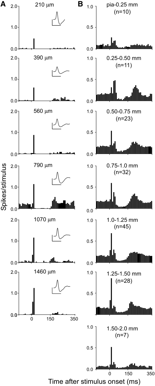
Consistent with previous results (Farkas et al. 1999), the MI responses to electrical stimulation consisted of a brief excitatory response that was followed by a long period of inhibition and then a secondary period of excitation. As shown by Fig. 2, the initial excitation gradually increased at successive depths of MI and reached a maximum at sites located 1.25 to 1.5 mm below the cortical surface. The population PSTHs in Fig. 2B suggest that the initial excitation might consist of two successive waves of activation, but these dual peaks were due to variations in the latency or onset of the first excitatory response. Hence, the latency of MI responses to electrical stimulation had a bimodal distribution in which most (n = 91) MI neurons responded within 20 ms of stimulus onset, but some (n = 40) did not respond until 30–50 ms after stimulus onset.
FIG. 2.
Responses of MI neurons to electrical stimulation (2 ms biphasic pulse, 250–300 μA) of the contralateral whisker pad. A: PSTHs of neurons encountered at specific depths in a single penetration through MI. Mean neuronal waveforms shown in insets; scales: 500 μV, 1 ms. B: mean PSTHs for the responsive MI neurons recorded in each depth range. Trials: 50; bin widths: 10 ms.
We also performed a laminar analysis of the MI responses to mechanical whisker deflections. Groups of whiskers were repetitively deflected at frequencies of 2, 5, and 8 Hz on each trial. Because the interval between each frequency lasted 1,000 ms, the first stimulus administered in the 5- and 8-Hz blocks was equivalent to a 1-Hz stimulus. Likewise, because the intertrial interval and subsequent prestimulus period lasted about 2,000 ms, the first stimulus in the 2-Hz block was equivalent to a stimulus rate of 0.5 Hz. Therefore responses to the first stimulus in each block of frequencies were grouped together (i.e., 0.5–1 Hz). By comparison, responses produced by the last seven stimuli in each frequency block indicate the effects of stimulation at 2, 5, or 8 Hz.
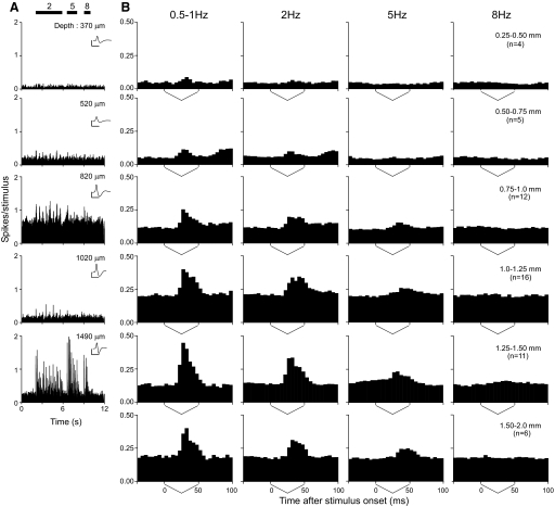
The magnitude of the MI responses to mechanical stimulation varied as a function of cortical depth. As shown in Fig. 3, MI responses to whisker deflections were not reliably observed until the electrodes reached a depth of 750 μm and maximum responses were recorded at depths between 1.25 and 1.5 mm (see Fig. 3B). At these depths, most MI neurons responded to whisker deflections at ≤2 Hz, but responded noticeably less when the frequency increased to 5 or 8 Hz.
FIG. 3.
Responses of MI neurons to repeated mechanical deflections of the contralateral whiskers. A: PSTHs of neurons encountered at specific depths in a single penetration through MI cortex. Multiple whiskers were deflected simultaneously at 2, 5, and 8 Hz as indicated by horizontal bars. Trials: 100; bin widths: 10 ms; waveform scales: 500 μV, 1 ms. B: mean population response produced by each stimulus cycle as a function of cortical depth and stimulus frequency. The responses to the first stimulus in the 2-, 5-, and 8-Hz sequences were grouped together to calculate the mean responses for 0.5–1 Hz; mean responses shown at 2, 5, and 8 Hz represent the effects of the last 7 stimuli in each block of frequencies. Stimulus duration indicated by the sawtooth profile below each PSTH. Bin widths: 5 ms.
The relationship between cortical depth and MI responsiveness during electrical and mechanical stimulation is summarized in Fig. 4. For both types of stimulation, the proportion of responsive neurons was greatest at depths <1.0 mm (Fig. 4A). Because electrical stimulation had a bimodal latency distribution (see preceding text), the effects of cortical depth on response magnitudes and latencies were analyzed only for neurons that responded within 20 ms of stimulus onset. This ensured that we determined which layers in MI cortex received the most rapid whisker inputs. Furthermore, many MI neurons did not respond to mechanical stimulation at 5 or 8 Hz, and the laminar comparison of response magnitudes and latencies was based only on MI responses to the first stimulus in each trial (i.e., an effective frequency of 0.5 Hz). Mean response magnitudes for electrical and mechanical stimulation were greatest for neurons recorded at depths between 1.0 and 1.5 mm (see Fig. 4B). An ANOVA confirmed that cortical depth was a significant factor in MI responsiveness during mechanical (F = 2.58, P < 0.05) and electrical stimulation (F = 2.35, P < 0.05).
FIG. 4.
Laminar analysis of MI responsiveness to electrical and mechanical whisker stimulation. A: MI neurons responsive to electrical (hatched) and mechanical (solid) whisker stimulation expressed as a percentage of total neurons sampled in each depth range. B: laminar distribution of response magnitudes of MI neurons to electrical and mechanical stimulation. C: laminar distribution of MI response latencies to electrical and mechanical whisker stimulation. Response magnitudes and latencies for mechanical stimulation were based on stimulation at 0.5 Hz. Brackets indicate SE.
Our laminar analysis of the MI responses, however, failed to reveal a significant effect of cortical depth on response latency for both types of stimulation (Fig. 4C). Thus an ANOVA on the effect of cortical depth on response latencies during electrical stimulation (n = 81) was insignificant (F = 1.82, P = 0.10). Similarly, the mean response latency to mechanical stimulation was shortest for neurons recorded at depths of 1.25 to 1.5 mm (see Fig. 4C), but this result was not statistically significant (F = 2.03, P = 0.10).
Comparison of MI and SI responses
Mechanical stimulation with a moving window screen at 2, 5, and 8 Hz was used to compare neuronal responses recorded simultaneously in MI and SI barrel cortex of five rats. For this analysis, SI and MI neuronal discharges were amplified and passed through an audio monitor to identify neurons that responded to all three frequencies of stimulation. Thus a total of 8 barrel neurons, 14 septal neurons, and 19 MI neurons were recorded that satisfied this criterion. Based on the temporal duration of the isolated neuronal waveforms (Bruno and Simons 2002; Zhang and Alloway 2004), neurons in which the total duration of depolarization and hyperpolarization exceeded 1.0 ms were classified as regular-spiking units (RSUs), whereas the rest were classified as fast-spiking units (FSUs). All 19 neurons recorded from MI cortex were RSUs. Of the 8 neurons recorded in the layer IV barrels, 5 were RSUs and 3 were FSUs. Among the 14 neurons recorded in the layer IV septa, 12 were RSUs and only 2 were classified as FSUs. These results are summarized in Table 1.
TABLE 1.
Location of regular- and fast-spiking units
| Brain Region | Total Number of Neurons | Regular-Spiking Units (RSUs) | Fast-Spiking Units (FSUs) |
|---|---|---|---|
| SI layer IV barrels | 8 | 5 | 3 |
| SI layer IV septa | 14 | 12 | 2 |
| MI | 19 | 19 | 0 |
In these experiments, the MI electrode was always placed at cortical depths of ≥1.0 mm because MI neurons at these depths received the strongest sensory inputs (see Fig. 4). The SI neurons were recorded at depths ranging from 400 to 950 μm, which corresponds to layer IV. Receptive field mapping was used to characterize the SI neurons. Those in which a principal whisker was identified were tentatively classified as being in barrels, whereas those that responded equally to multiple whiskers were classified as being in septa. Such classifications were confirmed by histological examination of electrode tracks and microlesions. Layer IV was chosen because the barrels and septa receive dense inputs from VPM and POm, respectively (Chmielowska et al. 1989; Koralek et al. 1988; Lu and Lin 1993).
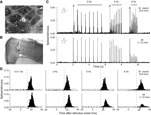
Representative examples of SI neurons recorded simultaneously with MI responses are shown in Figs. 5 and 6. Regardless of whether the SI neuron was in a barrel or the septa, response latencies were always shorter than those of simultaneously recorded MI neurons. All neurons showed some increases in response latencies as stimulus frequency increased, but the increases were much greater for the MI and SI septal neurons than for the neurons in the SI barrels. In Fig. 5, for example, the response latencies of the barrel neuron increased from 12 ms at the lowest stimulus frequency (0.5–1 Hz) to 15 ms at the highest frequency (8 Hz). Because this particular neuron was located on the edge of a barrel, it may have received inputs from adjacent septal areas. Indeed, septal neurons in layer IV have been shown to project to adjacent barrel neurons (Kim and Ebner 1999). By comparison, the MI response latency increased from 14 to 22 ms for the same change in stimulus frequency. The septal neuron illustrated in Fig. 6 showed an increase in latency from 10 to 16 ms as stimulus frequency increased from 0.5–1 to 8 Hz. In addition, the MI neuron illustrated in Fig. 6 showed a similar increase in latency, shifting from 16 ms at 0.5–1 Hz to 24 ms at 8 Hz.
FIG. 5.
Simultaneous responses of SI barrel and MI neurons during 300 trials of mechanical whisker deflections at 2, 5, and 8 Hz. A: tangential section through layer IV of SI barrel cortex shows a recording site (asterisk) on the edge of barrel C1. Scale: 250 μm. B: coronal section from the same animal shows the MI recording site (arrowhead). Scale: 500 μm. C: PSTHs of the SI and MI neuronal responses; electrode recording depths indicated in parentheses. Bin widths: 10 ms; waveform scales: 1 mV, 1 ms. Horizontal bars indicate duration of stimulus at 2, 5, and 8 Hz. Arrows indicate stimulus presentations at an effective frequency of 0.5–1 Hz. D: mean responses of the same neurons to each stimulation frequency. Horizontal dashed lines indicate 99% confidence intervals; bin widths: 1 ms. Vertical dashed lines indicate the onset latency of the barrel neuron; arrowheads indicate MI onset latency.
FIG. 6.
Simultaneous responses of SI septal and MI neurons shown as in Fig. 5. A: recording site in the SI septa. Scale: 250 μm. B: recording site in MI cortex. Scale: 500 μm. C: PSTHs of the SI and MI neuronal responses. Bin widths: 10 ms; waveform scales: 1 mV, 1 ms. D: mean responses to each stimulation frequency. Bin widths: 1 ms.
These findings are reminiscent of reports indicating that response latencies in POm and its SI cortical targets become longer with increasing frequency of whisker stimulation (Ahissar et al. 2001; Sosnik et al. 2001). Given that MI receives substantial inputs from POm (Aldes 1988; Alloway et al. 2004; Chakrabarti and Alloway 2006; Cicirata et al. 1986; Hoffer and Alloway 2001), we compared the mean response latencies of barrel, septal, and MI neurons as a function of different stimulus frequencies. A summary of this analysis for SI barrel (n = 8), septal (n = 14), and MI (n = 19) neurons is shown in Fig. 7. For each stimulus frequency analyzed, the MI neurons always responded after the SI neurons. Furthermore, the mean increase in response latencies of barrel neurons, as stimulus frequency increased from low (0.5–1 Hz) to high (8 Hz), was only 1.63 ± 1.48 ms (mean ± SE), which was much smaller than the increases in latency observed in the septal (4.61 ± 2.13 ms) or MI (5.5 ± 1.72 ms) neurons. ANOVA confirmed that the frequency of stimulation had a significant effect on the latency of responses recorded in MI (F = 9.17; P < 0.0001) and SI septa (F = 3.35; P < 0.05), but these frequency effects were not significant for SI neurons recorded in the layer IV barrels (F = 1.74; P = 0.18).
FIG. 7.
Mean response latencies of neurons in SI barrels (n = 8), septa (n = 14), and MI (n = 19) at different frequencies of mechanical whisker stimulation.
Cross-correlation analysis of SI and MI responses
In addition to latency comparisons, the SI and MI neurons recorded simultaneously during mechanical stimulation were also subjected to cross-correlation analysis. A total of 8 barrel neurons were recorded with 5 MI neurons to yield 8 barrel–MI neuron pairs for cross-correlation analysis. The unequal neuronal sample in each region occurred because we used two electrodes to record simultaneously from two separate SI barrels in one of the five animals and we were able to isolate two different neuronal waveforms on one of these SI electrodes. By comparison, 14 septal neurons were recorded simultaneously with 14 MI neurons. In some cases, however, two neurons were recorded on a single electrode in MI and/or SI, thereby producing 22 septa–MI neuron pairs for cross-correlation analysis. Each of the 36 neurons subjected to cross-correlation analysis displayed significant responses to all 24 stimulus presentations (8 stimuli at 3 frequencies). Cross-correlation analysis of stimulus-induced responses was conducted only on discharges occurring within 10–60 ms of each whisker deflection onset. Analysis of spontaneous activity was based on an equal number of 50-ms segments recorded during the prestimulus period (see methods).
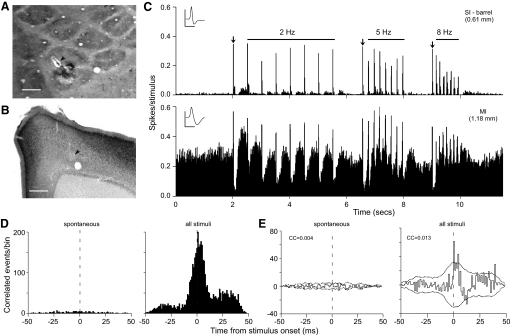
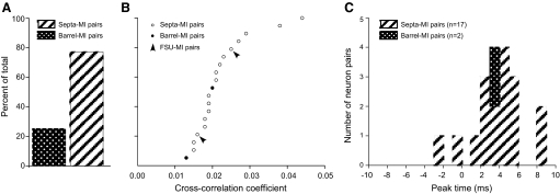
Of the 8 barrel–MI neuron pairs subjected to cross-correlation analysis, only 2 (25%) showed coordinated activity that exceeded the 99% confidence intervals in both the raw and smoothed shift-corrected CCGs. In both of these neuron pairs, the barrel neuron was an RSU. One of these neuron pairs is shown in Fig. 8. As this figure indicates, the tallest peak in the CCG was located to the right of time 0, which indicates that the MI discharges were most likely to follow the SI discharges. The cross-correlation coefficient for this particular neuron pair was <2% (see Fig. 8E).
FIG. 8.
Example of weak coordination between a barrel neuron and an MI neuron. A: tangential section through layer IV of SI barrel cortex shows the recording site (arrowhead) in barrel D1. Scale: 250 μm. B: coronal section from the same animal shows the MI recording site (arrowhead). Scale: 500 μm. C: PSTHs of SI and MI responses recorded at specific depths. Bin widths: 10 ms; waveform scales: 1 mV, 1 ms. D: raw cross-correlograms (CCGs) show changes in MI activity as a function of SI barrel discharges at time 0, during the prestimulus spontaneous period and subsequent whisker stimulation periods at 2, 5, and 8 Hz. The second CCG (all stimuli) is based on responses to all 24 stimulus presentations. E: shift-corrected CCGs for the same neuron pair based on spontaneous and all stimulus-induced discharges. Dotted lines indicate 99% confidence intervals. CC, correlation coefficient; bin widths: 1 ms.
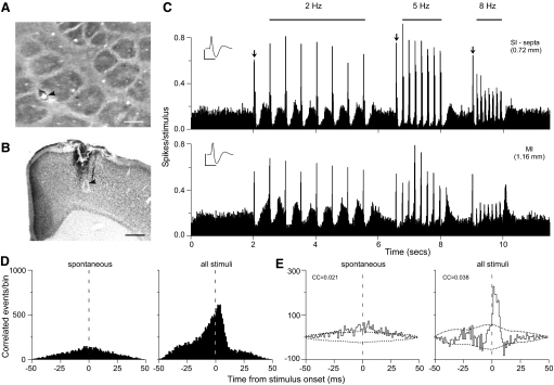
By comparison, 17 of the 22 (77.3%) septa–MI neuron pairs exhibited significant correlations. In 15 of these septa–MI neuron pairs, the septal neuron was an RSU; in the remaining 2 neuron pairs, the septal neuron was an FSU. As illustrated by the example in Fig. 9, layer IV septal neurons show high amounts of coordination with the MI response. For this septal–MI neuron pair, correlated activity was observed during both spontaneous and stimulus-induced activity, but the cross-correlation coefficient was higher during whisker stimulation (see Fig. 9E). Whereas the CCG for spontaneous activity was characterized by a broad shallow peak, the CCG for stimulus-induced activity had a tall, narrow peak located to the right of time 0. Hence, SI and MI activity was loosely synchronized in the absence of whisker stimulation and whisker deflections caused a noticeable increase in the number of time-locked SI and MI events.
FIG. 9.
Example of coordination between a septal neuron and an MI neuron shown as in Fig. 8. A: recording site in the SI septa. Scale: 250 μm. B: recording site in MI cortex. Scale: 500 μm. C: PSTHs of the SI and MI neuronal responses. Bin widths: 10 ms; waveform scales: 1 mV, 1 ms. D: raw CCGs show spontaneous and stimulus-induced coordination of SI septal neuron and the MI neuron. E: shift-corrected CCGs for the same neuron pair. Bin widths: 1 ms.
A summary of the strength and timing of SI–MI coordination during whisker stimulation appears in Fig. 10. Only 25% of the barrel–MI neuron pairs showed correlated activity, but nearly 80% of the septa–MI neuron pairs had correlated activity. A goodness-of-fit test indicated that this association was significant (χ2 = 6.90, P < 0.01). The correlation coefficients of the 19 neuron pairs (17 septa–MI and 2 barrel–MI) that displayed significant amounts of coordination are shown in a cumulative distribution plot in Fig. 10B. Whereas >50% of the septa–MI neuron pairs had correlation coefficients that varied from 2 to 5%, the barrel–MI neuron pair with the strongest coordination had a correlation coefficient that was only 2%. The distribution of the CCG peak times indicates that 14 of these 19 neuron pairs had coordination patterns in which the MI neuron was most likely to discharge 2–6 ms after the SI neuron (see Fig. 10C).
FIG. 10.
Quantitative summary of barrel–MI and septa–MI coordination. A: percentage of total barrel–MI and septa–MI neuron pairs with significant stimulus-induced correlations. B: cumulative frequency distribution of the cross-correlation coefficients of coordinated barrel–MI and septa–MI neuron pairs. The 2 neuron pairs that contained a fast-spiking unit (FSU) are indicated with arrowheads. C: peak times of shift-corrected CCGs constructed from all 24 stimulus periods.
Cross-correlation analysis also revealed changes in the pattern of septa–MI coordination as a function of stimulus frequency. To assess the effects of stimulus frequency on the strength and temporal precision of neural coordination, we analyzed only those neuron pairs that exhibited significant CCG peaks for each frequency block. A total of six such neuron pairs showed significant amounts of correlated activity for all four stimulus frequencies. In all six neuron pairs, the SI neuron was located in the layer IV septa. As shown in Fig. 11, these six neuron pairs were used to construct population CCGs for different stimulus frequencies. The CCGs for 0.5–1 Hz were computed only from the responses to the first stimulus in each frequency block. The CCGs for spontaneous activity were computed using seven disconnected 50-ms periods of neural activity recorded during the 2,000-ms prestimulus portion of each trial. Each CCG was normalized with respect to the maximum correlated activity for each neuron pair as a function of each stimulus, and then were averaged to yield the population CCGs in Fig. 11.
FIG. 11.
Effect of stimulus frequency on septa–MI coordination. Top and bottom rows depict the raw and shifted-corrected CCGs, respectively, for 6 neuronal pairs that displayed significant correlations during each frequency. Each CCG was constructed by normalizing each neuron pair to the maximum frequency response of pair, dividing by the number of stimuli, and then summing across neuron pairs to produce a population CCG. Each CCG lists the mean correlation coefficient. Dotted lines indicate 99% confidence intervals; bin widths: 1 ms.
The peaks of the population CCGs for each stimulus frequency exceeded the 99% confidence intervals, but the population CCG for spontaneous activity revealed only minor sporadic fluctuations above the confidence limits. The peaks for each of the frequency-specific CCGs, however, were noticeably taller than the weak coordination displayed in the spontaneous CCG. Furthermore, the mean correlation coefficient for each frequency-specific CCG was greater than the mean correlation coefficient for spontaneous activity.
As shown by both the raw and shift-corrected CCGs in Fig. 11, the tallest peaks of correlated activity always occurred 1–3 ms after time 0, irrespective of stimulus frequency. Some aspects of septa–MI coordination, however, were modulated by increasing the stimulus frequency. At low frequencies (≤2 Hz), the raw CCGs (top panels) had tall asymmetric peaks in which the majority of the near-coincident events were located just to the right of time 0. When the whiskers were stimulated at 8 Hz, however, the raw CCG contained a broad peak centered around time 0, indicating that the neurons were loosely synchronized. Hence, this frequency-dependent shift in temporal relationship suggests that the serial relationship between SI septa and MI neural activity during low frequencies of whisker stimulation is replaced by greater amounts of synchronous septa–MI discharges when stimulus frequency increases to 8 Hz.
DISCUSSION
We used multiple approaches to compare sensory responses in the SI and MI whisker regions in the anesthetized rat. Inactivation of somatosensory cortex reduced whisker-related responses in MI by about 80%, an indication of the importance of SI for transmitting sensory information to MI. The strongest MI responses were located at cortical depths of ≥1.0 mm and these responses always occurred after neuronal responses in SI barrel cortex. Furthermore, MI neurons exhibited increases in response latency as stimulus frequency increased, especially from 5 to 8 Hz. A similar increase in response latencies was observed in the SI septa, but this pattern was virtually absent in the SI barrels. Consistent with our previous hypothesis (Alloway 2008), cross-correlation analysis revealed that MI neurons are more strongly coordinated with the SI septa than with the SI barrels. We also observed changes in the temporal distribution of correlated activity in the septa and MI as stimulus frequencies increased to 8 Hz.
Paralemniscal pathway transmits sensory information to MI
Some data suggest that the lemniscal and paralemniscal processing streams extract and convey separate aspects of whisker-related information (Yu et al. 2006). Lemniscal regions, such as the nucleus VPM and the layer IV barrels, are characterized by a high degree of topography and finely tuned receptive fields that should enable encoding of detailed textural information (Alloway 2008; Chiaia et al. 1991b; Yu et al. 2006). By contrast, neurons in the POm nucleus have large receptive fields and may depend on corticothalamic feedback to exhibit maximum responsiveness to whisker stimulation (Chiaia et al. 1991b; Diamond et al. 1992b). Some reports indicate that this paralemniscal nucleus responds to higher rates of whisker stimulation with increasingly longer response latencies and this pattern has prompted the hypothesis that the rate of activity in POm may encode the frequency of self-generated whisker movements (Ahissar et al. 2000; Sosnik et al. 2001). One recent study, however, disputes this view (Masri et al. 2008).
Several findings in the present study suggest that whisker-related responses in MI cortex depend on inputs from the paralemniscal circuits. Consistent with the phase-locked loop hypothesis (Ahissar et al. 2000), responses in MI cortex showed increases in response latencies when stimulus frequencies increased to 8 Hz. We also observed a similar increase in the response latencies of the SI septal neurons. In addition, MI discharges were strongly correlated with neuronal discharges in the layer IV septa but not with neuronal activity in the barrels. This is remarkable because the neurons in the layer IV septa do not project to MI (Alloway et al. 2004). Instead, the SI projections to MI originate from extragranular neurons that are vertically aligned with the layer IV septa (Alloway et al. 2004). Furthermore, we also observed significant interactions between MI and septal FSUs, which probably represent inhibitory interneurons. Presumably, the SI neurons that actually project to MI would show even stronger amounts of SI–MI coordination than we reported here.
Our results indicate that coordinated activity between SI and MI can be observed among neurons that are not monosynaptically connected but are tightly embedded in the same neural circuit. The septa–MI coordination undoubtedly reflects the flow of information transmitted by SI neurons that are vertically aligned with the septa and have long-range projections to the MI whisker region. Relevant to our finding that coordination may appear among neurons that are not directly connected, similar results have been demonstrated in earlier primate studies. Thus somatosensory area 3b and motor area 4 in the primate are not directly interconnected (Jones et al. 1978), but may exhibit synchronized responses during manipulative tasks that require attention (Murthy and Fetz 1996a,b). Although we did not observe much coordination between MI and the SI barrels, it is conceivable that even the barrels might become coordinated with MI during states in which the rat is attentive.
Neural substrate for sensorimotor transformation
The afferent and efferent connections of MI cortex suggest that it uses sensory information from the whiskers to modulate the motor commands that control whisker movements. Descending projections from MI cortex terminate in brain stem regions that control facial nerve activity (Grinevich et al. 2005; Hattox et al. 2002). Consistent with this, intracranial microstimulation of MI produces short-latency whisker twitches (Brecht et al. 2004; Gioanni and Lamarche 1985; Hall and Lindholm 1974). In fact, prolonged stimulation of some caudal sites in MI can elicit sustained oscillatory movements of the whiskers (Haiss and Schwarz 2005).
Afferent whisker information may reach MI cortex by three possible routes. Projections from somatosensory cortex (SI and SII) represent a corticocortical route by which the somatosensory system may convey whisker information to MI (Alloway et al. 2004; Hoffer et al. 2003; Izraeli and Porter 1995). The other two routes involve the nucleus POm of the thalamus, which projects directly to the MI whisker region (Aldes 1988; Cicirata et al. 1986; Hoffer and Alloway 2001). Thus the SpVi nucleus projects to POm (Erzurumlu and Killackey 1980; Veinante et al. 2000) and the projection from POm represents the shortest route for conveying whisker information directly to MI (i.e., SPVi to POm to MI). Some data, however, indicate that POm responses are greatly reduced if SI is inactivated (Diamond et al. 1992a). Hence, optimal activation of POm and its projections to MI may depend on corticothalamic inputs from somatosensory cortex (Killackey and Sherman 2003).
Our data indicate that neurons in the deep layers of MI cortex (1.0 mm and deeper) displayed the best responses to peripheral whisker stimulation. Large pyramidal neurons in the deep layers of MI cortex project to a wide variety of subcortical nuclei (Gao and Zheng 2004; Groos et al. 1978; McGeorge and Faull 1989) and many of these neurons receive direct inputs from SI cortex in rats (Porter 1996). More recently we have shown that projections from SI barrel cortex terminate in all layers of the MI whisker region, but coronal reconstructions of these projection patterns indicate that labeling density is somewhat higher in the deeper layers of MI (see Fig. 4 in Hoffer et al. 2005). Thus MI responsiveness in the deep layers appears consistent with anatomical data indicating denser SI inputs to these layers.
Contributions of POm and somatosensory cortex
In the present study, reversible inactivation of somatosensory cortex reduced the magnitude of MI responses to whisker stimulation by 80–85%. Presumably, the small remaining response that persists in MI during inactivation of SI and SII represents whisker information that ascends the paralemniscal pathway and is conveyed directly to MI via synaptic relays in the nucleus POm. Consistent with this view, a previous report showed that ablation of somatosensory cortex did not completely block the MI response produced by electrical stimulation of the peripheral whisker pad (Farkas et al. 1999).
The large reduction in MI responsiveness during inactivation of somatosensory cortex does not mean that the paralemniscal pathway and its synaptic targets in POm play a minor role in transmitting whisker information to MI cortex. Cortical inactivation, which interferes with two of the three sensory routes to MI, indicates that the POm–MI route by itself has little influence on MI responsiveness, but the contribution of each of the three routes may depend on dynamic interactions that are not easily parsed by the changes that occur when one part of the circuit is inoperative. As whisker stimulation increases to higher frequencies, our data indicate that the raw timing of septal and MI discharges shifts from a serial to a more synchronous relationship. The increase in SI–MI synchronization at higher stimulus frequencies suggests that the direct projections from POm may exert a greater influence on MI as the frequency of whisker stimulation increases.
In summary, our results and previous studies suggest that the paralemniscal pathway has a strong influence on whisker-related responses in MI cortex. In addition to direct projections from POm to MI (Aldes 1988; Chakrabarti and Alloway 2006; Cicirata et al. 1986; Hoffer and Alloway 2001), the SI projections to MI originate from columns of neurons aligned with the septa (Alloway et al. 2004; Chakrabarti and Alloway 2006; Olavarria et al. 1984) and the layer IV septa receive their thalamic inputs largely from POm (Chmielowska et al. 1989; Koralek et al. 1988; Lu and Lin 1993). Analysis of local projections in SI has revealed a columnar organization that facilitates interactions among populations of septa-related neurons (Alloway 2008; Kim and Ebner 1999; Shepherd and Svoboda 2005). Collectively, these facts indicate that MI has strong functional connections with POm and the SI septa, both of which represent parts of the paralemniscal pathway.
Technical considerations
A previous study of sensory signals in MI reported a “weak sinusoidal modulation of the spike rate” that represented the fundamental frequencies of whisker movements across a range of SI stimulus frequencies (Kleinfeld et al. 2002). In our experiments, however, we observed clear responses at 2, 5, and 8 Hz. In contrast to the previous study in which rats were awake, our rat preparation was deeply anesthetized. When we reduced anesthesia, the rate of spontaneous activity in MI increased to levels that interfered with the detection of response latencies and other comparisons between MI and SI. Thus our use of anesthesia may account for this difference.
GRANTS
This work was supported by National Institute of Neurological Disorders and Stroke Grants NS-37532 and NS-052689.
Acknowledgments
We thank A. Petticoffer for technical assistance with some of the animal experiments.
Present address of M. Zhang: Department of Neuroscience and Pharmacology, University of Copenhagen, Blegdamsvej 3, DK-2200 Copenhagen, Denmark.
The costs of publication of this article were defrayed in part by the payment of page charges. The article must therefore be hereby marked “advertisement” in accordance with 18 U.S.C. Section 1734 solely to indicate this fact.
REFERENCES
- Aertsen et al. 1989.Aertsen AMHJ, Gerstein GL, Habib MK, Palm G. Dynamics of neuronal firing correlation: modulation of “effective connectivity.” J Neurophysiol 61: 900–917, 1989. [DOI] [PubMed] [Google Scholar]
- Ahissar and Kleinfeld 2003.Ahissar E, Kleinfeld D. Closed-loop neuronal computations: focus on vibrissa somatosensation in rat. Cereb Cortex 13: 53–62, 2003. [DOI] [PubMed] [Google Scholar]
- Ahissar et al. 2001.Ahissar E, Sosnik R, Bagdasarian K, Haidarliu S. Temporal frequency of whisker movement. II. Laminar organization of cortical representations. J Neurophysiol 86: 354–367, 2001. [DOI] [PubMed] [Google Scholar]
- Ahissar et al. 2000.Ahissar E, Sosnik R, Haidarliu S. Transformation from temporal to rate coding in a somatosensory thalamocortical pathway. Nature 406: 302–306, 2000. [DOI] [PubMed] [Google Scholar]
- Ahissar and Zacksenhouse 2001.Ahissar E, Zacksenhouse M. Temporal and spatial coding in the rat vibrissal system. Prog Brain Res 130: 75–87, 2001. [DOI] [PubMed] [Google Scholar]
- Aldes 1988.Aldes LD Thalamic connectivity of rat somatic motor cortex. Brain Res Bull 20: 333–348, 1988. [DOI] [PubMed] [Google Scholar]
- Alloway 2008.Alloway KD Information processing streams in rodent barrel cortex: the differential functions of barrel and septal circuits. Cereb Cortex 18: 979–989, 2008. [DOI] [PubMed] [Google Scholar]
- Alloway et al. 1993.Alloway KD, Johnson MJ, Wallace MB. Thalamocortical interactions in the somatosensory system: interpretations of latency and cross-correlation analyses. J Neurophysiol 70: 892–908, 1993. [DOI] [PubMed] [Google Scholar]
- Alloway et al. 2004.Alloway KD, Zhang M, Chakrabarti S. Septal columns in rodent barrel cortex: functional circuits for modulating whisking behavior. J Comp Neurol 480: 299–309, 2004. [DOI] [PubMed] [Google Scholar]
- Alloway et al. 2002.Alloway KD, Zhang M, Dick SH, Roy SA. Pervasive synchronization of local neural networks in the secondary somatosensory cortex of cats during focal cutaneous stimulation. Exp Brain Res 147: 227–242, 2002. [DOI] [PubMed] [Google Scholar]
- Armstrong-James and Callahan 1991.Armstrong-James M, Callahan CA. Thalamo-cortical processing of vibrissal information in the rat. II. Spatiotemporal convergence in the thalamic ventroposterior medial nucleus (VPm) and its relevance to generation of receptive fields of S1 cortical “barrel” neurons. J Comp Neurol 303: 211–224, 1991. [DOI] [PubMed] [Google Scholar]
- Armstrong-James and Fox 1987.Armstrong-James M, Fox K. Spatiotemporal convergence and divergence in the rat S1 “barrel” cortex. J Comp Neurol 263: 265–281, 1987. [DOI] [PubMed] [Google Scholar]
- Bartho et al. 2002.Bartho P, Freund TF, Acsady L. Selective GABAergic innervation of thalamic nuclei from zona incerta. Eur J Neurosci 16: 999–1014, 2002. [DOI] [PubMed] [Google Scholar]
- Berg and Kleinfeld 2003.Berg RW, Kleinfeld D. Vibrissa movement elicited by rhythmic electrical microstimulation to motor cortex in the aroused rat mimics exploratory whisking. J Neurophysiol 90: 2950–2963, 2003. [DOI] [PubMed] [Google Scholar]
- Brecht et al. 2004.Brecht M, Krauss A, Muhammad S, Sinai-Esfahani L, Bellanca S, Margrie TW. Organization of rat vibrissa motor cortex and adjacent areas according to cytoarchitectonics, microstimulation, and intracellular stimulation of identified cells. J Comp Neurol 479: 360–373, 2004. [DOI] [PubMed] [Google Scholar]
- Brecht and Sakmann 2002.Brecht M, Sakmann B. Dynamic representation of whisker deflection by synaptic potentials in spiny stellate and pyramidal cells in the barrels and septa of layer 4 rat somatosensory cortex. J Physiol 543: 49–70, 2002. [DOI] [PMC free article] [PubMed] [Google Scholar]
- Bruno and Simons 2002.Bruno RM, Simons DJ. Feedforward mechanisms of excitatory and inhibitory cortical receptive fields. J Neurosci 22: 10966–10975, 2002. [DOI] [PMC free article] [PubMed] [Google Scholar]
- Castro-Alamancos 2006.Castro-Alamancos MA Vibrissa myoclonus (rhythmic retractions) driven by resonance of excitatory networks in motor cortex. J Neurophysiol 96: 1691–1698, 2006. [DOI] [PubMed] [Google Scholar]
- Chakrabarti and Alloway 2006.Chakrabarti S, Alloway KD. Differential origin of projections from SI barrel cortex to the whisker representations in SII and MI. J Comp Neurol 198: 624–636, 2006. [DOI] [PubMed] [Google Scholar]
- Chapin and Lin 1984.Chapin JK, Lin CS. Mapping the body representation in the SI cortex of anesthetized and awake rats. J Comp Neurol 229: 199–213, 1984. [DOI] [PubMed] [Google Scholar]
- Chiaia et al. 1991a.Chiaia NL, Rhoades RW, Bennett-Clarke CA, Fish SE, Killackey HP. Thalamic processing of vibrissal information in the rat. I. Afferent input to the medial ventral posterior and posterior nuclei. J Comp Neurol 314: 201–216, 1991a. [DOI] [PubMed] [Google Scholar]
- Chiaia et al. 1991b.Chiaia NL, Rhoades RW, Fish SE, Killackey HP. Thalamic processing of vibrissal information in the rat. II. Morphological and functional properties of medial ventral posterior nucleus and posterior nucleus neurons. J Comp Neurol 314: 217–236, 1991b. [DOI] [PubMed] [Google Scholar]
- Chmielowska et al. 1989.Chmielowska J, Carvell G, Simons DJ. Spatial organization of thalamocortical and corticothalamic projection system in the rat SmI barrel cortex. J Comp Neurol 285: 325–338, 1989. [DOI] [PubMed] [Google Scholar]
- Cicirata et al. 1986.Cicirata F, Angaut P, Cioni M, Serapide MF, Papale A. Functional organization of thalamic projections to the motor cortex. An anatomical and electrophysiological study in the rat. Neuroscience 19: 81–99, 1986. [DOI] [PubMed] [Google Scholar]
- Cramer and Keller 2006.Cramer NP, Keller A. Cortical control of a whisking central pattern generator. J Neurophysiol 96: 209–217, 2006. [DOI] [PMC free article] [PubMed] [Google Scholar]
- Diamond et al. 1992a.Diamond ME, Armstrong-James M, Budway MJ, Ebner FF. Somatic sensory responses in the rostral sector of the posterior group (POm) and in the ventral posterior medial nucleus (VPM) of the rat thalamus: dependence on the barrel field cortex. J Comp Neurol 319: 66–84, 1992a. [DOI] [PubMed] [Google Scholar]
- Diamond et al. 1992b.Diamond ME, Armstrong-James M, Ebner FF. Somatic sensory responses in the rostral sector of the posterior group (POm) and in the ventral posterior medial nucleus (VPM) of the rat thalamus. J Comp Neurol 318: 462–476, 1992b. [DOI] [PubMed] [Google Scholar]
- Donoghue and Wise 1982.Donoghue JP, Wise SP. The motor cortex of the rat: cytoarchitecture and microstimulation mapping. J Comp Neurol 212: 76–88, 1982. [DOI] [PubMed] [Google Scholar]
- Eggermont 1992.Eggermont JJ Neural interaction in cat primary auditory cortex: dependence on recording depth, electrode separation, and age. J Neurophysiol 68: 1216–1228, 1992. [DOI] [PubMed] [Google Scholar]
- Erzurumlu and Killackey 1980.Erzurumlu RS, Killackey HP. Diencephalic projections of the subnucleus interpolaris of the brainstem trigeminal complex in the rat. Neuroscience 5: 1891–1901, 1980. [DOI] [PubMed] [Google Scholar]
- Farkas et al. 1999.Farkas T, Kis Z, Toldi J, Wolff JR. Activation of the primary motor cortex by somatosensory stimulation in adult rats is mediated mainly by associational connections from the somatosensory cortex. Neuroscience 90: 353–361, 1999. [DOI] [PubMed] [Google Scholar]
- Gao et al. 2001.Gao P, Bermejo R, Zeigler HP. Whisker deafferentation and rodent whisking patterns: behavioral evidence for a central pattern generator. J Neurosci 21: 5374–5380, 2001. [DOI] [PMC free article] [PubMed] [Google Scholar]
- Gao et al. 2003.Gao P, Hattox AM, Jones LM, Keller A, Zeigler HP. Whisker motor cortex ablation and whisker movement patterns. Somatosens Mot Res 20: 191–198, 2003. [DOI] [PubMed] [Google Scholar]
- Gao and Zheng 2004.Gao WJ, Zheng ZH. Target-specific differences in somatodendritic morphology of layer V pyramidal neurons in rat motor cortex. J Comp Neurol 476: 174–185, 2004. [DOI] [PubMed] [Google Scholar]
- Gerstein and Perkel 1972.Gerstein GL, Perkel DH. Mutual temporal relationships among neuronal spike trains. Statistical techniques for display and analysis. Biophys J 5: 453–473, 1972. [DOI] [PMC free article] [PubMed] [Google Scholar]
- Gioanni and Lamarche 1985.Gioanni Y, Lamarche M. A reappraisal of rat motor cortex organization by intracortical microstimulation. Brain Res 344: 49–61, 1985. [DOI] [PubMed] [Google Scholar]
- Grinevich et al. 2005.Grinevich V, Brecht M, Osten P. Monosynaptic pathway from rat vibrissa motor cortex to facial motor neurons revealed by lentivirus-based axonal tracing. J Neurosci 25: 8250–8258, 2005. [DOI] [PMC free article] [PubMed] [Google Scholar]
- Groos et al. 1978.Groos WP, Ewing LK, Carter CM, Coulter JD. Organization of corticospinal neurons in the cat. Brain Res 143: 393–419, 1978. [DOI] [PubMed] [Google Scholar]
- Haiss and Schwarz 2005.Haiss F, Schwarz C. Spatial segregation of different modes of movement control in the whisker representation of rat primary motor cortex. J Neurosci 25: 1579–1587, 2005. [DOI] [PMC free article] [PubMed] [Google Scholar]
- Hall and Lindholm 1974.Hall RD, Lindholm EP. Organization of motor and somatosensory neocortex in the albino rat. Brain Res 66: 23–38, 1974. [Google Scholar]
- Hattox et al. 2002.Hattox AM, Priest CA, Keller A. Functional circuitry involved in the regulation of whisker movements. J Comp Neurol 442: 266–276, 2002. [DOI] [PMC free article] [PubMed] [Google Scholar]
- Hoffer and Alloway 2001.Hoffer ZS, Alloway KD. Organization of corticostriatal projections from the vibrissal representations in the primary motor and somatosensory cortical areas of rodents. J Comp Neurol 439: 87–103, 2001. [DOI] [PubMed] [Google Scholar]
- Hoffer et al. 2003.Hoffer ZS, Hoover JE, Alloway KD. Sensorimotor corticocortical projections from rat barrel cortex have an anisotropic organization that facilitates integration of inputs from whiskers in the same row. J Comp Neurol 466: 525–544, 2003. [DOI] [PubMed] [Google Scholar]
- Izraeli and Porter 1995.Izraeli R, Porter LL. Vibrissal motor cortex in the rat: connections with the barrel field. Exp Brain Res 104: 41–54, 1995. [DOI] [PubMed] [Google Scholar]
- Johnson and Alloway 1996.Johnson MJ, Alloway KD. Cross-correlation analysis reveals laminar differences in thalamo-cortical interactions in the somatosensory system. J Neurophysiol 75: 1444–1457, 1996. [DOI] [PubMed] [Google Scholar]
- Jones et al. 1978.Jones EG, Coulter JD, Hendry SH. Intracortical connectivity of architectonic fields in the somatic sensory, motor and parietal cortex of monkeys. J Comp Neurol 181: 291–347, 1978. [DOI] [PubMed] [Google Scholar]
- Killackey and Sherman 2003.Killackey HP, Sherman SM. Corticothalamic projections from the rat primary somatosensory cortex. J Neurosci 23: 7381–7384, 2003. [DOI] [PMC free article] [PubMed] [Google Scholar]
- Kim and Ebner 1999.Kim U, Ebner FF. Barrels and septa: separate circuits in rat barrels field cortex. J Comp Neurol 408: 489–505, 1999. [PubMed] [Google Scholar]
- Kleinfeld et al. 2002.Kleinfeld D, Sachdev RN, Merchant LM, Jarvis MR, Ebner FF. Adaptive filtering of vibrissa input in motor cortex of rat. Neuron 34: 1021–1034, 2002. [DOI] [PubMed] [Google Scholar]
- Koralek et al. 1988.Koralek KA, Jensen KF, Killackey HP. Evidence for two complementary patterns of thalamic input to the rat somatosensory cortex. Brain Res 463: 346–351, 1988. [DOI] [PubMed] [Google Scholar]
- Land and Simons 1985.Land PW, Simons DJ. Cytochrome oxidase staining in the rat SmI barrel cortex. J Comp Neurol 238: 225–235, 1985. [DOI] [PubMed] [Google Scholar]
- Lavallee et al. 2005.Lavallee P, Urbain N, Dufresne C, Bokor H, Acsady L, Deschênes M. Feedforward inhibitory control of sensory information in higher order thalamic nuclei. J Neurosci 25: 7489–7498, 2005. [DOI] [PMC free article] [PubMed] [Google Scholar]
- Lu and Lin 1993.Lu SM, Lin RC. Thalamic afferents of the rat barrel cortex: a light- and electron-microscopic study using Phaseolus vulgaris leucoagglutinin as an anterograde tracer. Somatosens Mot Res 10: 1–16, 1993. [DOI] [PubMed] [Google Scholar]
- Masri et al. 2008.Masri R, Bezdudnaya T, Trageser JC, Keller A. Encoding of stimulus frequency and sensor motion in the posterior medial thalamic nucleus. J Neurophysiol (January 30, 2008). doi: 10.1152/jn.01322.2007. [DOI] [PMC free article] [PubMed]
- McGeorge and Faull 1989.McGeorge AJ, Faull RL. The organization of the projection from the cerebral cortex to the striatum in the rat. Neuroscience 29: 503–537, 1989. [DOI] [PubMed] [Google Scholar]
- Murthy and Fetz 1996a.Murthy VN, Fetz EE. Oscillatory activity in sensorimotor cortex of awake monkeys: synchronization of local field potentials and relation to behavior. J Neurophysiol 76: 3949–3967, 1996a. [DOI] [PubMed] [Google Scholar]
- Murthy and Fetz 1996b.Murthy VN, Fetz EE. Synchronization of neurons during local field potential oscillations in sensorimotor cortex of awake monkeys. J Neurophysiol 76: 3968–3982, 1996b. [DOI] [PubMed] [Google Scholar]
- Neafsey et al. 1986.Neafsey EJ, Bold EL, Haas G, Hurley-Gius KM, Quirk G, Sievert CF, Terreberry RR. The organization of the rat motor cortex: a microstimulation mapping study. Brain Res 396: 77–96, 1986. [DOI] [PubMed] [Google Scholar]
- Olavarria et al. 1984.Olavarria J, Van Sluyters RC, Killackey HP. Evidence for the complementary organization of callosal and thalamic connections with rat somatosensory cortex. Brain Res 291: 364–368, 1984. [DOI] [PubMed] [Google Scholar]
- Peschanski 1984.Peschanski M Trigeminal afferents to the diencephalon in the rat. Neuroscience 12: 465–467, 1984. [DOI] [PubMed] [Google Scholar]
- Pierret et al. 2000.Pierret T, Lavallee P, Deschênes M. Parallel streams for the relay of vibrissal information through thalamic barreloids. J Neurosci 20: 7455–7462, 2000. [DOI] [PMC free article] [PubMed] [Google Scholar]
- Porter 1996.Porter LL Somatosensory input into pyramidal tract neurons in rodent motor cortex. Neuroreport 7: 2309–2315, 1996. [DOI] [PubMed] [Google Scholar]
- Rhoades et al. 1987.Rhoades RW, Belford GR, Killackey HP. Receptive field properties of rat ventral posterior medial neurons before and after selective kainic acid lesions of the trigeminal brain stem complex. J Neurophysiol 57: 1577–1600, 1987. [DOI] [PubMed] [Google Scholar]
- Roy and Alloway 1999.Roy SA, Alloway KD. Synchronization of local neural networks in the somatosensory cortex: a comparison of stationary and moving stimuli. J Neurophysiol 81: 999–1013, 1999. [DOI] [PubMed] [Google Scholar]
- Shepherd and Svoboda 2005.Shepherd GM, Svoboda K. Laminar and columnar organization of ascending excitatory projections to layer 2/3 pyramidal neurons in rat barrel cortex. J Neurosci 25: 5670–5679, 2005. [DOI] [PMC free article] [PubMed] [Google Scholar]
- Sosnik et al. 2001.Sosnik R, Haidarliu S, Ahissar E. Temporal frequency of whisker movement. I. Representations in brain stem and thalamus. J Neurophysiol 86: 339–353, 2001. [DOI] [PubMed] [Google Scholar]
- Trageser and Keller 2004.Trageser JC, Keller A. Reducing the uncertainty: gating of peripheral inputs by zona incerta. J Neurosci 24: 8911–8915, 2004. [DOI] [PMC free article] [PubMed] [Google Scholar]
- Urbain and Deschênes 2007.Urbain N, Deschênes M. Motor cortex gates vibrissal responses in a thalamocortical projection pathway. Neuron 56: 714–725, 2007. [DOI] [PubMed] [Google Scholar]
- Veinante et al. 2000.Veinante P, Jacquin MF, Deschênes M. Thalamic projections from the whisker-sensitive regions of the spinal trigeminal complex in the rat. J Comp Neurol 420: 233–243, 2000. [DOI] [PubMed] [Google Scholar]
- Weiss and Keller 1994.Weiss DS, Keller A. Specific patterns of intrinsic connections between representation zones in the rat motor cortex. Cereb Cortex 4: 205–214, 1994. [DOI] [PubMed] [Google Scholar]
- Wong-Riley 1979.Wong-Riley M Changes in the visual system of monocularly sutured or enucleated cats demonstrable with cytochrome oxidase histochemistry. Brain Res 171: 11–28, 1979. [DOI] [PubMed] [Google Scholar]
- Yu et al. 2006.Yu C, Derdikman D, Haidarliu S, Ahissar E. Parallel thalamic pathways for whisking and touch signals in the rat. PLoS Biol 4: 819–825, 2006. [DOI] [PMC free article] [PubMed] [Google Scholar]
- Zacksenhouse and Ahissar 2006.Zacksenhouse M, Ahissar E. Temporal decoding by phase-locked loops: unique features of circuit-level implementations and their significance for vibrissal information processing. Neural Comput 18: 1611–1636, 2006. [DOI] [PubMed] [Google Scholar]
- Zhang and Alloway 2004.Zhang M, Alloway KD. Stimulus-induced intercolumnar synchronization of neuronal activity in rat barrel cortex: a laminar analysis. J Neurophysiol 92: 1464–1478, 2004. [DOI] [PubMed] [Google Scholar]
- Zhang and Alloway 2006.Zhang M, Alloway KD. Intercolumnar synchronization of neuronal activity in rat barrel cortex during patterned airjet stimulation: a laminar analysis. Exp Brain Res 169: 311–325, 2006. [DOI] [PubMed] [Google Scholar]


