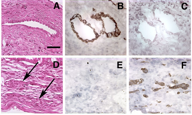
Figure 3.
Histological and immunohistochemical analysis of growth under the renal capsule. Growths arising from the implantation of 2 × 10⁵ MLE-ßgal cells and 5 × 10⁵ PE-L-1 cells (A to C) or PE-B-1 cells (D to F) were harvested after 60 days, and 5 μm sections were stained with hematoxylin and eosin (A and D). Sections were also immunostained with antibodies to cytokeratin 8 (B and E) or cytokeratin 5 (C and F). Arrows in D point out cords of epitheloid cells. Bar in A equals 50 μm. All panels have the same magnification.

