

Abstract
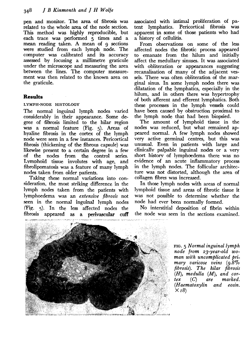
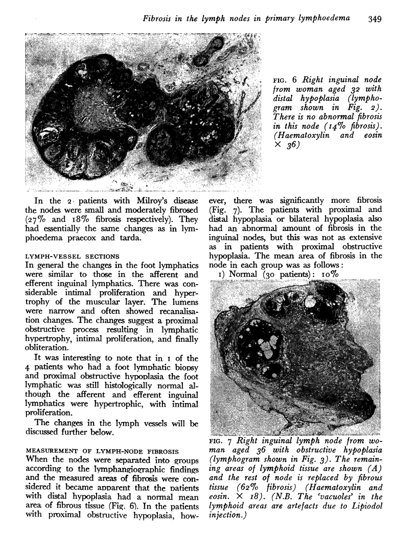
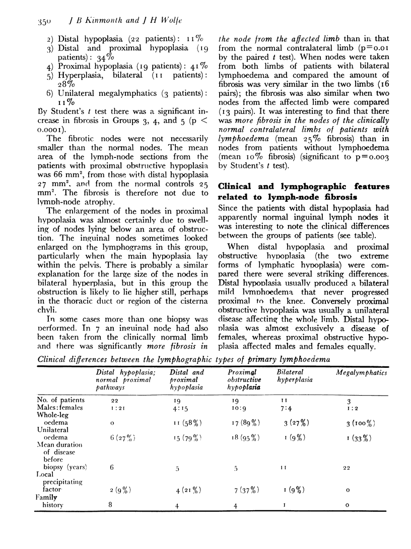
There are many different clinical and lymphographic groups of patients with primary lymphoedema. Improved lymphographic techniques have emphasised the importance of changes in the nodes as well as in the lymph vessels. A systematic histological study has been made of nodes removed during therapeutic operations or investigations on patients with primary lymphoedema. Many nodes showed a marked fibrotic process. This, in its distribution in the node and its histological appearance, was quite different from that which might have arisen from attacks of infection and inflammation. The majority of patients had no clinical history of such attacks. It may be regarded as primary fibrosis in the nodes. Associated clinical features suggest strong genetic or familial factors in its aetiology. Many of the changes found in the lymph vessels may follow obstructive effects from fibrosis in the nodes. The histological findings have been related to the clinical and lymphographic features in different types of primary lymphoedema. The degree of fibrosis and its distribution have important bearings on the prognosis, clinical course, and treatment of the patient.
Full text
PDF
Images in this article
Selected References
These references are in PubMed. This may not be the complete list of references from this article.
- Boggon R. P., Palfrey A. J. The microscopic anatomy of human lymphatic trunks. J Anat. 1973 Apr;114(Pt 3):389–405. [PMC free article] [PubMed] [Google Scholar]
- Hurst P. A., Kinmonth J. B., Rutt D. L. A gut and mesentery pedicle for bridging lymphatic obstruction: experimental studies. J Cardiovasc Surg (Torino) 1978 Nov-Dec;19(6):589–596. [PubMed] [Google Scholar]
- Kinmonth J. B., Hurst P. A., Edwards J. M., Rutt D. L. Relief of lymph obstruction by use of a bridge of mesentery and ileum. Br J Surg. 1978 Dec;65(12):829–833. doi: 10.1002/bjs.1800651202. [DOI] [PubMed] [Google Scholar]