

Abstract
The pathophysiologic basis for multiple myeloma (MM) has been attributed to the dysregulation of various paracrine or autocrine growth factor loops and to perturbations in several signal transduction pathways including IKK/NF-κB. The aim of the present study was to investigate the effect of a pharmaceutical IKK2 inhibitor, the anilinopyrimidine derivative AS602868, on the in vitro growth of 14 human MM cell lines (HMCL) and primary cells from 13 patients. Results show that using HMCL or primary MM cells, AS602868 induces a clear dose-dependent inhibition of MM cell growth, which is the result of a simultaneous induction of apoptosis and inhibition of the cell cycle progression. Combination of AS602868 with suboptimal doses of melphalan or Velcade showed an additive effect in growth inhibition of HMCL. AS602868 also induced apoptosis of primary myeloma cells. Importantly, AS602868 does not alter the survival of other bone marrow mononuclear cells (CD138−) co-cultured with primary MM (CD138+) cells, except on CD34+ hematopoietic stem cells. The results demonstrate the important role of NF-κB in maintaining survival of MM cells and suggest that a pharmacological inhibition of the NF-κB pathway by the AS602868 IKK2 inhibitor can efficiently kill myeloma cell lines and primary myeloma cells and therefore might represent an innovative approach for treating MM patients.
Keywords: Antineoplastic Agents, pharmacology, Antineoplastic Agents, Alkylating, pharmacology, Apoptosis, drug effects, Boronic Acids, pharmacology, Cell Division, drug effects, Cell Line, Tumor, Cell Survival, drug effects, Enzyme Inhibitors, pharmacology, Humans, I-kappa B Kinase, antagonists & inhibitors, Melphalan, pharmacology, Multiple Myeloma, physiopathology, NF-kappa B, drug effects, Pyrazines, pharmacology, Pyrimidines, pharmacology
Keywords: Multiple myeloma, NF-kappa B, IKK2 inhibitor, apoptosis, cell cycle
Introduction
Multiple myeloma (MM) is a B cell neoplasia, covering more than 10 % of total hematologic malignancies, which currently remains associated with a poor prognosis. This pathology is mainly characterized by the accumulation of clonal malignant plasma cells in the bone marrow.
Several transduction pathways are constitutively activated in patients’ primary multiple myeloma cells (MMC), particularly the Janus kinase (JAK)/signal transducer and activator of transduction 3 (STAT3) (Bharti, et al 2004; Catlett-Falcone, et al 1999; De Vos, et al 2000), the phosphatidylinositol-3 kinase (PI-3K)/Akt (Hsu, et al 2001), the mitogen-activated protein kinase (MAPK)/extracellular signal-regulated kinase (ERK) (Giuliani, et al 2004), Wnt/βcatenin(Derksen, et al 2004) and the nuclear factor kappa-B (NF-κB) pathways (Berenson, et al 2001; Bharti, et al 2003; Bharti, et al 2004; Mitsiades, et al 2002a; Ni, et al 2001). These pathways are activated by interactions with the microenvironment, plasma factors and a variety of MM growth factors (MGFs) (De Vos, et al 2006). The main MGFs are interleukin (IL)-6 and IL-6 receptor (Gaillard, et al 1997; Zhang, et al 1992), insulin-like growth factor-1 (IGF-1) (Ferlin, et al 2000), B-cell activating factor (BAFF)/a proliferation–inducing ligand (APRIL) (Moreaux, et al 2005; Moreaux, et al 2004), hepatocyte growth factor (HGF) (Derksen, et al 2003), the Wnt family (Derksen, et al 2004), the epidermal growth factor (EGF) family (Mahtouk, et al 2006; Mahtouk, et al 2005; Mahtouk, et al 2004), IL-10 (Gu, et al 1996), IFN-alpha (Ferlin-Bezombes, et al 1998), IL-15 (Hjorth-Hansen, et al 1999) and IL-21 (Brenne, et al 2002). Using human myeloma cell lines (HMCL), it was shown that serum and MGF cell starvation can downregulate these pathways (De Vos, et al 2006; Jourdan, et al 2000). Of note, no genetic alterations were shown to target these transduction pathways in patients with MM (Fonseca, et al 2004).
The NF-κB family includes NF-κB1 (or p50), RelA (or p65), c-Rel, NF-κB2 (or p52) and RelB proteins that constitute dimeric transcription factors that are triggered by the canonical NF-κB pathway (p50, p65 and/or c-Rel) or an alternative pathway (p52, RelB) (Karin and Lin 2002; Viatour, et al 2005). The canonical pathway implies the recruitment of adaptors proteins which is followed by the recruitment and activation of the IκB kinase (IKK) complex which includes IKKα (or IKK1) and IKKβ (or IKK2) kinases and a scaffold protein named IKKγ (or Nemo). In the cytoplasm, NF-κB associates with the inhibitory protein IκB. The IKK complex induces the phosphorylation of IκBα, which is ubiquitinated and degraded, allowing the migration of NF-κB to the nucleus and the transcription of target genes. In the alternative pathway, the IKK complex includes only IKKα homodimers and is Nemo-independent. In HMCL or primary myeloma cells, a constitutive activation of the canonical pathway was only investigated.
We show here that AS602868, a specific inhibitor of IKK2, induces growth arrest and apoptosis of HMCL and inhibits the survival of primary MMC. These results suggest that the AS602868 IKK2 inhibitor used alone or in combination with conventional drug therapies could be of therapeutic value for patients with myeloma.
Materials and methods
Cytokines and reagents
Human recombinant IL-6 was purchased from AbCys SA (Paris, France) and TNF-α from Peprotech (Rocky Hill, NJ, USA). AS602868 was provided by Serono International SA (Geneva, Switzerland). AS602868 is a specific ATP-competitive inhibitor of IKK2 which has been shown to block phosphorylation of IkB and subsequent NF-κB activation in various cell lines (Frelin, et al 2003) and to induce apoptosis of human acute myeloid leukaemia cells (Frelin, et al 2005). A stock solution of AS602868 was prepared in DMSO. For each experiment using AS602868. the control culture medium was culture medium supplemented with 0.33 % (v/v) DMSO. This was the highest DMSO concentration reached with the highest AS602868 concentration used in the experiments. This 0.33 % (v/v) DMSO concentration was not toxic for the 14 HMCLs or primary myeloma cells. Phycoerythrin-conjugated anti-CD34 and anti-CD138 monoclonal antibodies (mAbs) were obtained from BD Biosciences (San Jose, CA). Anti-IκBα and anti-Phospho-IκBα were obtained from Cell Signalling Technology (Beverly, MA).
HMCL
The IL-6-dependent myeloma cell lines XG-1, XG-2, XG-3, XG-4, XG-6, XG-7, XG-11, XG-12, XG-13, XG-14, XG-19 and XG-20 were obtained in our laboratory from 12 patients with terminal disease(Rebouissou, et al 1998; Tarte, et al 1999; Zhang, et al 1994). L363 and RPMI8226 myeloma cell lines were obtained from ATCC (Rockville, MD, USA). Cells were free of mycoplasma contamination as assayed using the Boehringer Mannheim kit of detection (Mannheim, Germany). All the XG cell lines, excepted XG-14, were routinely cultured with 2 ng/ml IL-6 in RPMI 1640 medium supplemented with 10% foetal calf serum (PCS), XG-14 was cultured in X-VIVO 20 medium (Biowhittaker, Walkersville, MD) supplemented with 2 ng/ml IL-6. L363 and RPMI8226 grew autonomously in RPMI 1640, 10% PCS.
Cell proliferation assay
The cells were washed twice, incubated in RPMI 1640 with 10% PCS for 3 hours at 37°C and washed again. Cells were then cultured for 4 days in 96 well flat-bottomed microtiter plates at 5 × 10³, 104 or 2 × 10⁴ cells/well, depending on the cell line used, in RPMI 1640/10% PCS and 2 ng/ml IL-6 with the indicated concentrations of inhibitors or drugs to be tested. Cultures were made in triplicate. Eight hours before the end of culture, 0.5 μCi of [3H]-thymidine (specific activity: 25 Ci/mM, Amersham Pharmacia Biotech, Orsay, France) was added per well and [3H]-thymidine incorporation determined as previously described (Mahtouk, et al 2004).
Short-term culture of primary myeloma cells
Bone marrow samples were collected from 13 myeloma patients (6 patients at diagnosis and 7 relapsing patients) after informed consent was given. The bone marrow mononuclear cells were isolated by centrifugation on Ficoll-hypaque medium. Mononuclear cells (5 × 10⁵ cells/ml) were cultured for 5 days in RPMI 1640/10 % FCS/2 ng/ml IL-6, without (control) or with the indicated concentration of AS602868 inhibitor. The median IC10, IC50 and IC90 concentrations of the inhibitor determined with the HMCL were used. The number of total viable cells was determined in each culture group by trypan blue staining at day 5 of culture. The cells were then stained with anti-CD138, which labels only plasma cells (Costes, et al 1999) and anti-CD34 antibodies and analysed with a FACScan flow cytometer using Cell Quest software (Becton Dickinson, Moutain View, CA, USA). The relative fractions of viable CD138 negative, CD138 and CD34 positive cells were determined and the number of cells calculated for each cell population.
Western blot analysis
HMCL were IL-6 starved for 3 hours in RPMI1640/2 % PCS, cultured with or without AS602868 (10 μM) for 30 minutes and then stimulated for 15 minutes with or without TNF-α (10 ng/ml). Cells were lysated in 10 mM Hepes (pH 7.9), 10 mM KCl, 0.1 mM EDTA, 0.1 mM EGTA, 0.1% NP-40, 100 μM Na3VO4, 1mM DTT, 100 μg/ml Pefabloc, 20 mM β-glycerophosphate, 10 mM p-nitrophenolphosphate (PNPP), 10 μg/mL aprotinin, 10 mM NaF, 1 μg/mL leupeptin, 0.5 mM PMSF and 0.5 μg/mL pepstatin. Aliquots containing 50 μg of total protein were resolved in 12% sodium dodecyl sulfate-polyacrylamide by gel electrophoresis (SDS-PAGE) and transferred to a nitrocellulose membrane (Schleicher and Schuell, Dassel, Germany). Membranes were blocked for 2h at room temperature in 140 mM NaCl, 3 mM KCl, 25 mM Tris-HCl (pH 7.4), 0.1% tween 20 (TBS-T), 5% non-fat milk), then incubated for 1h at room temperature with primary antibodies. The primary antibodies were visualized with goat anti-mouse (Biorad SA, Yvry Sur Seine, France) peroxydase-conjugated antibodies (at 1:10 000 dilution in TBS-T) using an enhanced chemiluminescence detection system. The membranes were stripped twice with 100 mM Glycin (pH 2.2), 0.1% NP 40 and 1% SDS for 30 minutes at room temperature.
Real-time RT-PCR
Total RNA was converted to cDNA using the Superscript II reverse transcriptase (RT; Invitrogen, Cergy Pontoise, France). The assays-on-demand primers and probes and the TaqMan Universal Master Mix were used according to the manufacturer’s instructions (Applied Biosystems, Foster City, CA, USA). Measurement of gene expression was performed using the ABI Prism 7000 Sequence Detection System. Quantitative PCR analysis was completed using ABI PRISM 7000 SDS Software. Ct values were collected for glyceraldehyde-3-phosphate deshydrogenase (GAPDH) and the genes of interest during log phase of the cycle. Gene of interest levels were normalized to GAPDH for each sample (δCt = Ct gene of interest − Ct GAPDH) and compared with the values obtained for a known positive control using the following formula 100/2δδCt where δδCt = δCt unknown − δCt positive control.
Apoptotic cell detection assay
Apoptotic cells were detected by using fluorescein isothiocyanate-labeled annexin V (FITC-annexin-V, Boehringer Mannheim). Annexin V has a high affinity for phosphatidylserine present on the outer cytoplasmic membrane of apoptotic cells (Vermes, et al 1995). Cells were washed, labelled with FITC-annexin-V according to the manufacturer’s recommendations and analyzed with a FACScan flow cytometer using Cell Quest software (Becton Dickinson).
Cell cycle analysis
The cell cycle distribution of the cell lines was assessed by flow cytometry, using propidium iodide (PI) and bromodeoxyuridine (BrdU) double-staining. Thirty minutes before stopping the culture, BrdU (10 μM) was added to the cultures, and then the cells were collected by centrifugation, washed twice in phosphate buffer saline (PBS) and fixed in 70% ethanol for 20 minutes at room temperature. After two washes with PBS, cells were resuspended in 50 μl of 3 N HCl/0.5% Tween 20 and incubated 20 minutes at 20°C to denature the DMA. The cells were then recovered by centrifugation, resuspended in 250 μl of 10 mM sodium tetraborate to neutralize the reaction, washed twice with PBS, 0.05% Tween 20, and incubated with 20 μl of anti-BrdU-FITC monoclonal antibody (BD Biosciences). After two additional washes, the cells were resuspended in 500 μl of PBS, 0.05% Tween 20 containing 10 μg/ml PI. The fluorescence of FL1-H (BrdU) and FL2-A (PI) were analyzed on a FACScan flow cytometer using Cell Quest software (Becton Dickinson).
Statistical analysis
Statistical comparisons were made with the non- parametric Mann-Whitney test or the Student t-test. The minimal level of significance was P < .05.
Results
AS602868 IKK2 inhibitor blocks NF-κB activation in human myeloma cell lines (HMCL)
In order to visualise the inhibitory effects of AS602868 on NF-κB activation in myeloma cells, two HMCL (XG-6 and XG-14) were first starved of IL-6 in low serum conditions to downregulate constitutive NF-κb activation, then stimulated with TNF-α. Phosphorylation of IκBα was assessed by Western blot analysis. As shown in Fig 1A, TNF-α induces a phosphorylation of IκBα in both HMCL which is completely abrogated by AS602868 IKK2 inhibitor. IL-6-induced STAT3 and ERK1/2 phosphorylation was not inhibited by AS602868 in either cell line (data not shown). Using real-time quantitative RT-PCR, we show that TNF-α induced an upregulation in the expression of a panel of NF-κB-inducible genes in XG-6 and XG-14 cells and that AS602868 strongly inhibited the TNF-α effect (Fig 1B).
Fig 1. Inhibition of NF-kB activation in HMCL by AS602868 IKK2 inhibitor.
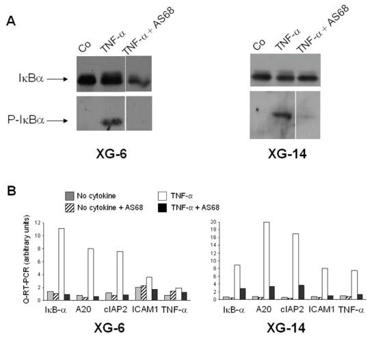
(A) Western blot analysis of IκBα activation. XG-6 and XG-14 cell lines were IL-6-starved in culture medium with 2 % FCS for 3 hours in order to reduce the constitutive NF-κB activation. Then, cells were pre-treated for 30 min_with or without AS602868 (AS68) (10 μM) before stimulation with TNF-α (10 ng/ml) for 15 min. Cells were harvested for Western blot analysis. Blots were probed with anti-phospho-IκBα and anti-IκBα antibodies.
(B) Gene expression analysis by quantitative RT-PCR (Q-RT-PCR). XG-6 and XG-14 HMCL were IL-6-starved to reduce constitutive NF-κB activation and pre-treated with or without AS602868 as in (A) before stimulation with TNF-α (10 ng/ml) for one hour. The relative gene RNA level was determined as described in Materials and Methods. The “1” value was assigned for each cell line to the control before pre-treatment with AS602868.
AS602868IKK2 inhibitor blocks the proliferation of human myeloma cell lines
Effects of increasing concentrations of AS602868 IKK2 inhibitor on the proliferation of 12 IL-6-dependent and 2 autonomously growing myeloma cell lines were examined. Results obtained with 10 representative cell lines are shown in Fig 2. The AS602868 concentrations yielding 50% (IC50) and 90% (IC90) inhibition of the proliferation of each cell line are indicated in Table 1. The AS602868 IKK2 inhibitor yielded a full inhibition of 13 out of 14 HMCL (IC90 was not reached for the RPMI8226 cell line) (Fig 2, Table 1). The median IC50 was 2.65 μM, ranging from 0.28 (XG-14) to 8.30 μM (RPMI8226) and the median IC90 was 8.79 μM, ranging from 0.76 (XG-14) to 14.43μM(XG-11).
Fig 2. Inhibition of the HMCL proliferation by AS602868 IKK2 inhibitor.
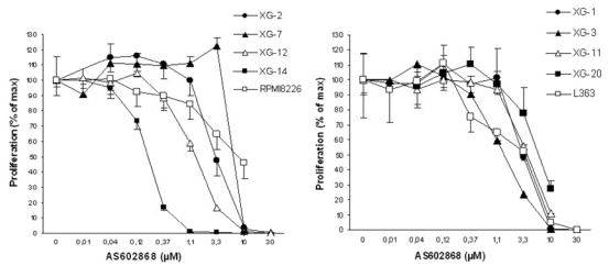
HMCL were cultured for 4 days in the presence of graded concentrations of AS602868 IKK2 inhibitor. Mean values of [3H]-thymidine incorporation were determined in triplicate culture wells and results are expressed as percentages of the mean proliferation without inhibitor (control culture medium). Results are those of one representative experiment out of a minimum of two for each cell line. The mean thymidine incorporation without AS602868 inhibitor was 32823 cpm for XG-2, 100434 for XG-7, 102505 for XG-12, 35294 for XG-14, 56981 for RPMI8226, 38677 for XG-1, 127328 for XG-3, 22309 for XG-11, 23535 for XG-20 and 61951 for L363.
Table 1. Inhibition of HMCL proliferation bv AS602868 IKK2 inhibitor.
HMCL were cultured for 4 days in the presence of graded concentrations of IKK2 inhibitor. Proliferation was assayed by thymidine incorporation. The IC50 and IC90 concentrations of AS602868 IKK2 inhibitor (concentrations yielding respectively 50% and 90% inhibition of proliferation) were determined for each cell line. Results are the mean of at least two separate experiments. NR = not reached.
| AS602868 IKK2 inhibitor (μM)
|
||
|---|---|---|
| HMCL | IC50 | IC90 |
| XG-14 | 0.28 | 0.76 |
| XG-12 | 1.29 | 4.53 |
| XG-3 | 1.68 | 6.14 |
| L363 | 2.06 | 9.76 |
| XG-6 | 2.27 | 6.11 |
| XG-19 | 2.44 | 9.03 |
| XG-4 | 2.53 | 8.79 |
| XG-1 | 2.78 | 7.58 |
| XG-2 | 3.04 | 8.22 |
| XG-13 | 3.32 | 9.06 |
| XG-11 | 4.21 | 14.43 |
| XG-20 | 6.43 | 14.07 |
| XG-7 | 6.70 | 9.65 |
| RPMI8226 | 8.30 | NR |
| Median | 2.65 | 8.79 |
AS602868 IKK2 inhibitor induces apoptosis and blocks cell cycle progression of HMCL
To assess the effects of the IKK2 inhibitor on apoptosis and cell cycle of MM cells, the two cell lines determined to be the most sensitive to AS602868 (XG-3 and XG-12) were selected and cultured with graded inhibitor concentrations. We chose concentrations inducing 10, 50 or 90 % of growth inhibition of the cell line in a previous experiment. As indicated in Table 2, AS602868 induced a dose-dependent increase in apoptosis, as assayed by annexin V staining, and a reduction in entry to the S phase of the cell cycle. These results indicate that the inhibition of the HMCL growth by AS602868 IKK2 inhibitor is the result of a simultaneous induction of apoptosis and blockade of the cell cycle.
Table 2. AS602868 induces apoptosis and blockade of entry in the S phase of the cell cycle resulting in the inhibition of the cell growth of HMCL.
HMCL were cultured without (control culture medium) or with AS602868 IKK2 inhibitor (0.35, 1.68 and 6.14 μM for XG-3 and 0.24, 1.29 and 4.53 μM for XG-12). These concentrations were those inducing 10%, 50% and 90% inhibition of the cell line proliferation in a previous experiment. The percentage of Annexin V+ cells, of cells in the S phase of the cell cycle and the cell counts were assayed at day 3 of culture. Results are the mean ± SD of three separate experiments. Statistical analyses were carried out using a Student’s t test for pairs.
| AS602868 IKK2 inhibitor
|
|||||
|---|---|---|---|---|---|
| Co
|
0.35 μM | 1.68μM | 6.14 μM | ||
| XG-3 | Annexin V % | 23.6 ± 3.3 | 25.4 ± 2.9 | 31.0 ±0.6* | 55.8 ±11.1 * |
| S phase % | 37.5 ±6.1 | 34.7 ± 6.3 | 30.2 ±6.1 ** | 16.6 ±6.1 ** | |
| Cell count ×106/ml | 0.60 ±0.16 | 0.53 ±0.11 | 0.40 ± 0.06 * | 0.20 ± 0.07 * | |
| Co
|
0.24 μM | 1.29μM | 4.53 μM | ||
| XG-12 | Annexin V % | 22.5 ±5.1 | 22.7 ±1.0 | 32.3 ± 7.2 ** | 70.0 ± 9.5 ** |
| S phase % | 40.2 ± 0.7 | 38.1 ±1.4 | 36.4 ±1.9* | 20.0 ± 7.6 ** | |
| Cell count ×106/ml | 0.57 ±0.17 | 0.57 ±0.11 | 0.43 ±0.10 | 0.19 ±0.07* | |
Significantly higher or lower values than control (P < .05);
significantly higher or lower values than control (P< .01).
AS602868IKK2 inhibitor inhibits the survival of primary myeloma cells
The bone marrow environment has been shown to play an essential role in the survival of primary myeloma cells. To assess the effects of AS602868 on primary myeloma cells interacting with cells of the bone marrow environment, we established cultures of bone marrow mononuclear cells from 13 patients, 6 patients with newly-diagnosed myeloma and 7 patients with relapsing myeloma. At day 5 of culture, cells were counted, the fraction of viable myeloma cells (CD138+ cells) (Jourdan, et al 1998), non-myeloma cells (CD138− cells) and hematopoietic progenitors (CD34+ cells) were determined by flow cytometry, and the cell number of each population was calculated. As shown in Fig 3, AS602868 induced a dose-dependent inhibition of the survival of myeloma cells in both categories of patients with a median IC50 of 10 μM. Interestingly, AS602868 induced apoptosis of primary myeloma cells without affecting the survival of other bone marrow cells, except that of hematopoietic progenitors (Fig 3).
Fig 3. AS602868 IKK2 inhibitor inhibits the cell survival of primary myeloma cells in culture bone marrow.
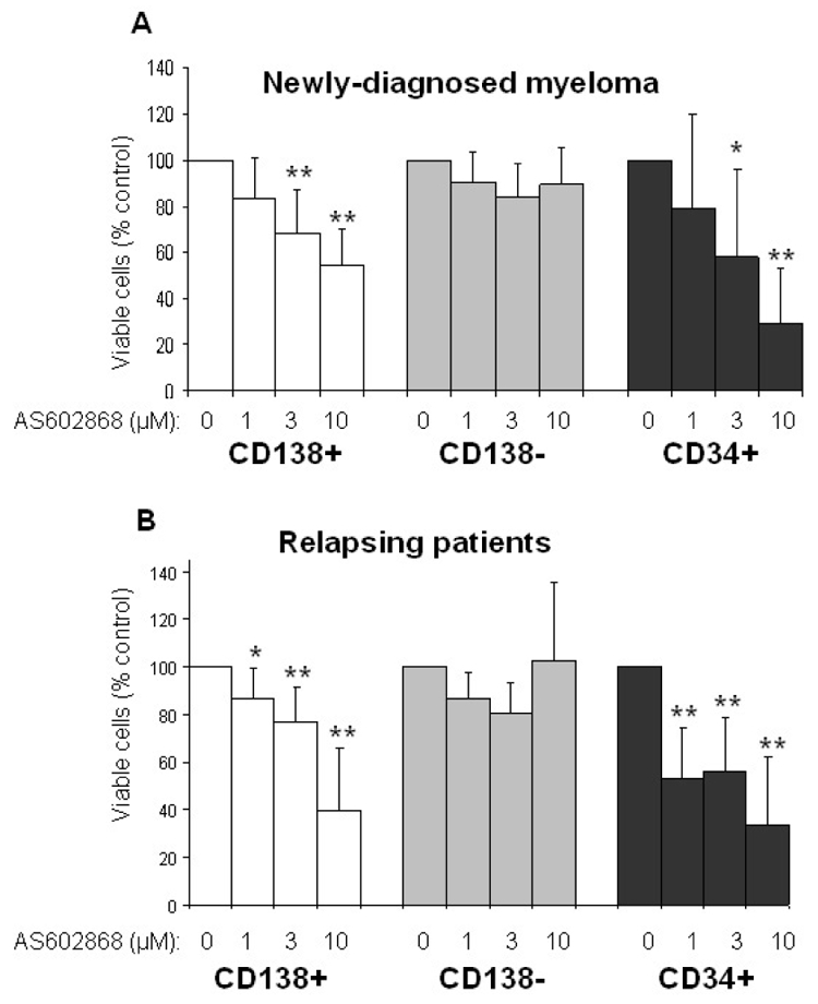
Bone marrow mononuclear cells from 6 newly-diagnosed myeloma (A) and 7 relapsing patients (B) were isolated as described in Material and methods and cultured without (control culture medium) or with graded concentrations of AS602868 IKK2 inhibitor. At day 5 of culture, the number of viable cells in each group of culture was assessed by trypan blue exclusion, the cells were stained with anti-CD138 and anti-CD34 antibodies and the percentage of CD138 and CD34 cells determined by flow cytometry. The number of CD138+, CD138− and CD34+ viable cells was then calculated. Results are expressed as the relative number of viable cells from each cell subpopulation obtained with AS602868 compared to that obtained in the corresponding subpopulation in the presence of control culture medium. Statistical analyses were carried out using a Student’s t test for pairs. * Significantly lower values than control (P < .05); ** significantly lower values than control (P < .01).
AS602868 increases the cytotoxicity of melphalan or Velcade
In order to investigate the effect of AS602868 association with melphalan or Velcade, two current drugs in myeloma, we chose four HMCL with a different sensitivity to the IKK2 inhibitor. The concentrations of melphalan yielding 10% (IC10) or 50% (IC50) inhibition of the HMCL proliferation ranged respectively from 0.11 to 0.73 μM and from 0.46 to 2.47 μM depending on the cell lines (data not shown). For Velcade, the IC10 ranged from 0.0014 to 0.0049 μM and the IC50 from 0.0022 to 0.0084 μM. Then, we determined the sensitivity to IKK2 inhibitor of the HMCL cultured in presence of their melphalan or Velcade respective IC10 and IC50 concentrations. A representative experiment is shown in Fig 4. AS602868 increased the cytotoxicity induced by melphalan or Velcade. Noteworthy, the dose sensitivity of myeloma cells to AS602868 was not changed by melphalan or Velcade (Fig 4 and data not shown).
Fig 4. Sensitivity of HMCL to AS602868 IKK2 inhibitor without or with IC10 or IC50 melphalan or Velcade.
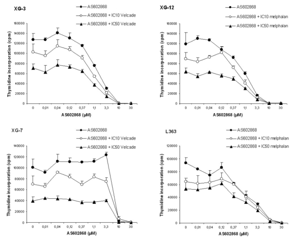
HMCL were cultured as indicated in Fig 2 in the presence of graded concentrations of AS602868, without (control culture medium) or with the respective IC10 and IC50 concentrations of melphalan or Velcade for each cell line. IC50 and IC90 for AS602868 without or with melphalan or Velcade were then deduced.
Discussion
A constitutive activation of the NF-κB pathway, in particular the activation of NF-κB and IKK and phosphorylation of IκBα, has been described in human myeloma cell lines and in primary myeloma cells, and plays an essential role in the survival and the growth of myeloma cells (Berenson, et al 2001; Bharti, et al 2003; Bharti, et al 2004; Mitsiades, et al 2002a; Ni, et al 2001; Sanda, et al 2005). We show here that a pharmacological inhibitor of IKK2, the anilinopyrimidine derivative AS602868, induces apoptosis and inhibits the proliferation of human myeloma cells. Our data are in agreement with previous studies showing that targeting NF-κB directly or upstream elements of the NF-κB pathway can induce myeloma cell apoptosis (Bharti, et al 2003; Bharti, et al 2004; Hideshima, et al 2006; Mitsiades, et al 2002a; Ni, et al 2001; Sanda, et al 2005). In acute myeloid leukaemia (AML) Frelin et al have shown that AS602868 blocked NF-κB activation and led to apoptosis of human primary AML cells (Frelin, et al 2005). The IC50 for AS602868 on AML cells ranged from 0.6 μM to 14 μM, these values are quite similar to those we observed on MM cells.
The survival of myeloma cells is strongly dependent of interactions with bone marrow environment (Gu, et al 2000). In particular, myeloma cell adhesion to bone marrow stromal cells (BMSC) induces NF-κB-dependent up-regulation of transcription of IL-6 (a major myeloma growth factor) by BMSC (Chauhan, et al 1996). Importantly, we show here that the AS602868 IKK2 inhibitor induces apoptosis in patients’ primary myeloma cells that were cultured together with their bone marrow environment cells. Our results indicate that hematopoietic progenitors (CD34+ cells) are targeted by AS602868. We may anticipate that treatment of patients with AS602868 will result in decreased hematopoietic cell counts, limiting the long term use of this inhibitor. It has been suggested that an increase in NF-κB activation could be responsible for tumor chemoresistance in MM, as in others cancers (Feinman, et al 1999; Ma, et al 2003; Mitsiades, et al 2002a; Wang, et al 1999). Indeed, using NF-κB inhibitor could revert or delay the development of chemoresistance to conventional drugs. We show here that addition of suboptimal AS602868 IKK2 inhibitor concentrations increases the growth inhibition induced by two major therapeutic myeloma drugs, the melphalan alkylating agent and the proteasome inhibitor PS341 (Velcade). Velcade, as dexamethasone or thalidomide, was shown to induce apoptosis and growth inhibition of myeloma cells in part via the inhibition of constitutive activation of NF-κB (Feinman, et al 1999; Hideshima, et al 2002; Mitsiades, et al 2002b). However, these drugs do not target NF-κB exclusively and their inhibitory effects cannot be totally attributed to the inhibition of NF-κB activation. Moreover, it is not clear if these drugs allow a complete or a partial and reversible inhibition of NF-κB activation. In the case of Velcade, this drug inhibits the NF-κB pathway by blocking IκB-α degradation, but this effect is not sufficient to explain its major antitumoral effects (Ma, et al 2003). Indeed, Velcade has also been shown to inhibit MAPK signalling of MM cells (Hideshima, et al 2001) and to induce proapoptotic/terminal unfolded protein response in tumoral cells (Obeng, et al 2006). Our results suggest that AS602868 IKK2 inhibitor can improve a partial inhibition of NF-κB activation induced by Velcade and strengthen the idea that treatments using a combination of conventional drugs with a specific NF-κB inhibitor could present an advantage to inhibit the myeloma cell growth.
In conclusion, this study shows that a pharmacological inhibition of the constitutive activation of the NF-κB pathway can block HMCL and primary myeloma cells growth and, in combination with conventional therapy, might represent an innovative approach for treating MM patients.
Acknowledgments
This work was supported by grants from the Ligue Nationale Contre Le Cancer (équipe labellisée), Paris, France.
References
- Berenson JR, Ma HM, Vescio R. The role of nuclear factor-kappaB in the biology and treatment of multiple myeloma. Seminars in Oncology. 2001;28:626–633. doi: 10.1016/s0093-7754(01)90036-3. [DOI] [PubMed] [Google Scholar]
- Bharti AC, Donato N, Singh S, Aggarwal BB. Curcumin (diferuloylmethane) down-regulates the constitutive activation of nuclear factor-kappa B and IkappaBalpha kinase in human multiple myeloma cells, leading to suppression of proliferation and induction of apoptosis. Blood. 2003;101:1053–1062. doi: 10.1182/blood-2002-05-1320. [DOI] [PubMed] [Google Scholar]
- Bharti AC, Shishodia S, Reuben JM, Weber D, Alexanian R, Raj-Vadhan S, Estrov Z, Talpaz M, Aggarwal BB. Nuclear factor-kappaB and STAT3 are constitutively active in CD138+ cells derived from multiple myeloma patients, and suppression of these transcription factors leads to apoptosis. Blood. 2004;103:3175–3184. doi: 10.1182/blood-2003-06-2151. [DOI] [PubMed] [Google Scholar]
- Brenne AT, Ro TB, Waage A, Sundan A, Borset M, Hjorth-Hansen H. Interleukin-21 is a growth and survival factor for human myeloma cells. Blood. 2002;99:3756–3762. doi: 10.1182/blood.v99.10.3756. [DOI] [PubMed] [Google Scholar]
- Catlett-Falcone R, Landowski TH, Oshiro MM, Turkson J, Levitzki A, Savino R, Ciliberto G, Moscinski L, Fernandez-Luna JL, Nunez G, Dalton WS, Jove R. Constitutive activation of Stat3 signaling confers resistance to apoptosis in human U266 myeloma cells. Immunity. 1999;10:105–115. doi: 10.1016/s1074-7613(00)80011-4. [DOI] [PubMed] [Google Scholar]
- Chauhan D, Uchiyama H, Akbarali Y, Urashima M, Yamamoto K, Libermann TA, Anderson KC. Multiple myeloma cell adhesion-induced interleukin-6 expression in bone marrow stromal cells involves activation of NF-kappa b. Blood. 1996;87:1104–1112. [PubMed] [Google Scholar]
- Costes V, Magen V, Legouffe E, Durand L, Baldet P, Rossi JF, Klein B, Brochier J. The Mi15 monoclonal antibody (anti-syndecan-1) is a reliable marker for quantifying plasma cells in paraffin-embedded bone marrow biopsy specimens. Human Pathology. 1999;30:1405–1411. doi: 10.1016/s0046-8177(99)90160-0. [DOI] [PubMed] [Google Scholar]
- De Vos J, Hose D, Reme T, Tarte K, Moreaux J, Mahtouk K, Jourdan M, Goldschmidt H, Rossi JF, Cremer FW, Klein B. Microarray-based understanding of normal and malignant plasma cells. Immunological Review. 2006;210:86–104. doi: 10.1111/j.0105-2896.2006.00362.x. [DOI] [PMC free article] [PubMed] [Google Scholar]
- De Vos J, Jourdan M, Tarte K, Jasmin C, Klein B. JAK2 tyrosine kinase inhibitor tyrphostin AG490 downregulates the mitogen-activated protein kinase (MAPK) and signal transducer and activator of transcription (STAT) pathways and induces apoptosis in myeloma cells. British Journal of Haematology. 2000;109:823–828. doi: 10.1046/j.1365-2141.2000.02127.x. [DOI] [PubMed] [Google Scholar]
- Derksen PW, de Gorter DJ, Meijer HP, Bende RJ, van Dijk M, Lokhorst HM, Bloem AC, Spaargaren M, Pals ST. The hepatocyte growth factor/Met pathway controls proliferation and apoptosis in multiple myeloma. Leukemia. 2003;17:764–774. doi: 10.1038/sj.leu.2402875. [DOI] [PubMed] [Google Scholar]
- Derksen PW, Tjin E, Meijer HP, Klok MD, MacGillavry HD, van Oers MH, Lokhorst HM, Bloem AC, Clevers H, Nusse R, van der Neut R, Spaargaren M, Pals ST. Illegitimate WNT signaling promotes proliferation of multiple myeloma cells. Proceedings of the National Academy of Sciences of the United States of America. 2004;101:6122–6127. doi: 10.1073/pnas.0305855101. [DOI] [PMC free article] [PubMed] [Google Scholar]
- Feinman R, Koury J, Thames M, Barlogie B, Epstein J, Siegel DS. Role of NF-kappaB in the rescue of multiple myeloma cells from glucocorticoid-induced apoptosis by bcl-2. Blood. 1999;93:3044–3052. [PubMed] [Google Scholar]
- Ferlin-Bezombes M, Jourdan M, Liautard J, Brochier J, Rossi JF, Klein B. IFN-alpha is a survival factor for human myeloma cells and reduces dexamethasone-induced apoptosis. Journal of Immunology. 1998;161:2692–2699. [PubMed] [Google Scholar]
- Ferlin M, Noraz N, Hertogh C, Brochier J, Taylor N, Klein B. Insulin-like growth factor induces the survival and proliferation of myeloma cells through an interleukin-6-independent transduction pathway. British Journal of Haematology. 2000;111:626–634. doi: 10.1046/j.1365-2141.2000.02364.x. [DOI] [PubMed] [Google Scholar]
- Fonseca R, Barlogie B, Bataille R, Bastard C, Bergsagel PL, Chesi M, Davies FE, Drach J, Greipp PR, Kirsch IR, Kuehl WM, Hernandez JM, Minvielle S, Pilarski LM, Shaughnessy JD, Jr, Stewart AK, Avet-Loiseau H. Genetics and cytogenetics of multiple myeloma: a workshop report. Cancer Research. 2004;64:1546–1558. doi: 10.1158/0008-5472.can-03-2876. [DOI] [PubMed] [Google Scholar]
- Frelin C, Imbert V, Griessinger E, Loubat A, Dreano M, Peyron JF. AS602868, a pharmacological inhibitor of IKK2, reveals the apoptotic potential of TNF-alpha in Jurkat leukemic cells. Oncogene. 2003;22:8187–8194. doi: 10.1038/sj.onc.1206963. [DOI] [PubMed] [Google Scholar]
- Frelin C, Imbert V, Griessinger E, Peyron AC, Rochet N, Philip P, Dageville C, Sirvent A, Hummelsberger M, Berard E, Dreano M, Sirvent N, Peyron JF. Targeting NF-kappaB activation via pharmacologic inhibition of IKK2-induced apoptosis of human acute myeloid leukemia cells. Blood. 2005;105:804–811. doi: 10.1182/blood-2004-04-1463. [DOI] [PubMed] [Google Scholar]
- Gaillard JP, Liautard J, Klein B, Brochier J. Major role of the soluble interleukin-6/interleukin-6 receptor complex for the proliferation of interleukin-6-dependent human myeloma cell lines. European Journal of Immunology. 1997;27:3332–3340. doi: 10.1002/eji.1830271232. [DOI] [PubMed] [Google Scholar]
- Giuliani N, Lunghi P, Morandi F, Colla S, Bonomini S, Hojden M, Rizzoli V, Bonati A. Downmodulation of ERK protein kinase activity inhibits VEGF secretion by human myeloma cells and myeloma-induced angiogenesis. Leukemia. 2004;18:628–635. doi: 10.1038/sj.leu.2403269. [DOI] [PubMed] [Google Scholar]
- Gu ZJ, Costes V, Lu ZY, Zhang XG, Pitard V, Moreau JF, Bataille R, Wijdenes J, Rossi JF, Klein B. Interleukin-10 is a growth factor for human myeloma cells by induction of an oncostatin M autocrine loop. Blood. 1996;88:3972–3986. [PubMed] [Google Scholar]
- Gu ZJ, Vos JD, Rebouissou C, Jourdan M, Zhang XG, Rossi JF, Wijdenes J, Klein B. Agonist anti-gp130 transducer monoclonal antibodies are human myeloma cell survival and growth factors. Leukemia. 2000;14:188–197. doi: 10.1038/sj.leu.2401632. [DOI] [PubMed] [Google Scholar]
- Hideshima T, Chauhan D, Richardson P, Mitsiades C, Mitsiades N, Hayashi T, Munshi N, Dang L, Castro A, Palombella V, Adams J, Anderson KC. NF-kappa B as a therapeutic target in multiple myeloma. Journal of Biological Chemistry. 2002;277:16639–16647. doi: 10.1074/jbc.M200360200. [DOI] [PubMed] [Google Scholar]
- Hideshima T, Neri P, Tassone P, Yasui H, Ishitsuka K, Raje N, Chauhan D, Podar K, Mitsiades C, Dang L, Munshi N, Richardson P, Schenkein D, Anderson KC. MLN120B, a novel IkappaB kinase beta inhibitor, blocks multiple myeloma cell growth in vitro and in vivo. Clinical Cancer Research. 2006;12:5887–5894. doi: 10.1158/1078-0432.CCR-05-2501. [DOI] [PubMed] [Google Scholar]
- Hideshima T, Richardson P, Chauhan D, Palombella VJ, Elliott PJ, Adams J, Anderson KC. The proteasome inhibitor PS-341 inhibits growth, induces apoptosis, and overcomes drug resistance in human multiple myeloma cells. Cancer Research. 2001;61:3071–3076. [PubMed] [Google Scholar]
- Hjorth-Hansen H, Waage A, Borset M. Interleukin-15 blocks apoptosis and induces proliferation of the human myeloma cell line OH-2 and freshly isolated myeloma cells. British Journal of Haematology. 1999;106:28–34. doi: 10.1046/j.1365-2141.1999.01510.x. [DOI] [PubMed] [Google Scholar]
- Hsu J, Shi Y, Krajewski S, Renner S, Fisher M, Reed JC, Franke TF, Lichtenstein A. The AKT kinase is activated in multiple myeloma tumor cells. Blood. 2001;98:2853–2855. doi: 10.1182/blood.v98.9.2853. [DOI] [PubMed] [Google Scholar]
- Jourdan M, De Vos J, Mechti N, Klein B. Regulation of Bcl-2-family proteins in myeloma cells by three myeloma survival factors: interleukin-6, interferon-alpha and insulin-like growth factor 1. Cell Death and Differentiation. 2000;7:1244–1252. doi: 10.1038/sj.cdd.4400758. [DOI] [PMC free article] [PubMed] [Google Scholar]
- Jourdan M, Ferlin M, Legouffe E, Horvathova M, Liautard J, Rossi JF, Wijdenes J, Brochier J, Klein B. The myeloma cell antigen syndecan-1 is lost by apoptotic myeloma cells. Br J Haematol. 1998;100:637–646. doi: 10.1046/j.1365-2141.1998.00623.x. [DOI] [PubMed] [Google Scholar]
- Karin M, Lin A. NF-kappaB at the crossroads of life and death. Nature Immunology. 2002;3:221–227. doi: 10.1038/ni0302-221. [DOI] [PubMed] [Google Scholar]
- Ma MH, Yang HH, Parker K, Manyak S, Friedman JM, Altamirano C, Wu ZQ, Borad MJ, Frantzen M, Roussos E, Neeser J, Mikail A, Adams J, Sjak-Shie N, Vescio RA, Berenson JR. The proteasome inhibitor PS-341 markedly enhances sensitivity of multiple myeloma tumor cells to chemotherapeutic agents. Clinical Cancer Research. 2003;9:1136–1144. [PubMed] [Google Scholar]
- Mahtouk K, Cremer FW, Reme T, Jourdan M, Baudard M, Moreaux J, Requirand G, Fiol G, De Vos J, Moos M, Quittet P, Goldschmidt H, Rossi JF, Hose D, Klein B. Heparan sulphate proteoglycans are essential for the myeloma cell growth activity of EGF-family ligands in multiple myeloma. Oncogene. 2006 doi: 10.1038/sj.onc.1209699. [DOI] [PMC free article] [PubMed] [Google Scholar]
- Mahtouk K, Hose D, Reme T, De Vos J, Jourdan M, Moreaux J, Fiol G, Raab M, Jourdan E, Grau V, Moos M, Goldschmidt H, Baudard M, Rossi JF, Cremer FW, Klein B. Expression of EGF-family receptors and amphiregulin in multiple myeloma. Amphiregulin is a growth factor for myeloma cells. Oncogene. 2005;24:3512–3524. doi: 10.1038/sj.onc.1208536. [DOI] [PMC free article] [PubMed] [Google Scholar]
- Mahtouk K, Jourdan M, De Vos J, Hertogh C, Fiol G, Jourdan E, Rossi JF, Klein B. An inhibitor of the EGF receptor family blocks myeloma cell growth factor activity of HB-EGF and potentiates dexamethasone or anti-IL-6 antibody-induced apoptosis. Blood. 2004;103:1829–1837. doi: 10.1182/blood-2003-05-1510. [DOI] [PMC free article] [PubMed] [Google Scholar]
- Mitsiades N, Mitsiades CS, Poulaki V, Chauhan D, Richardson PG, Hideshima T, Munshi N, Treon SP, Anderson KC. Biologic sequelae of nuclear factor-kappaB blockade in multiple myeloma: therapeutic applications. Blood. 2002a;99:4079–4086. doi: 10.1182/blood.v99.11.4079. [DOI] [PubMed] [Google Scholar]
- Mitsiades N, Mitsiades CS, Poulaki V, Chauhan D, Richardson PG, Hideshima T, Munshi NC, Treon SP, Anderson KC. Apoptotic signaling induced by immunomodulatory thalidomide analogs in human multiple myeloma cells: therapeutic implications. Blood. 2002b;99:4525–4530. doi: 10.1182/blood.v99.12.4525. [DOI] [PubMed] [Google Scholar]
- Moreaux J, Cremer FW, Reme T, Raab M, Mahtouk K, Kaukel P, Pantesco V, De Vos J, Jourdan E, Jauch A, Legouffe E, Moos M, Fiol G, Goldschmidt H, Rossi JF, Hose D, Klein B. The level of TACI gene expression in myeloma cells is associated with a signature of microenvironment dependence versus a plasmablastic signature. Blood. 2005;106:1021–1030. doi: 10.1182/blood-2004-11-4512. [DOI] [PMC free article] [PubMed] [Google Scholar]
- Moreaux J, Legouffe E, Jourdan E, Quittet P, Reme T, Lugagne C, Moine P, Rossi JF, Klein B, Tarte K. BAFF and APRIL protect myeloma cells from apoptosis induced by interleukin 6 deprivation and dexamethasone. Blood. 2004;103:3148–3157. doi: 10.1182/blood-2003-06-1984. [DOI] [PMC free article] [PubMed] [Google Scholar]
- Ni H, Ergin M, Huang Q, Qin JZ, Amin HM, Martinez RL, Saeed S, Barton K, Alkan S. Analysis of expression of nuclear factor kappa B (NF-kappa B) in multiple myeloma: downregulation of NF-kappa B induces apoptosis. British Journal of Haematology. 2001;115:279–286. doi: 10.1046/j.1365-2141.2001.03102.x. [DOI] [PubMed] [Google Scholar]
- Obeng EA, Carlson LM, Gutman DM, Harrington WJ, Jr, Lee KP, Boise LH. Proteasome inhibitors induce a terminal unfolded protein response in multiple myeloma cells. Blood. 2006;107:4907–4916. doi: 10.1182/blood-2005-08-3531. [DOI] [PMC free article] [PubMed] [Google Scholar]
- Rebouissou C, Wijdenes J, Autissier P, Tarte K, Costes V, Liautard J, Rossi JF, Brochier J, Klein B. A gp130 interleukin-6 transducer-dependent SCID model of human multiple myeloma. Blood. 1998;91:4727–4737. [PubMed] [Google Scholar]
- Sanda T, Iida S, Ogura H, Asamitsu K, Murata T, Bacon KB, Ueda R, Okamoto T. Growth inhibition of multiple myeloma cells by a novel IkappaB kinase inhibitor. Clinical Cancer Research. 2005;11:1974–1982. doi: 10.1158/1078-0432.CCR-04-1936. [DOI] [PubMed] [Google Scholar]
- Tarte K, Zhang XG, Legouffe E, Hertog C, Mehtali M, Rossi JF, Klein B. Induced expression of B7-1 on myeloma cells following retroviral gene transfer results in tumor-specific recognition by cytotoxic T cells. Journal of Immunology. 1999;163:514–524. [PubMed] [Google Scholar]
- Vermes I, Haanen C, Steffens-Nakken H, Reutelingsperger C. A novel assay for apoptosis. Flow cytometric detection of phosphatidylserine expression on early apoptotic cells using fluorescein labelled Annexin V. Journal of Immunological Methods. 1995;184:39–51. doi: 10.1016/0022-1759(95)00072-i. [DOI] [PubMed] [Google Scholar]
- Viatour P, Merville MP, Bours V, Chariot A. Phosphorylation of NF-kappaB and IkappaB proteins: implications in cancer and inflammation. Trends in Biochemical Sciences. 2005;30:43–52. doi: 10.1016/j.tibs.2004.11.009. [DOI] [PubMed] [Google Scholar]
- Wang CY, Cusack JC, Jr, Liu R, Baldwin AS., Jr Control of inducible chemoresistance: enhanced anti-tumor therapy through increased apoptosis by inhibition of NF-kappaB. Nature Medicine. 1999;5:412–417. doi: 10.1038/7410. [DOI] [PubMed] [Google Scholar]
- Zhang XG, Bataille R, Widjenes J, Klein B. Interleukin-6 dependence of advanced malignant plasma cell dyscrasias. Cancer. 1992;69:1373–1376. doi: 10.1002/1097-0142(19920315)69:6<1373::aid-cncr2820690612>3.0.co;2-1. [DOI] [PubMed] [Google Scholar]
- Zhang XG, Gaillard JP, Robillard N, Lu ZY, Gu ZJ, Jourdan M, Boiron JM, Bataille R, Klein B. Reproducible obtaining of human myeloma cell lines as a model for tumor stem cell study in human multiple myeloma. Blood. 1994;83:3654–3663. [PubMed] [Google Scholar]