

Abstract
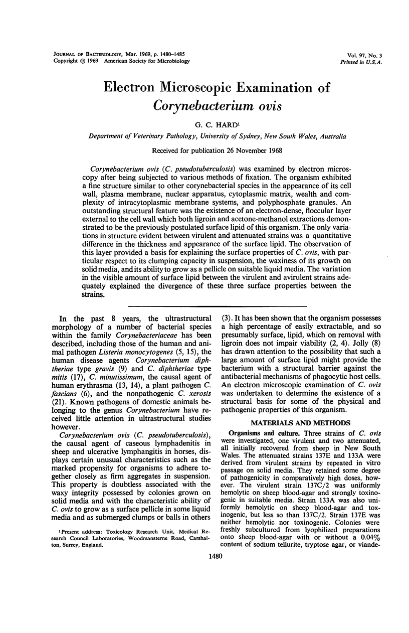
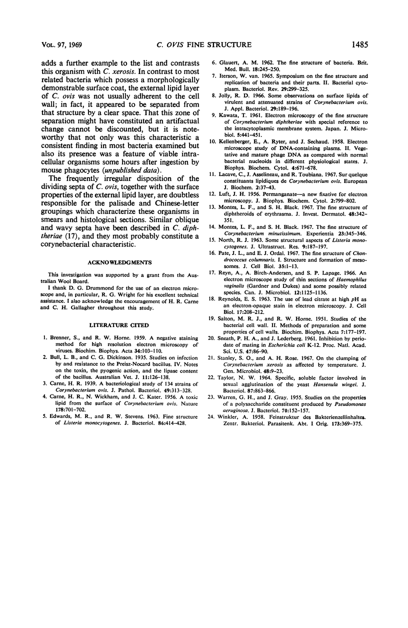
Corynebacterium ovis (C. pseudotuberculosis) was examined by electron microscopy after being subjected to various methods of fixation. The organism exhibited a fine structure similar to other corynebacterial species in the appearance of its cell wall, plasma membrane, nuclear apparatus, cytoplasmic matrix, wealth and complexity of intracytoplasmic membrane systems, and polyphosphate granules. An outstanding structural feature was the existence of an electron-dense, floccular layer external to the cell wall which both ligroin and acetone-methanol extractions demonstrated to be the previously postulated surface lipid of this organism. The only variations in structure evident between virulent and attenuated strains was a quantitative difference in the thickness and appearance of the surface lipid. The observation of this layer provided a basis for explaining the surface properties of C. ovis, with particular respect to its clumping capacity in suspension, the waxiness of its growth on solid media, and its ability to grow as a pellicle on suitable liquid media. The variation in the visible amount of surface lipid between the virulent and avirulent strains adequately explained the divergence of these three surface properties between the strains.
Full text
PDF


Images in this article


Selected References
These references are in PubMed. This may not be the complete list of references from this article.
- BRENNER S., HORNE R. W. A negative staining method for high resolution electron microscopy of viruses. Biochim Biophys Acta. 1959 Jul;34:103–110. doi: 10.1016/0006-3002(59)90237-9. [DOI] [PubMed] [Google Scholar]
- CARNE H. R., KATER J. C., WICKHAM N. A toxic lipid from the surface of Corynebacterium ovis. Nature. 1956 Sep 29;178(4535):701–702. doi: 10.1038/178701a0. [DOI] [PubMed] [Google Scholar]
- EDWARDS M. R., STEVENS R. W. FINE STRUCTURE OF LISTERIA MONOCYTOGENES. J Bacteriol. 1963 Sep;86:414–428. doi: 10.1128/jb.86.3.414-428.1963. [DOI] [PMC free article] [PubMed] [Google Scholar]
- GLAUERT A. M. The fine structure of bacteria. Br Med Bull. 1962 Sep;18:245–250. doi: 10.1093/oxfordjournals.bmb.a069988. [DOI] [PubMed] [Google Scholar]
- KELLENBERGER E., RYTER A., SECHAUD J. Electron microscope study of DNA-containing plasms. II. Vegetative and mature phage DNA as compared with normal bacterial nucleoids in different physiological states. J Biophys Biochem Cytol. 1958 Nov 25;4(6):671–678. doi: 10.1083/jcb.4.6.671. [DOI] [PMC free article] [PubMed] [Google Scholar]
- LUFT J. H. Permanganate; a new fixative for electron microscopy. J Biophys Biochem Cytol. 1956 Nov 25;2(6):799–802. doi: 10.1083/jcb.2.6.799. [DOI] [PMC free article] [PubMed] [Google Scholar]
- Lacave C., Asselineau J., Toubiana R. Sur quelques constituants lipidiques de Corynebacterium ovis. Eur J Biochem. 1967 Jul;2(1):37–43. doi: 10.1111/j.1432-1033.1967.tb00102.x. [DOI] [PubMed] [Google Scholar]
- Montes L. F., Black S. H. The fine structure of Corynebacterium minutissimum. Experientia. 1967 May 15;23(5):345–346. doi: 10.1007/BF02144506. [DOI] [PubMed] [Google Scholar]
- Montes L. F., Black S. H. The fine structure of diphtheroids of erythrasma. J Invest Dermatol. 1967 Apr;48(4):342–351. [PubMed] [Google Scholar]
- NORTH R. J. SOME STRUCTURAL ASPECTS OF LISTERIA MONOCYTOGENES. J Ultrastruct Res. 1963 Oct;59:187–197. doi: 10.1016/s0022-5320(63)80001-5. [DOI] [PubMed] [Google Scholar]
- Pate J. L., Ordal E. J. The fine structure of Chondrococcus columnaris. I. Structure and formation of mesosomes. J Cell Biol. 1967 Oct;35(1):1–13. doi: 10.1083/jcb.35.1.1. [DOI] [PMC free article] [PubMed] [Google Scholar]
- REYNOLDS E. S. The use of lead citrate at high pH as an electron-opaque stain in electron microscopy. J Cell Biol. 1963 Apr;17:208–212. doi: 10.1083/jcb.17.1.208. [DOI] [PMC free article] [PubMed] [Google Scholar]
- Reyn A., Birch-Andersen A., Lapage S. P. An electron microscope study of thin sections of Haemophilus vaginalis (Gardner and Dukes) and some possibly related species. Can J Microbiol. 1966 Dec;12(6):1125–1136. doi: 10.1139/m66-154. [DOI] [PubMed] [Google Scholar]
- SALTON M. R. J., HORNE R. W. Studies of the bacterial cell wall. II. Methods of preparation and some properties of cell walls. Biochim Biophys Acta. 1951 Jul;7(2):177–197. doi: 10.1016/0006-3002(51)90017-0. [DOI] [PubMed] [Google Scholar]
- Sneath P. H., Lederberg J. INHIBITION BY PERIODATE OF MATING IN ESCHERICHIA COLI K-12. Proc Natl Acad Sci U S A. 1961 Jan;47(1):86–90. doi: 10.1073/pnas.47.1.86. [DOI] [PMC free article] [PubMed] [Google Scholar]
- TAYLOR N. W. SPECIFIC, SOLUBLE FACTOR INVOLVED IN SEXUAL AGGLUTINATION OF THE YEAST HANSENULA WINGEI. J Bacteriol. 1964 Apr;87:863–866. doi: 10.1128/jb.87.4.863-866.1964. [DOI] [PMC free article] [PubMed] [Google Scholar]
- WARREN G. H., GRAY J. Studies on the properties of a polysaccharide constituent produced by Pseudomonas aeruginosa. J Bacteriol. 1955 Aug;70(2):152–157. doi: 10.1128/jb.70.2.152-157.1955. [DOI] [PMC free article] [PubMed] [Google Scholar]
- WINKLER A. Feinstruktur des Bakterienzellinhaltes. Zentralbl Bakteriol Orig. 1958 Dec;173(5-6):369–375. [PubMed] [Google Scholar]
- van Iterson W. Symposium on the fine structure and replication of bacteria and their parts. II. Bacterial cytoplasm. Bacteriol Rev. 1965 Sep;29(3):299–325. doi: 10.1128/br.29.3.299-325.1965. [DOI] [PMC free article] [PubMed] [Google Scholar]