Abstract
Background
Precordial ECG electrode positioning was standardised in the early 1940s. However, it has been customary for the V3 to V6 electrodes to be placed under the left breast in women rather than in the correct anatomical positions relating to the 4th and 5th interspaces. For this reason, a comparison between the two approaches to chest electrode positioning in women was undertaken.
Methods
In total 84 women were recruited and ECGs recorded with electrodes in the correct anatomical position and also in the more commonly used positions under the breast. As a separate study, 299 healthy women were recruited to study normal limits of leads V3 to V6 recorded with electrodes in the correct anatomical positions and compare them with published normal limits with electrodes in the more commonly used locations.
Results
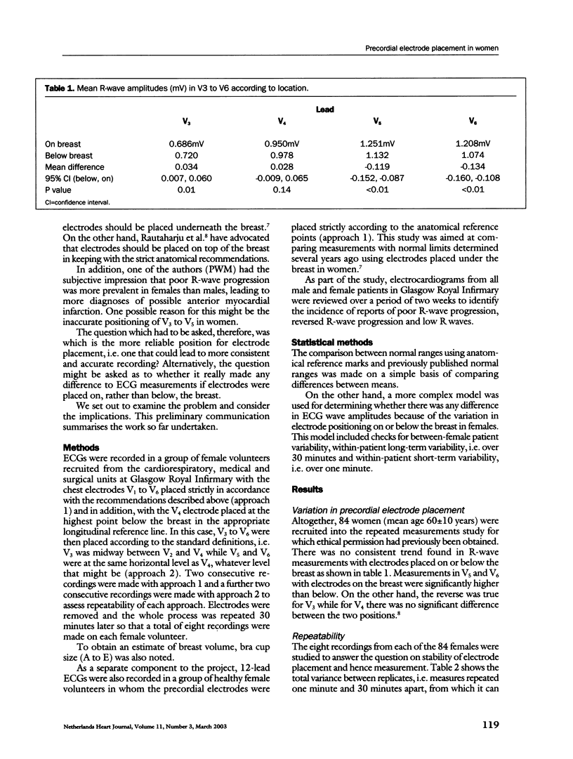
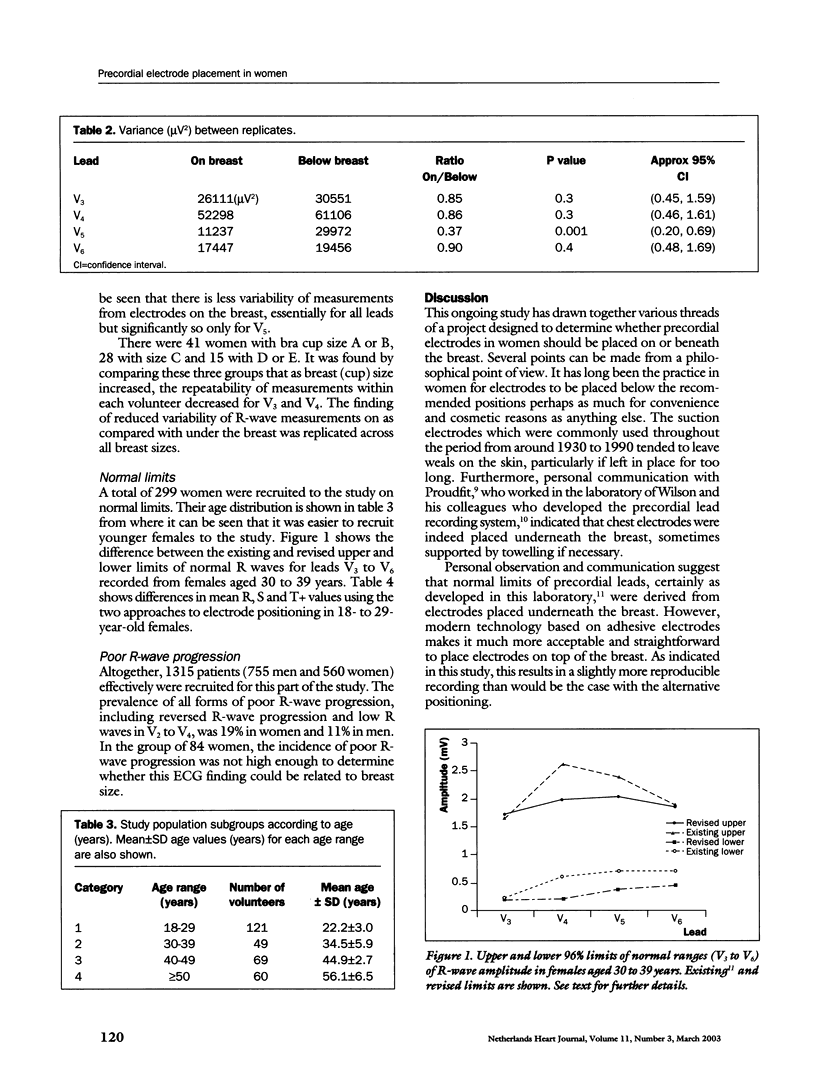
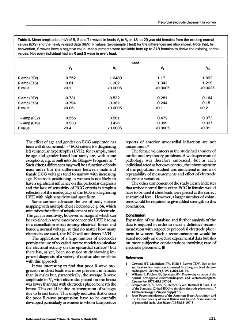
It was shown that there was less variability with electrodes in the correct anatomical positions and that there were significant differences between the new limits of normality compared with the old established limits.
Conclusion
Expansion of the database and further analysis of the data is required to make a definitive recommendation with respect to precordial electrode placement in women.
Keywords: EGG, precordial electrode, women, normal limits, ECG analysis
Full text
PDF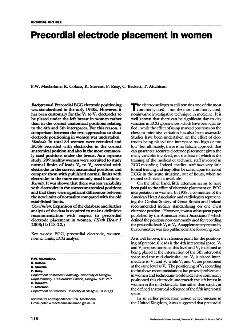
Selected References
These references are in PubMed. This may not be the complete list of references from this article.
- Cawood H. T., Macfarlane P. W., Hillis S., Lawrie T. D. Day to day and beat to beat variation in normal 3 orthogonal lead electrocardiograms. Br Heart J. 1974 Dec;36(12):1225–1230. doi: 10.1136/hrt.36.12.1225. [DOI] [PMC free article] [PubMed] [Google Scholar]
- Colaco R., Reay P., Beckett C., Aitchison T. C., Mcfarlane P. W. False positive ECG reports of anterior myocardial infarction in women. J Electrocardiol. 2000;33 (Suppl):239–244. doi: 10.1054/jelc.2000.20359. [DOI] [PubMed] [Google Scholar]
- Macfarlane P. W., Devine B., Latif S., McLaughlin S., Shoat D. B., Watts M. P. Methodology of ECG interpretation in the Glasgow program. Methods Inf Med. 1990 Sep;29(4):354–361. [PubMed] [Google Scholar]
- Ramanathan C., Rudy Y. Electrocardiographic imaging: II. Effect of torso inhomogeneities on noninvasive reconstruction of epicardial potentials, electrograms, and isochrones. J Cardiovasc Electrophysiol. 2001 Feb;12(2):241–252. doi: 10.1046/j.1540-8167.2001.00241.x. [DOI] [PubMed] [Google Scholar]
- Rautaharju P. M., Park L., Rautaharju F. S., Crow R. A standardized procedure for locating and documenting ECG chest electrode positions: consideration of the effect of breast tissue on ECG amplitudes in women. J Electrocardiol. 1998 Jan;31(1):17–29. doi: 10.1016/s0022-0736(98)90003-6. [DOI] [PubMed] [Google Scholar]
- Willems J. L., Poblete P. F., Pipberger H. V. Day-to-day variation of the normal orthogonal electrocardiogram and vectorcardiogram. Circulation. 1972 May;45(5):1057–1064. doi: 10.1161/01.cir.45.5.1057. [DOI] [PubMed] [Google Scholar]


