

Abstract
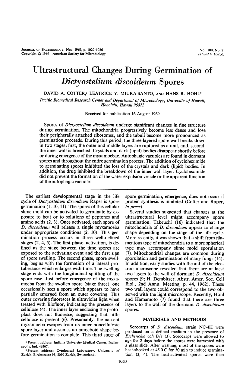
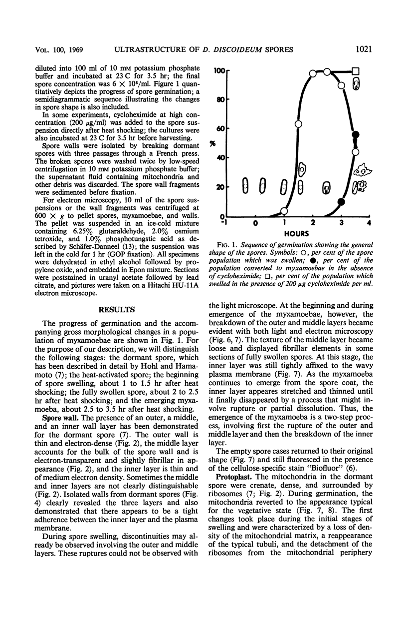
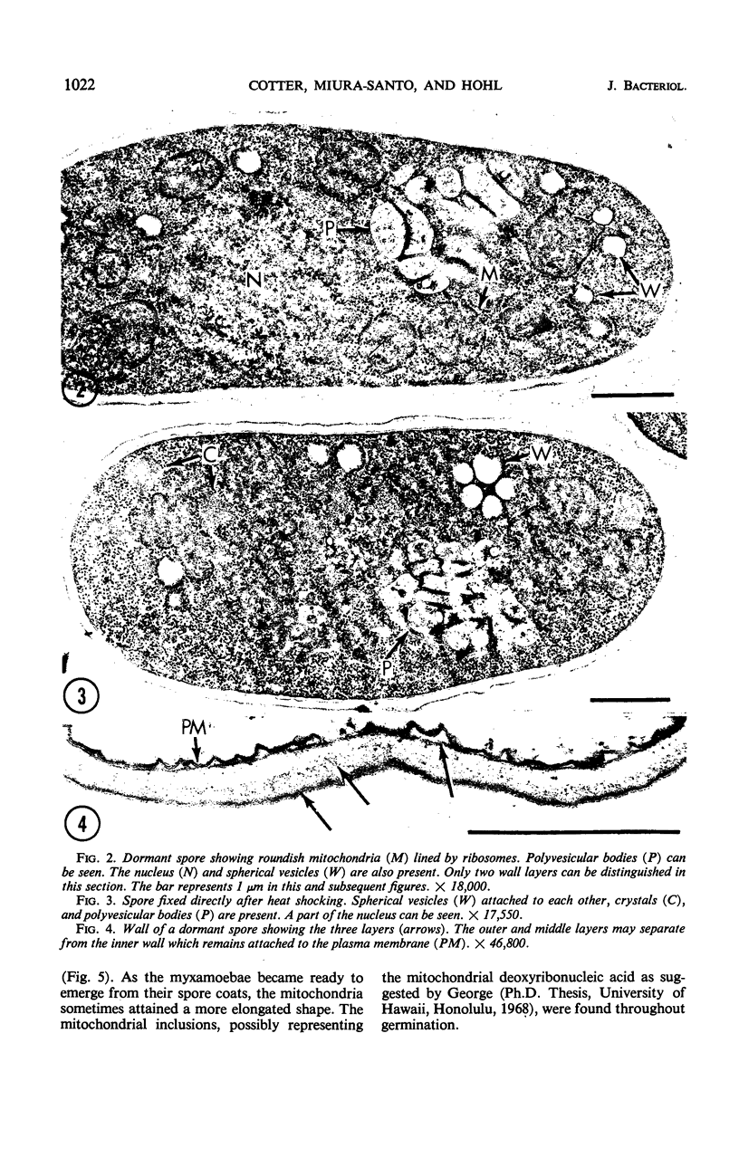
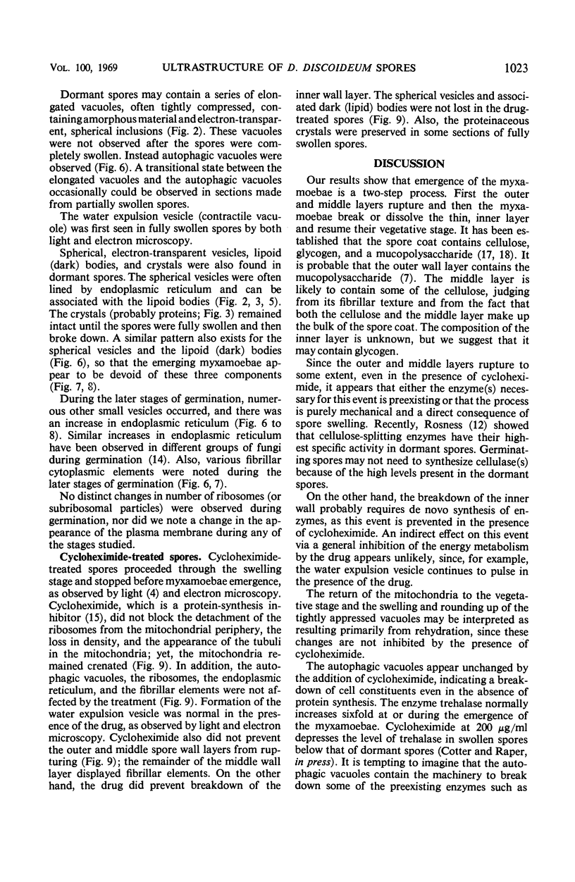
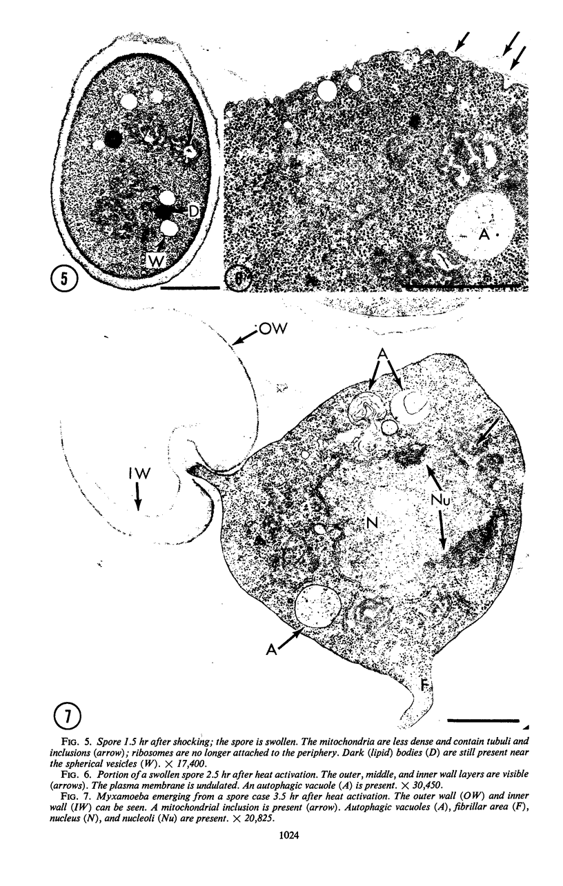
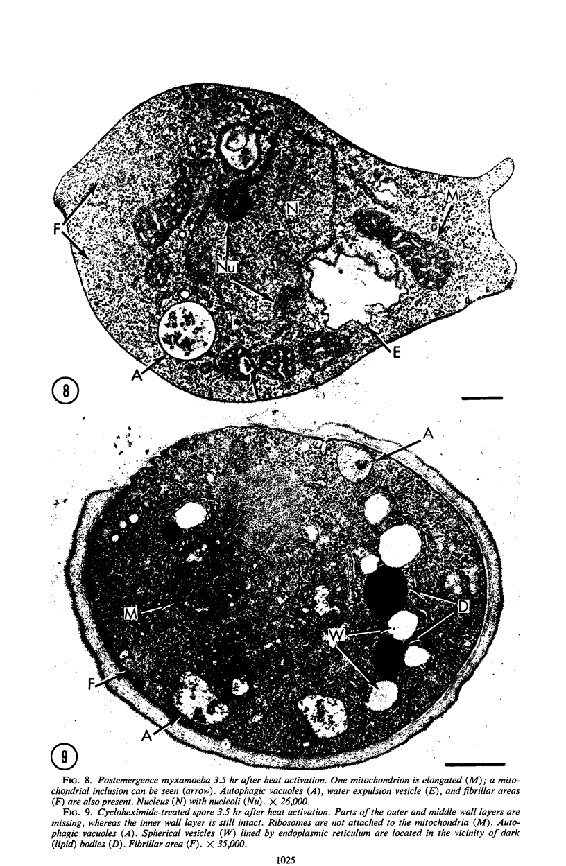
Spores of Dictyostelium discoideum undergo significant changes in fine structure during germination. The mitochondria progressively become less dense and lose their peripherally attached ribosomes, and the tubuli become more pronounced as germination proceeds. During this period, the three-layered spore wall breaks down in two stages: first, the outer and middle layers are ruptured as a unit, and, second, the inner wall is breached. Crystals and dark (lipid) bodies disappear shortly before or during emergence of the myxamoebae. Autophagic vacuoles are found in dormant spores and throughout the entire germination process. The addition of cycloheximide to germinating spores inhibited the loss of the crystals and dark (lipid) bodies. In addition, the drug inhibited the breakdown of the inner wall layer. Cycloheximide did not prevent the formation of the water expulsion vesicle or the apparent function of the autophagic vacuoles.
Full text
PDF
Images in this article
Selected References
These references are in PubMed. This may not be the complete list of references from this article.
- Cotter D. A., Raper K. B. Factors affecting the rate of heat-induced spore germination in Dictyostelium discoideum. J Bacteriol. 1968 Jul;96(1):86–92. doi: 10.1128/jb.96.1.86-92.1968. [DOI] [PMC free article] [PubMed] [Google Scholar]
- Cotter D. A., Raper K. B. Properties of germinating spores of Dictyostelium discoideum. J Bacteriol. 1968 Nov;96(5):1680–1689. doi: 10.1128/jb.96.5.1680-1689.1968. [DOI] [PMC free article] [PubMed] [Google Scholar]
- Cotter D. A., Raper K. B. Spore germination in Dictyostelium discoideum. Proc Natl Acad Sci U S A. 1966 Sep;56(3):880–887. doi: 10.1073/pnas.56.3.880. [DOI] [PMC free article] [PubMed] [Google Scholar]
- Cotter D. A., Raper K. B. Spore germination in strains of Dictyostelium discoideum and other members of the Dictyosteliaceae. J Bacteriol. 1968 Nov;96(5):1690–1695. doi: 10.1128/jb.96.5.1690-1695.1968. [DOI] [PMC free article] [PubMed] [Google Scholar]
- Harrington B. J., Raper K. B. Use of a fluorescent brightener to demonstrate cellulose in the cellular slime molds. Appl Microbiol. 1968 Jan;16(1):106–113. doi: 10.1128/am.16.1.106-113.1968. [DOI] [PMC free article] [PubMed] [Google Scholar]
- Hohl H. R., Hamamoto S. T. Ultrastructure of spore differentiation in Dictyostelium: the prespore vacuole. J Ultrastruct Res. 1969 Mar;26(5):442–453. doi: 10.1016/s0022-5320(69)90050-1. [DOI] [PubMed] [Google Scholar]
- Rosness A. Cellulolytic enzymes during morphogenesis in Dictyostelium discoideum. J Bacteriol. 1968 Sep;96(3):639–645. doi: 10.1128/jb.96.3.639-645.1968. [DOI] [PMC free article] [PubMed] [Google Scholar]
- Schäfer-Danneel S. Strukturelle und funktionelle Voraussetzungen für die Bewegung von Amoeba proteus. Z Zellforsch Mikrosk Anat. 1967;78(4):441–462. [PubMed] [Google Scholar]
- TAKEUCHI I. The correlation of cellular changes with succinic dehydrogenase and cytochrome oxidase activities in the development of the cellular slime molds. Dev Biol. 1960 Aug;2:343–366. doi: 10.1016/0012-1606(60)90021-x. [DOI] [PubMed] [Google Scholar]
- WHITE G. J., SUSSMAN M. Polysaccharides involved in slimemold development. I. Water-soluble glucose polymer (s). Biochim Biophys Acta. 1963 Jul 16;74:173–178. doi: 10.1016/0006-3002(63)91355-6. [DOI] [PubMed] [Google Scholar]