

Abstract
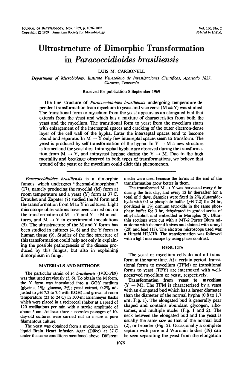
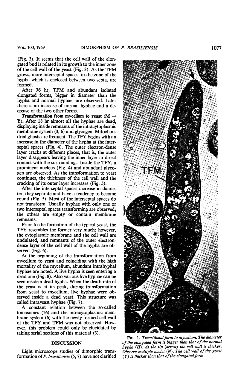
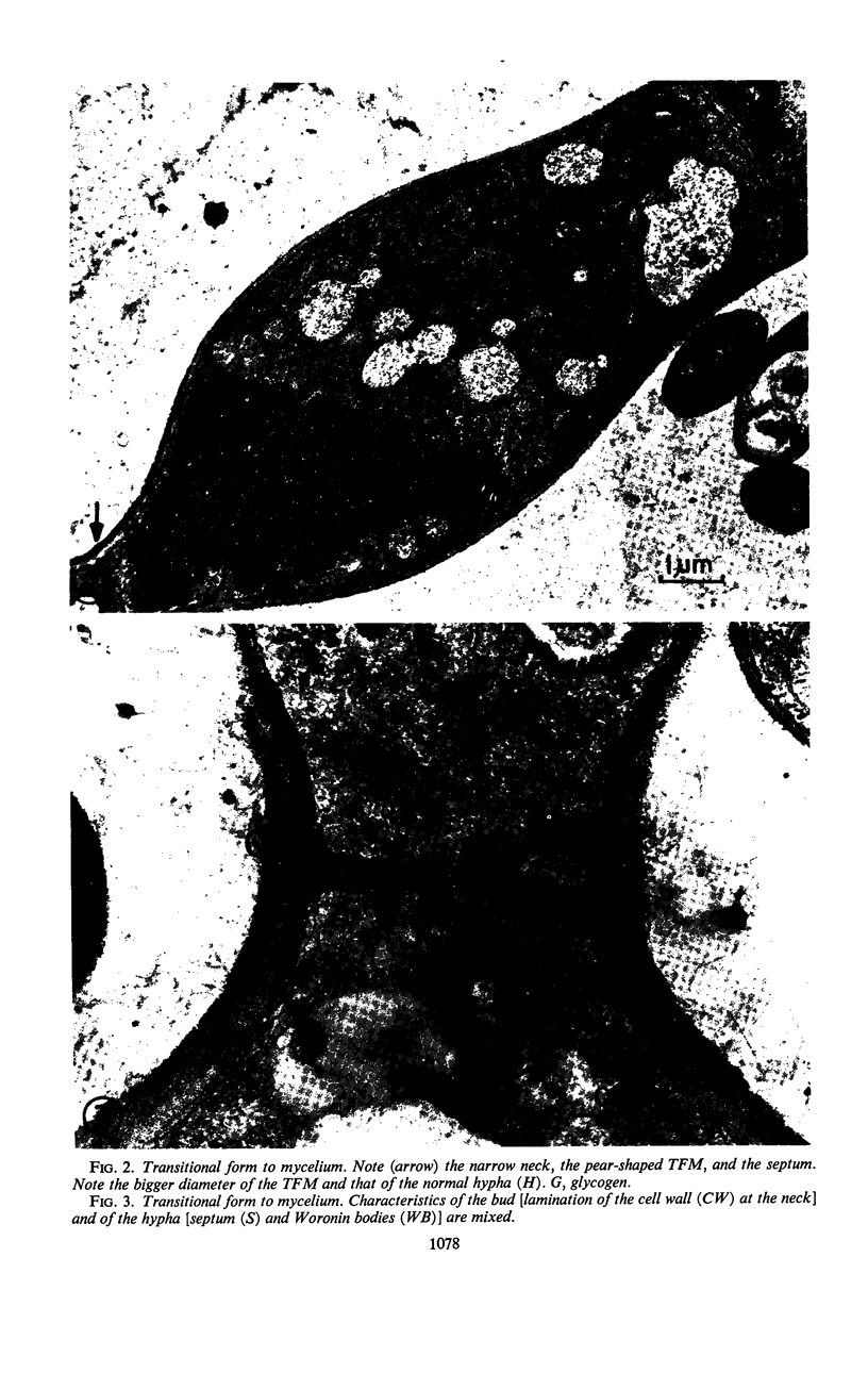
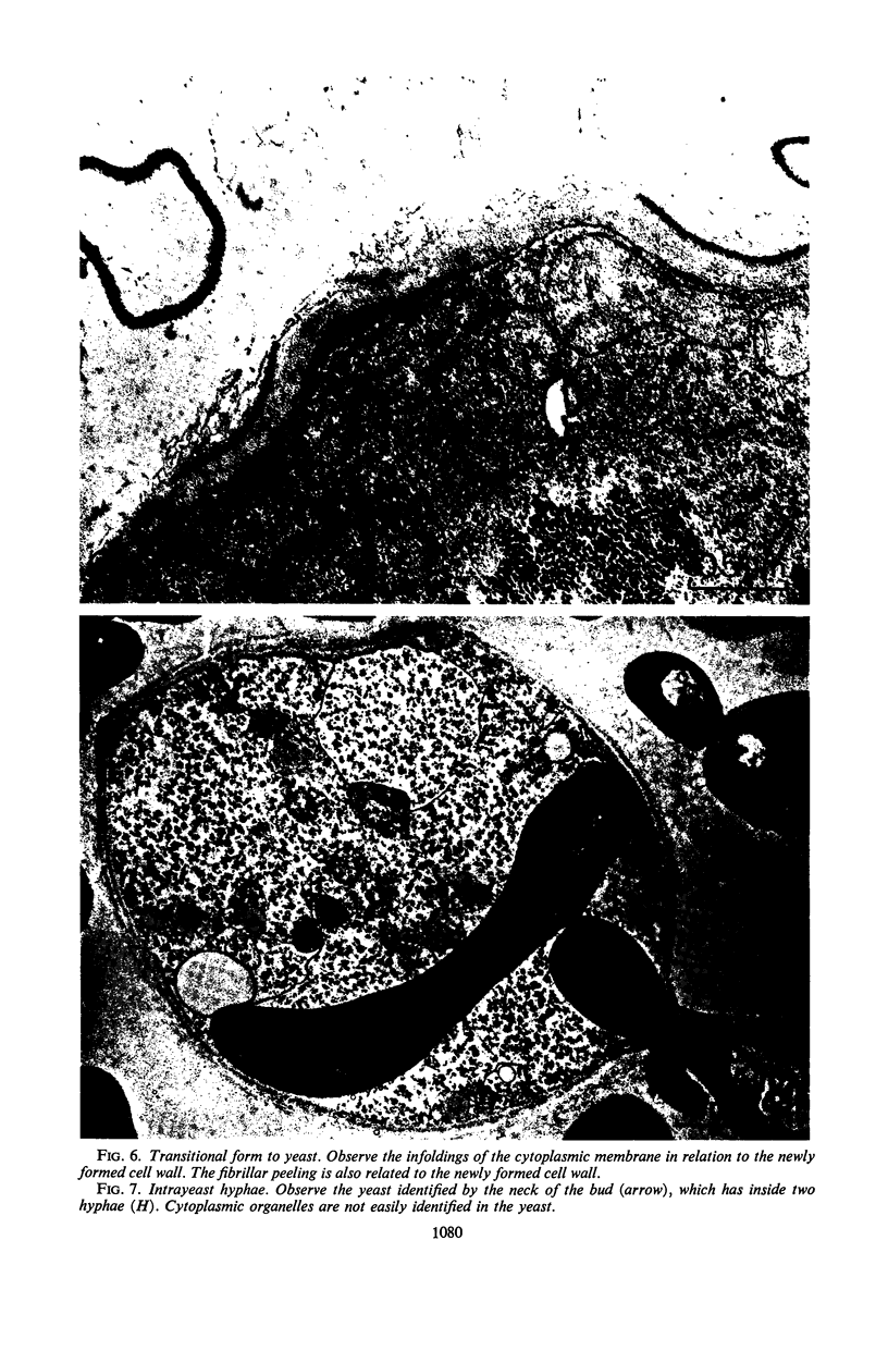
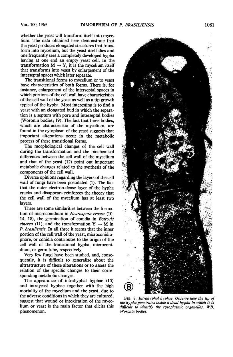
The fine structure of Paracoccidioides brasiliensis undergoing temperature-dependent transformation from mycelium to yeast and vice versa (M ⇌ Y) was studied. The transitional form to mycelium from the yeast appears as an elongated bud that extends from the yeast and which has a mixture of characteristics from both the yeast and the mycelium. The transitional form to yeast from the mycelium starts with enlargement of the interseptal spaces and cracking of the outer electron-dense layer of the cell wall of the hypha. Later the interseptal spaces tend to become round and separate. In M → Y only few interseptal spaces seem to transform. The yeast is produced by self-transformation of the hypha. In Y → M a new structure is formed and the yeast dies. Intrahyphal hyphae are observed during the transformation from M → Y, and intrayeast hyphae during the Y → M. Due to the high mortality and breakage observed in both types of transformations, we believe that wound of the yeast or the mycelium could elicit this phenomenon.
Full text
PDF
Images in this article
Selected References
These references are in PubMed. This may not be the complete list of references from this article.
- CARBONELL L. M., POLLAK L. ULTRAESTRUCTURA DEL PARACOCCIDIOIDES BRASILIENSIS EN CULTIVOS DE LA FASE LEVADURIFORME. Mycopathol Mycol Appl. 1963 Jun 15;19:184–204. doi: 10.1007/BF02051247. [DOI] [PubMed] [Google Scholar]
- CARBONELL L. M., RODRIGUEZ J. TRANSFORMATION OF MYCELIAL AND YEAST FORMS OF PARACOCCIDIOIDES BRASILIENSIS IN CULTURES AND IN EXPERIMENTAL INOCULATIONS. J Bacteriol. 1965 Aug;90:504–510. doi: 10.1128/jb.90.2.504-510.1965. [DOI] [PMC free article] [PubMed] [Google Scholar]
- Carbonell L. M. Cell wall changes during the budding process of Paracoccidioides brasiliensis and Blastomyces dermatitidis. J Bacteriol. 1967 Jul;94(1):213–223. doi: 10.1128/jb.94.1.213-223.1967. [DOI] [PMC free article] [PubMed] [Google Scholar]
- Carbonell L. M., Rodriguez J. Mycelial phase of Paracoccidioides brasiliensis and Blastomyces dermatitidis: an electron microscope study. J Bacteriol. 1968 Aug;96(2):533–543. doi: 10.1128/jb.96.2.533-543.1968. [DOI] [PMC free article] [PubMed] [Google Scholar]
- DROUHET E., ZAPATER R. C. Phase levure et phase filamenteuse de Paracoccidioides brasiliensis; étude des noyaux. Ann Inst Pasteur (Paris) 1954 Oct;87(4):396–403. [PubMed] [Google Scholar]
- FREEMAN J. A., SPURLOCK B. O. A new epoxy embedment for electron microscopy. J Cell Biol. 1962 Jun;13:437–443. doi: 10.1083/jcb.13.3.437. [DOI] [PMC free article] [PubMed] [Google Scholar]
- Furtado J. S., de Brito T., Freymuller E. The structure and reproduction of Paracoccidioides brasiliensis in human tissue. Sabouraudia. 1967 Feb;5(3):226–229. [PubMed] [Google Scholar]
- HAWKER L. E., HENDY R. J. AN ELECTRON-MICROSCOPE STUDY OF GERMINATION OF CONIDIA OF BOTRYTIS CINEREA. J Gen Microbiol. 1963 Oct;33:43–46. doi: 10.1099/00221287-33-1-43. [DOI] [PubMed] [Google Scholar]
- KARNOVSKY M. J. Simple methods for "staining with lead" at high pH in electron microscopy. J Biophys Biochem Cytol. 1961 Dec;11:729–732. doi: 10.1083/jcb.11.3.729. [DOI] [PMC free article] [PubMed] [Google Scholar]
- Kanetsuna F., Carbonell L. M., Moreno R. E., Rodriguez J. Cell wall composition of the yeast and mycelial forms of Paracoccidioides brasiliensis. J Bacteriol. 1969 Mar;97(3):1036–1041. doi: 10.1128/jb.97.3.1036-1041.1969. [DOI] [PMC free article] [PubMed] [Google Scholar]
- Lowry R. J., Durkee T. L., Sussman A. S. Ultrastructural studies of microconidium formation in neurospora crassa. J Bacteriol. 1967 Nov;94(5):1757–1763. doi: 10.4148/1941-4765.1976. [DOI] [PMC free article] [PubMed] [Google Scholar]
- Lowry R. J., Sussman A. S. Intra-hyphal hyphae in "clock" mutants of Neurospora. Mycologia. 1966 Jul-Aug;58(4):541–548. [PubMed] [Google Scholar]
- Oulevey-Matikian N., Turian G. Contrôle métabolique et aspects ultrastructuraux de la conidiation (macro-microconidies) de Neurospora crassa. Arch Mikrobiol. 1968;60(1):35–58. [PubMed] [Google Scholar]
- WATSON M. L. Staining of tissue sections for electron microscopy with heavy metals. J Biophys Biochem Cytol. 1958 Jul 25;4(4):475–478. doi: 10.1083/jcb.4.4.475. [DOI] [PMC free article] [PubMed] [Google Scholar]