Abstract
Infection is a devastating complication following total knee replacement (TKR). In the majority of cases, single- or two-stage revision has excellent results in eradicating infection and restoring function. Rarely, recurrent infection requires alternative treatments such as resection, amputation, or arthrodesis. A review of infections following TKR treated at two joint replacement centers identified 29 cases of resistant knee sepsis treated with a long intramedullary fusion nail. Clinical outcome and radiographs were reviewed at an average follow-up of 48 months (13–114). After the initial intramedullary arthrodesis union occurred in 24 of 29 patients (83%). The average time to fusion was 6 months (3–18 months). Failures included two cases of nail breakage, one of which subsequently achieved fusion following revision nailing, and three cases of recurrent infection requiring nail removal and permanent resection. At a minimum 2-year follow-up, 28% of the patients that achieved fusion complained of pain in the fused knee, 28% complained of ipsilateral hip pain, and two patients complained of contralateral knee pain. Four of the 25 fused patients (16%) remained nonambulatory after fusion, 17 required walking aids (68%) and only four ambulated unassisted. There was no association between age, number of previous procedures, the use of two-stage versus single stage technique, or infecting organism and failure of arthrodesis. Intramedullary arthrodesis is a viable treatment for refractory infection after TKR. Patients undergoing fusion should be informed of the potential for nonunion, recurrence of infection, pain in the ipsilateral extremity, and the long-term need for walking aids.
Introduction
Despite many recent advancements in surgical technique and implant design in total knee replacement (TKR), infection remains a significant source of failure. Excellent success rates have been reported for revision for infection [1, 2]; however, recurrent periprosthetic sepsis is a devastating problem, which frequently requires resection arthroplasty, arthrodesis, or amputation [3–7].
Few patients or surgeons are willing to agree to amputation, and arthrodesis has gained increased acceptance in the treatment of resistant periprosthetic sepsis about the knee. In recent series, septic complications following TKR have become the most common indication for knee arthrodesis [3, 5]. A variety of devices and techniques have been devised and implemented with the goals of achieving a solid knee fusion, yet the most effective technique for arthrodesis in the setting of infection remains controversial [3, 4, 8].
There are numerous reports in the literature with fusion rates ranging from 50 to 100% for a variety of techniques [9–11]. External fixation historically has been a very effective technique for arthrodesis, particularly in the face of infection [12]. However, after TKR severely compromised bone stock may require prolonged use of a fixator resulting in a high complication rate with a more variable outcome [8–10, 13]. Plating is less popular, as the suboptimal soft tissues of the multiply operated knee may compromise coverage of bulky hardware [14, 15]. Intramedullary devices have become increasingly popular, yet there are few large series in the literature reporting results for this technique exclusively in the management of failed TKR due to sepsis [16–20].
In this study, we review the results of intramedullary arthrodesis using a long nail performed at two centers in a series of 29 consecutive patients with knee sepsis after TKR. This is one of the largest series using modern intramedullary techniques in the management of failed septic TKR.
Materials and methods
A retrospective review of all patients requiring reoperation for sepsis after TKR identified 30 patients treated with arthrodesis using a long intramedullary nail. Medical records were reviewed including demographic data, operative reports, and preoperative and postoperative radiographs. Postoperative and follow-up evaluations were performed in the clinical setting including assessment of musculoskeletal pain in the extremities, tenderness at the fusion site and ambulation. In two cases, patients were unable to return for final follow-up examinations and telephone interviews were performed and patients referred to local institutions for radiographs, which were mailed to us. A subset of eight patients agreed to answer questionnaires consisting of visual analog scales for pain and function. Follow-up radiographs were reviewed for evidence of bridging trabeculae across the knee joint indicative of fusion on two views. All patients were followed at regular intervals until fusion and for a minimum of 1 year. One patient relocated and refused to return for follow-up and was excluded, leaving 29 patients with minimum follow-up of 1 year for analysis.
All patients were treated with a long curved nail extending from the hip or isthmus of the proximal femur to just above the ankle joint. Twenty-five of 29 patients were treated using a nonmodular titanium nail (Biomet Co, Warsaw, IN), whereas four patients were treated with a reamed modular nail (Neff nail, Zimmer Co, Warsaw, IN). In 25 cases, a two-stage technique was used with initial debridement and removal of the prosthesis, insertion of a cement spacer, and 4–6 weeks course of culture specific intravenous antibiotics followed by arthrodesis after resolution of the infection. In the remaining four patients, a single-stage procedure was performed because of significant medical comorbidities, a history of multiple debridements with component retention and a benign appearance of the tissues intraoperatively.
The average age of the patients was 67 years ranging from 35 to 82. The primary diagnosis was osteoarthritis in 21 patients, rheumatoid arthritis in two, lupus arthritis in two, Charcot arthropathy in one, posttraumatic arthritis in one and gout in one patient. Twelve of the patients were male and 17 female. The average number of previous procedures for all patients was 4.5 (2–10).
The indication for arthrodesis was septic failure of previous two-stage revision in 12 patients, septic primary TKR and extensor mechanism disruption in five patients, septic TKR after previous revision for nonseptic failure in one, and failed fusion with external fixation following septic primary TKR in one. In nine patients fusion was performed for septic primary TKR following multiple debridements with prosthesis retention. This occurred in patients at the extremes of age with either severe medical illness or the desire for a definitive procedure that would not risk future amputation.
Implants and surgical technique
The majority of arthrodeses (25 of 29) were performed with the use of a long cylindrical titanium nail (Biomet Co.) capable of accepting a single proximal and two distal interlocking screws.
The original, longitudinal incision was used for exposure followed by debridement and removal of retained spacers and components, along with excess scar tissue and any bone suspicious for osteomyelitis. The bone ends were freshened and prepared using resection guides so that cancellous bone was exposed on the opposing surfaces of the femur tibia, and the overall alignment of the limb was neutral to slight valgus. An intramedullary ball-tip guide wire was introduced distally into the tibial shaft to the plafond, and the canal was sequentially reamed until the cortex was engaged at the tibial isthmus. This canal width determined the size of the rod, and the tibial length was measured using the guide rod as a reference.
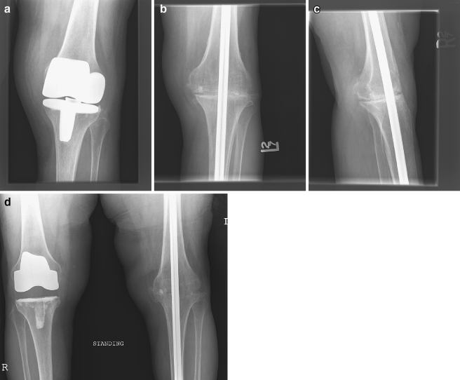
The ball tip guide wire was then passed retrograde into the femoral shaft until the tip contacted the piriformis recess. The femoral canal was reamed until it matched the size of the tibial reamer, and femoral length was measured using the guide rod at the piriformis fossa as a reference. Subtracting 1 cm from the combined length of the femur and tibial measurements determined the appropriate rod length. In a retrograde fashion, the guide wire was then tapped through the piriformis recess with a mallet. The guide wire was advanced until it could be easily palpated under the skin of the hip with the leg placed in an adducted position. A skin incision was made over the guide wire and dissection was carried down through the gluteal musculature to the piriformis recess. The recess was reamed to 1 mm larger than the final tibial and femoral reamer size. After the reaming, the appropriately sized curved nail was introduced antegrade into the femur with care being taken to insert the rod with the curve positioned anteromedially down the femoral shaft. The rod will then come through the tibia in valgus alignment with slight flexion at the knee, which is desirable. An axial load was placed on the proximal tibia against the distal end of the femur during rod insertion. Sometimes, the rod forced the anterior tibial flare forward making closure of the arthrotomy difficult. When this problem occurred, the surgeon modified the anterior flare with a reciprocating saw. Resected bone was then used as autograft at the fusion site, along with the patella. Proximal and distal locking was performed based on the discretion of the surgeon. When static locking was performed, the nail was routinely dynamized at 3 to 6 months postoperatively (Fig. 1).
Fig. 1.
A 69-year-old female with polymicrobial infection of a left TKR and chronic extensor mechanism dysfunction (A) was treated with two-stage intramedullary arthrodesis using a long-locked titanium nail. At 6 months there is evidence of early fusion with partial bridging callous present (B and C). The patient underwent dynamization with removal of the distal interlocks, and at 15 months a solid fusion with bridging trabeculae is present (D)
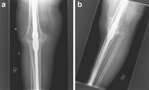
The modular Neff nail (Zimmer Co.) was utilized in four cases (Fig. 2). The nail is composed of two titanium-alloy components with longitudinally machined splines, which are inserted separately into the femur and tibia after reaming, and then joined at the level of the knee by a press-fit couple secured by multiple set screws. The nail is available in a single length and, therefore, must be cut intraoperatively with a high-speed burr to match the appropriate length of the femur and tibia. The modular nails were performed using similar preoperative and postoperative protocols. The complete surgical technique has been described previously [21].
Fig. 2.
A solid fusion of the knee after intramedullary arthrodesis with the modular Neff nail (Zimmer Co, Warsaw, IN)
Results
All 29 patients were available for minimum 1-year follow-up. One patient died of unrelated causes at 18 months, after achieving fusion at 1 year. The remaining 28 patients were available at minimum 2 years. Average final follow-up for the entire group was 48 months (13–114).
Radiographic fusion indicated by bridging bony trabeculae on AP and lateral views of the knee was present in 24 of the 29 patients following the index procedure at an average of 6 months (range 4 to 18 months), indicating a fusion rate of 83% following the initial arthrodesis procedure.
The most common infecting organism was Staph aureus, which was present in the cultures of 11 patients, including one case of methicillin resistance. Other infecting organisms included S. epidermidus in five cases, E. coli in one, Candida albicans in one and seven cases of polymicrobial infection. In two cases, the cultures remained negative despite gross purulence noted intraoperatively and in two cases culture data was unavailable as the patients were initially treated with planned chronic resection arthroplasty before referral for arthrodesis.
Four patients suffered from recurrent or persistent infection before achieving union. One patient with a persistent Candidal infection presented with nail breakage and was treated with revision nailing but remains ununited, likely with chronic infection. Three patients required nail removal and redebridement followed by resection arthroplasty for recurrent infection. In two cases, cultures were consistent with the prior prosthetic infection and in one case a new organism was detected at the time of resection. A fifth patient presented with aseptic nonunion and nail breakage and underwent revision intramedullary nailing and bone grafting and subsequently achieved fusion at 7.2 months, making the final fusion rate 86% in this series. None of the patients who had undergone a single-stage fusion underwent reoperation for recurrent sepsis or nonunion, and all went on to successful fusion.
An additional complication occurred in one patient with osteoporosis who sustained a nondisplaced ankle fracture at the distal interlock, which was treated successfully in a CAM walker.
Analysis of the failures demonstrated no significant correlation between age, infecting organism, or number of previous procedures. Two of the patients treated for recurrent infection were significantly immune-compromised including one patient with lupus and renal failure treated with chronic steroids and another patient with diabetes mellitus and cirrhosis.
Clinical follow-up demonstrated that 21 of 25 patients who achieved fusion returned to ambulation; however, a significant number of patients had limited walking tolerance and required the use of an assistive device. Eleven patients ambulated with the use of a cane, four with the use of a walker, and two patients returned to ambulation with crutches; however, one of these patients was able to ambulate for only a short time before becoming restricted to a wheelchair because of medical comorbidities. Four patients never returned to ambulation and required a wheelchair.
Seven of 29 (24%) of fused patients reported mild or moderate ipsilateral knee pain; in two cases this was attributed to neuropathic pain, which remained chronic. Pain in the ipsilateral hip was reported by seven patients (24%). Two patients reported ipsilateral ankle pain and two reported contralateral knee pain with radiographic evidence of osteoarthritis. The average leg length discrepancy was 3 cm (1–6 cm).
A subset of eight patients who achieved fusion agreed to answer a questionnaire composed of visual analog scales for pain and function. At an average follow-up of 42 months, the average score for pain was 30 out of 40 points, the average score for ambulation was eight out of 20 and the average score for function was 10 out of 20 points.
Discussion
Intramedullary arthrodesis remains a useful technique for the treatment of failed TKR secondary to infection, with a reported fusion rate of 83% in this series, with the majority of failures occurring secondary to recurrent fulminant infection. Fusion was ultimately achieved in 86% of patients after revision nailing in one patient with aseptic nonunion.
The benefits of intramedullary nailing include rigid load-sharing internal fixation, which allows immediate full weight-bearing and limited exposure and dissection, preserving and simplifying soft-tissue coverage [3, 22]. With the increased popularity of intramedullary fixation of femur fractures, surgeons have gained technical proficiency with intramedullary techniques, making this method increasingly appealing [4]. However, the use of an intramedullary internal fixation device in the setting of sepsis carries a significant risk of persistent infection with the potential for intramedullary spread and ultimate failure as demonstrated in four of 29 cases (14%) in this series.
Although these results are consistent with other published series for arthrodesis following infected TKR (Table 1), the significant number of complications requiring reoperation emphasizes the severity of this condition. In previous reports of intramedullary arthrodesis for septic TKR, rates of reoperation for recurrent infection have varied. Using a long nonmodular fusion nail, Crockerell recently reported a 10% incidence of reoperation for deep infection [19], whereas both Wilde and Bargiotas each reported a 17% rate of recurrence using similar devices [18, 23]. Whereas experience with modular intramedullary fusion devices is less extensive for septic TKR, the available literature indicates recurrence rates ranging from 0 to 12% [17, 20, 21]. Although there were no recurrences in this series in patients undergoing a single-stage procedure, a number of authors have recommended use of a two-stage technique including eradication of infection followed by nailing when using an intramedullary device [10, 17, 24]. Use of preoperative aspiration, lab tests such as CRP and ESR, intraoperative frozen section, and serial debridements may also be beneficial in avoiding recurrent infection and improving fusion [3].
Table 1.
Treatment of failed septic TKR with intramedullary arthrodesis
| Author | # of Knees | Technique | Fusion (%) | Interval (mo) | Complications/Reoperation (%) |
|---|---|---|---|---|---|
| Lai et al. | 31 | IM nail—short Huckstep | 93.5 | 5.2 | 10 |
| McQueen et al. | 26 | IM nail—Witchita | 100 | 3.9 | 15 |
| Waldman et al. | 21 | IM nail—Neff | 95 | 6.3 | 10 |
| Bargiotas et al. | 12 | IM nail—Biomet | 83 | 7 | 25 |
| Crockarell et al. | 10 | IM nail—Mayday (custom) | 100 | – | 10 |
| Panagiotopoulos | 9 | IM nail—Mixed | 89 | 6.5 | – |
Although most authors recommend a four to six week course of intravenous culture specific antibiotics as part of treatment protocols for both single-stage and two-stage procedures in the treatment of periprosthetic sepsis, postoperative antibiotic regimens are significantly more variable. Advocated by some authors [25, 26], the use of chronic suppressive oral antibiotics following surgical treatment of periprosthetic infection remains controversial. At an average 5 years, Rao et al. reported a success rate of 86% for chronic oral suppression after debridement and retention of septic total joint replacements [26]. In another series, medical complications were reported in greater than 20% of patients on long-term suppression for orthopedic implants [27]. After arthrodesis for infection, there is also little consensus on the use of long-term oral suppression and most published reports have been inconsistent in the use of chronic suppression postoperatively. Chronic oral suppression was not used routinely in our patients, but may have provided some benefit in lowering the rate of recurrent fulminant infection. Whereas there is insufficient evidence to recommend the routine use of chronic suppression following arthrodesis for sepsis, it should be considered in high-risk immune-compromised patients with higher virulence organisms.
Numerous authors have suggested that arthrodesis using external fixation is preferred for septic TKR, as the absence of a foreign body at the site of infection may reduce the risk of recurrence [8, 9]. In addition, Ilizarov and other modern frame designs may allow more accurate correction of deformity and maintenance of alignment. However, many surgeons and patients are hesitant to accept external fixation for a number of reasons. Fixators may be bulky and cumbersome for patients, and they frequently require prolonged application to achieve fusion [8, 10, 22]. Pin tract-infections are extremely common, and other more serious complications such as fracture of the femur or tibia through pin sites are reported with a significant frequency [8, 9]. Neurovascular structures are also at risk when numerous pins and complex fixation schemes are employed to achieve the stability necessary for fusion using these devices after failure of knee replacement surgery.
An analysis of results for the two different nail designs used in this series was not possible based on the limited number of modular nails used. A number of theoretical differences are apparent based on differences in design and surgical technique. The modular Neff nail has the advantage of requiring only a single incision at the knee joint and also does not utilize locking at the hip or ankle. Disadvantages to the use of this device include the need for intraoperative modification with a high-speed cutting tool, and the limited amount of compression that can be achieved across the fusion site upon coupling of the nail in situ. Although it was not required in this series, removal of the Neff nail after arthrodesis if necessary may be technically demanding, requiring one or more bone windows and removal in segments using metal cutting burrs [21]. Both nails are limited in their ability to correct deformity and restore valgus alignment, which is performed by careful insertion of the bowed portion of the nail.
The ultimate functional outcome after arthrodesis was significantly compromised for a number of patients in this series, with 17% of patients ultimately unable to ambulate and 84% of patients requiring assistive devices for locomotion. Previous studies have demonstrated a significant elevation in the energy cost associated with ambulation after arthrodesis of the knee [28]. These costs are particularly evident in elderly patients who may return to a period of ambulation only to become increasingly dependent as medical and other musculoskeletal comorbidities become more significant with aging. Alternative treatments such as transfemoral amputation are reported to carry an even greater energy cost for ambulation compared to arthrodesis, requiring double the oxygen consumption in one study [3]. However, comparative studies in similar compromised patients undergoing amputation for treatment of septic TKR are not available to determine which procedure provides the most lasting function and patient satisfaction.
Consistent with other reports was the incidence of ipsilateral hip and ankle pain in this series [3, 11, 19]. Arthrodesis increases the forces transmitted across adjacent joints, and patients should be informed of the potential for ipsilateral joint pain particularly in the setting of preexisting multifocal arthritis. Contralateral knee pain and osteoarthritis was present in two patients, who were resistant to undergo contralateral TKR.
A significant finding in this series was the occurrence of ipsilateral knee pain in seven patients (28%) despite radiographic fusion. Reports of pain after successful arthrodesis of the knee are varied. Morrey reported a 15% incidence of knee pain despite solid arthrodesis using a number of techniques after infected TKR [29]. Waldman reported a 33% incidence of pain in the ipsilateral knee after intramedullary arthrodesis in 21 patients [17], whereas Crockarell reported a 36% incidence of knee pain after intramedullary arthrodesis at an average follow-up of 7 years [19]. Our results are consistent with these reports. Reasons for pain include neuropathic pain and complex regional pain syndrome, a known complication in patients undergoing multiple surgeries, which occurred in two of our patients. Other potential reasons for pain include a residual focus of osteomyelitis, local injury to soft tissues from previous surgery or infection, muscle fatigue secondary to the altered anatomy, and subclinical or undetected delayed or nonunion. Patients undergoing arthrodesis should be informed of the possibility of continued mild to moderate knee pain following arthrodesis.
The strengths of our study include the large group of patients in our series, all with a diagnosis of infection following TKR. Weaknesses include the retrospective nature of the review. Only eight of the 29 patients agreed to answer questionnaires including visual analog scores, as many of the patients were elderly or significantly ill at final follow-up examination. Whereas some studies have reported modified knee outcome scores for patients with arthrodesis [18, 19], these scores have not been validated for this purpose and may poorly reflect the actual function and satisfaction of these patients.
Intramedullary arthrodesis remains an effective technique for arthrodesis following septic failure of TKR. Patients should be informed preoperatively and followed carefully for a number of potential complications including nonunion, recurrent infection, pain in the ipsilateral hip, knee and ankle, and limited walking tolerance requiring the use of assistive devices in the majority of patients. Severely compromised patients and patients with more virulent infectious organism such as fungus, MRSA, or polymicrobial infection may be at a higher risk for recurrent infection and failure, and should be treated aggressively and counseled on the potential for resection arthroplasty and amputation.
References
- 1.Segawa H, Tsukayama DT, Kyle RF, Becker DA, Gustilo RB (1999) Infection after total knee arthroplasty. A retrospective study of the treatment of eighty-one infections. J Bone Jt Surg Am 81:1434–1445 [DOI] [PubMed]
- 2.Windsor RE, Insall JN, Urs WK, Miller DV, Brause BD (1990) Two-stage reimplantation for the salvage of total knee arthroplasty complicated by infection. A further follow-up and refinement of indications. J Bone Jt Surg Am 72:272–277 [PubMed]
- 3.Conway JD, Mont MA, Bezwada HP (2004) Current concepts review: arthrodesis of the knee. J Bone Jt Surg Am 86:835–848 [DOI] [PubMed]
- 4.Weidel JD (2002) Salvage of infected total knee fusion: The last option. Clin Orthop 404:139–142 [DOI] [PubMed]
- 5.Hanssen AD, Trousdale RT, Osmon DR (1995) Patient outcome with reinfection following reimplantation for the infected total knee arthroplasty. Clin Orthop 321:55–67 [PubMed]
- 6.Damron TA, McBeath AA (1995) Arthrodesis following failed total knee arthroplasty: comprehensive review and meta-analysis of recent literature. Orthopedics 18:361–368 [DOI] [PubMed]
- 7.Wasielewski RC, Barden RM, Rosenberg AG (1996) Results of different surgical procedures on total knee arthroplasty infections. J Arthroplast 11:931–938 [DOI] [PubMed]
- 8.Salem KH, Keppler P, Kinzl L, Schmelz (2006) Hybrid external fixation for arthrodesis in knee sepsis. Clin Orthop May, Epub ahead of print [DOI] [PubMed]
- 9.Oostenbroek HJ, vanRoermund PM (2001) Arthrodesis of the knee after an infected arthroplasty using the Ilizarov method. J Bone Jt Surg Am 83:50–54 [DOI] [PubMed]
- 10.Knutson K, Hovelius L, Lindstrand A, Lidgren L (1984) Arthrodesis after failed knee arthroplasty. A nationwide multicenter investigation of 91 cases. Clin Orthop 191:202–211 [PubMed]
- 11.Lai KA, Shen WJ, Yang CY (1998) Arthrodesis with a short Huckstep nail as a salvage procedure for failed total knee arthroplasty. J Bone Jt Surg Am 80:380–388 [DOI] [PubMed]
- 12.Charnley J (1960) Arthrodesis of the knee. Clin Orthop 18:37–42
- 13.Rand JA, Bryan RS, Chao EY (1987) Failed total knee arthroplasty treated by arthrodesis of the knee using the Ace-Fischer apparatus. J Bone Jt Surg Am 69:39–45 [PubMed]
- 14.Lucas DB, Murray WR (1961) Arthrodesis of the knee by double-plating. J Bone Jt Surg Am 43:795–808
- 15.Nichols SJ, Landon GC, Tullos HS (1991) Arthrodesis with dual plates after failed total knee arthroplasty. J Bone Jt Surg Am 73:1020–1024 [PubMed]
- 16.Vlasak R, Gearen PF, Petty W (1995) Knee arthrodesis in the treatment of the failed total knee replacement. 321:138–144 [PubMed]
- 17.Waldeman BJ, Mont MA, Payman KR, Freiberg AA, Windsor RE, Sculco TP, Hungerford DS (1999) Infected total knee arthroplasty treated with arthrodesis using a modular nail. Clin Orthop 367:230–237 [PubMed]
- 18.Bargiotas K, Wohlrab D, Sewecke JJ, Lavigne G, DeMeo PJ, Sotereanos NG (2006) Arthrodesis of the knee with a long intramedullary nail following the failure of a total knee arthroplasty as the result of infection. J Bone Jt Surg Am 88:553–558 [DOI] [PubMed]
- 19.Crockarell JR, Mihalko MJ (2005) Knee arthrodesis using an intramedullary nail. J Arthroplast 20:703–708 [DOI] [PubMed]
- 20.McQueen DA, Cooke FW, Hahn DL (2006) Knee arthrodesis with the Wichita fusion nail. An outcome comparison. Clin Orthop 446:132–139 [DOI] [PubMed]
- 21.Arroyo JS, Garvin KL, Neff JR (1997) Arthrodesis of the knee with a modular titanium intramedullary nail. J Bone Jt Surg Am 79:26–35 [DOI] [PubMed]
- 22.MacDonald JH, Agarwal A, Lorei MP, Johanson NA, Freiberg AA (2006) Knee Arthrodesis 14:154–163 [DOI] [PubMed]
- 23.Wilde AH, Stearns KL (1989) Intramedullary fixation for arthrodesis of the knee after infected total knee arthroplsty. Clin Orthop 248:87–92 [PubMed]
- 24.Donley BG, Mathews LS, Kaufer H (1991) Arthrodesis of the knee with an intramedullary nail. J Bone Jt Surg Am 73:907–913 [PubMed]
- 25.Knutson K, Lindstrand A, Lindgren L (1985) Arthrodesis for failed total knee arthroplasty. J Bone Jt Surg Br 67:47–52 [DOI] [PubMed]
- 26.Rao N, Crossett LS, Sinha RK, Le Frock JL (2003) Long-term suppression of infection in total joint arthroplasty. Clin Orthop Relat Res 414:55–60 [DOI] [PubMed]
- 27.Segreti J, Nelson JA, Trenholme GM (1998) Prolonged suppressive antibiotic therapy for infected orthopedic prosthesis. Clin Infect Dis 27:714–716 [DOI] [PubMed]
- 28.Waters RL, Perry J, Antonelli D, Hislop H (1976) Energy cost of walking of amputees: the influence of the level of amputation. J Bone Jt Surg Am 58:42–46 [PubMed]
- 29.Morrey BF, Westholm F, Schoifet S, Rand JA, Bryan RS (1989) Long term results for various treatment options for infected total knee arthroplasty. Clin Orthop 248:120–128 [PubMed]
- 30.Panagiotopoulos E, Kouzelis A, Matzaroglou C, Saradis A, Lambiris E (2006) Intramedullary knee arthrodesis as a salvage procedure after failed total knee replacement. Int Orthop, Epub ahead of print [DOI] [PMC free article] [PubMed]


