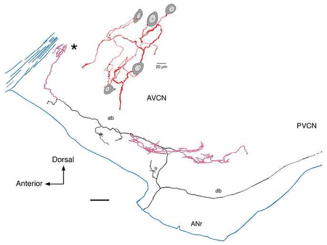
Figure 3.
Lateral view of a low SR fiber as it collateralizes (red) in the rostral and lateral small cell cap (CF=0.45 kHz; SR=1.2 s/s; Th=34 dB SPL). The collateral in the rostral AVCN (*) is enlarged to show en passant and terminal swellings that lie in close contact with small cells of various shapes (oval or polygonal). This collateral shows how small cells received multiple inputs from collateral branches from a single auditory nerve fiber and how a single collateral can distribute divergent terminals to multiple small cells. This figure was modified from figure 15 of Fekete et al., 1984 and figure 1B of Ryugo and Rouiller, 1988. An electron micrograph of this fiber forming an axosomatic contact with a small cell is shown in figure 1 of Rouiller et al., 1986. The scale bar is 0.2 mm.

