Abstract
The human polyomavirus JC (JCV) causes the central nervous system demyelinating disease progressive multifocal leukoencephalopathy. Previously, we showed that 40% of Caucasians in the United States excrete JCV in the urine as detected by PCR. We have now studied 68 Navaho from New Mexico, 25 Flathead from Montana, and 29 Chamorro from Guam. By using PCR amplification of a fragment of the VP1 gene, JCV DNA was detected in the urine of 45 (66%) Navaho, 14 (56%) Flathead, and 20 (69%) Chamorro. Genotyping of viral DNAs in these cohorts by cycle sequencing showed predominantly type 2 (Asian), rather than type 1 (European). Type 1 is the major type in the United States and Hungary. Type 2 can be further subdivided into 2A, 2B, and 2C. Type 2A is found in China and Japan. Type 2B is a subtype related to the East Asian type, and is now found in Europe and the United States. The large majority (56–89%) of strains excreted by Native Americans and Pacific Islanders were the type 2A subtype, consistent with the origin of these strains in Asia. These findings indicate that JCV infection of Native Americans predates contact with Europeans, and likely predates migration of Amerind ancestors across the Bering land bridge around 12,000–30,000 years ago. If JCV had already differentiated into stable modern genotypes and subtypes prior to first settlement, the origin of JCV in humans may date from 50,000 to 100,000 years ago or more. We conclude that JCV may have coevolved with the human species, and that it provides a convenient marker for human migrations in both prehistoric and modern times.
JCV is a human polyomavirus that causes a rare fatal brain infection of oligodendrocytes known as progressive multifocal leukoencephalopathy (PML) in immunocompromised individuals (1). PML occurs in about 5% of patients who are dying of AIDS. However, the infection rate is much higher. In a control group comprising general medical clinic patients without overt immunosuppression and healthy volunteers from Pennsylvania and Maryland, 40% excreted JCV in the urine (2).
JCV exists as five or more geographically based genotypes, which have been defined in the United States, Africa, and parts of Europe and Asia (2–7). The most common types of JCV in the United States are type 1 (European), type 2 (Asian), type 3 (African), and type 4 (United States) (2). JCV infection in the United States is still population associated, meaning that individuals of European ancestry excrete mainly type 1. The source of most type 2 strains in the United States is unknown, although both types 2A (Northeast Asia, including China and Japan) and 2B [presumably western Asian in origin for which the prototype is strain GS from Germany (8)] are found. To date, type 3 and another African genotype, type 6 [also termed type C (6)], have not been found in Americans of European origin. The population of origin of type 4 is unknown. Type 4 has been observed only in the United States (2), predominantly in African Americans (unpublished data), and may represent recombination between a type 1 strain and a short fragment of a type 3 strain (9). [A strain tentatively defined as type 5 has been reclassified as a subtype of type 3 (7), and a new genotype has not yet been assigned to type 5.]
In Native Americans and Pacific Islanders, the genotyping of JCV has the potential to reveal the origins and relationships of these people of presumed Asiatic descent. We hypothesized that Native Americans would carry an East Asian form of the virus, as would inhabitants of Oceania. Accordingly, we studied urine from the Navaho, the Flathead, and the Chamorro indigenous peoples.
People of the Navaho nation, who represent the largest Native American group in the United States, are today found primarily in New Mexico, Arizona, and Utah. They speak a language of the Athabaskan group, which is also spoken in the northwestern part of Canada. For this reason the Navaho are thought to have migrated to the southwestern United States approximately 1,000 years ago.
The Flathead people are a small group in the Salishan linguistic family who originally inhabited southwestern Montana. There was extensive white settlement among them and intermarriage beginning in the 1850s (10). In the 1870s, the Flathead agreed to leave their aboriginal homelands and move to their present location in central Montana.
The Chamorro are the indigenous people of Guam, which is in the western Pacific Ocean. Geographically, Guam is a part of the Mariana Islands group within Micronesia and was held by Spain for more than 300 years beginning in 1565. The Chamorro, Guam’s largest ethnic group, were likely part of an ancient migration from Asia via the larger islands near the coast onto the distant small islands of the Pacific Ocean. In addition to Spanish admixture, there has been a significant Filipino contribution to the present-day Chamorro. For more detailed information on the Chamorro and on the epidemiology of neurological disease in the Marianas, see ref. 11.
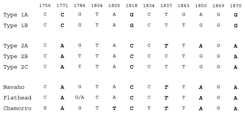
The majority of the JCV strains obtained from these three population groups belong to the Asian genotype (type 2A) or a variant of it. The Chamorro type 2A strains are distinct from those of Native Americans (and East Asians) at position 1805 in the VP1 gene. This is consistent with the Chamorro having a separate and distinct origin and prehistory. These findings strongly support the origin of two Native American populations and a Pacific Island population in Asia.
MATERIALS AND METHODS
Urine Specimens.
Native American probands were outpatients attending general medical clinics in New Mexico or Montana. Urine specimens from Chamorro were collected at the National Institute of Neurological Disorders and Stroke Research Center on Guam in 1980 and stored at −20°C until extracted as outlined below. Demographic features are presented in Table 1.
Table 1.
Age and gender of Native American and Pacific Islander cohorts
| Cohort | Gender | No. tested | Age range, yr | Median |
|---|---|---|---|---|
| Navaho | M | 24 | 12–83 | 50 |
| F | 44 | 12–78 | 39 | |
| Total | 68 | 45 | ||
| Flathead | M | 7 | 19–65 | 44 |
| F | 12 | 31–88 | 52 | |
| Unknown | 6 | 59–63 | 61 | |
| Total | 25 | 57 | ||
| Chamorro | M | 21 | 44–68 | 55 |
| F | 8 | 26–63 | 54 | |
| Total | 29 | 55 |
The control population of 270 individuals from the United States included general clinic patients and healthy volunteers from Pennsylvania and Maryland, as well as cohorts of HIV-infected individuals and multiple sclerosis (MS) patients from California, as described previously (12, 13). These cohorts included representative numbers of African Americans and Hispanic Americans. A European control group from Hungary consisted of 30 MS patients and 30 paired controls (14).
The method for specimen preparation was described previously (2, 9, 13). Briefly, urine (10–15 ml) was centrifuged at 4300 × g for 10 min. The cell pellet was resuspended in 10 ml of PBS and recentrifuged and the supernatant was discarded. Cells were suspended in 100 μl of digestion buffer containing proteinase K, incubated at 55°C overnight in a water bath, and boiled for 10 min.
PCR.
Initially, JCV DNA was detected by PCR amplification of a 129-bp region of the VP1 gene using primers JLP-1 and JLP-4 (12). This fragment of the JCV genome includes four typing sites that distinguish the four major JCV genotypes. Subtypes of type 1 are defined at positions 1843 and 1850 (2, 13). Other samples were analyzed with primers JLP-15 (nucleotides 1710–1734, 5′-ACAGTGTGGCCAGAATTCCACTACC-3′) and JLP-16 (nucleotides 1924–1902, 5′-TAAAGCCTCCCCCCCAACAGAAA-3′), which amplify a 215-bp fragment in the same region. The longer fragment amplified by the JLP-15 and JLP-16 primer pair provides additional typing sites at positions 1756, 1771, and 1786. Other primers included JTP-5 (nucleotides 5621–5642, 5′-CTTTGTTTGGCTGCTACAGTA-3′) and JTP-6 (nucleotides 3896–3877, 5′-GCCTTAAGGAGCATGACTTT-3′), which amplify a 276-bp fragment in the portion of the T-antigen gene encoding the zinc-finger motif. This region is the site of a mutation changing a Gln codon to Leu at amino acid 301 (7). Following an initial denaturation at 94°C for 30 sec, the 50-cycle, two-step PCR program included 1 min for annealing and elongation at 63°C, followed by 1 min for denaturation at 94°C. Reactions were performed by using UlTma DNA polymerase with 3′-5′ proofreading activity (Perkin–Elmer Cetus) in a standard PCR buffer containing 1.5 mM MgCl2.
Amplification of the Entire JCV Genome.
The entire JCV genome was amplified and sequenced as described previously (7, 15).
Cycle Sequencing.
Gel-purified PCR products were cycle sequenced directly by using the Excel kit (Epicentre Technologies, Madison, WI) with the same primers used for DNA amplification end labeled with [33P]ATP (Amersham). The initial denaturation of 1 min at 95°C was followed by 30 cycles of 30 sec at 95°C for denaturation and 1 min at 63°C for annealing and elongation. Products were electrophoresed for 90 or 180 min on a 6% polyacrylamide gel containing 50% (wt/vol) urea (National Diagnostics, Atlanta). Thereafter, gels were fixed with 12% (vol/vol) methanol and 10% acetic acid, transferred to 3MM chromatography paper (Whatman), dried under vacuum, and exposed to Bio-Max X-ray film (Kodak) for 14 to 48 hr.
Genotype Determination.
JCV genotypes were identified in the VP1 gene fragment as described previously (2, 9, 12). Subtypes of type 1 and type 2 were defined as illustrated in Fig. 1.
Figure 1.
Sequence of Native American and Chamorro JCV type 2A compared with previously defined types and subtypes in the VP1 typing fragment. These sites are in the 215-bp fragment amplified by primers JTP-15 and JTP-16. types 1 and 2 are distinguished at sites 1771, 1818, and 1870 (bold). Subtyping sites are located at positions 1843 and 1850 (type 1), or at 1837 and 1850 (type 2) (italic). type 2C is defined by “T” and “G” at these subtyping sites, but may not be an independent group. The large majority of Navaho (41/46), Flathead (9/16), and Chamorro (17/20) strains are type 2A. All 17 Chamorro type 2A strains have a variant “T” at position no. 1805. The invariant positions listed here define types 3, 4, and 6 (not shown). H = A, T, or C; Y = C or T.
Statistical Methods and Software.
JCV type distribution in paired cohorts was analyzed in a 2 × 2 contingency table using the χ2 statistic with the Yates correction for continuity (Jandel sigmastat program, San Rafael, CA). If any cell of the contingency table contained less than five expected observations, the Fisher exact test was applied. Sequence relationships of the entire genomes were analyzed with the 8-Unix version of the GCG programs (Genetic Computer Group, Madison, WI). Primer design used the oligo program version 5.0 (NBI, Plymouth, MN).
RESULTS
JCV excretion among Navaho is presented as a function of gender and age in Table 2. Males had a higher frequency of excretion than females. This may in part be attributable to the higher median age of the males.
Table 2.
Excretion of JCV among Navaho as a function of age and gender
| Gender | Age, yr
|
Total | % positive | ||||||
|---|---|---|---|---|---|---|---|---|---|
| 10–19 | 20–29 | 30–39 | 40–49 | 50–59 | 60–69 | ≥70 | |||
| M, no. positive/total | 2/3 | 1/2 | 2/4 | 2/2 | 7/7 | 3/3 | 2/3 | 19/24 | 79 |
| F, no. positive/total | 4/8 | 10/15 | 3/9 | 5/7 | 0/0 | 2/2 | 2/3 | 26/44 | 59 |
| Total, no. positive/total | 6/11 | 11/17 | 5/13 | 7/9 | 7/7 | 5/5 | 4/6 | 45/68 | 66 |
In Table 3 the percentages of individuals excreting JCV are given, as are the number of double infections and the total number of JCV strains detected. Among the Navaho, Flathead, and Chamorro, the JCV excretion frequency of 56% to 69% was significantly higher than that of 40% to 45% observed among the controls (P < 0.001).
Table 3.
Excretion of JCV in indigenous and control cohorts
| Group | Total no. | No. positive | % positive | No. double infections | Total strains |
|---|---|---|---|---|---|
| Navaho | 68 | 45 | 66 | 1 | 46 |
| Flathead | 25 | 14 | 56 | 2 | 16 |
| Chamorro | 29 | 20 | 69 | 0 | 20 |
| Total | 122 | 79 | 65 | 3 | 82 |
| PA & MD | 107 | 43 | 40 | 2 | 45 |
| HIV | 71 | 32 | 45 | 0 | 32 |
| MS | 92 | 40 | 43 | 2 | 42 |
| Total | 270 | 115 | 43 | 4 | 119 |
| Hungarian | 60 | 25 | 42 | 0 | 25 |
PA & MD, Pennsylvania and Maryland.
The JCV genotype distinctions assigned here in the VP1 gene fragment (Fig. 1) have now been validated by the sequencing of 18 complete genomes (unpublished data). Almost all of the JCV type 2A strains found in Navaho and Flathead people appear to be identical to prototype 2A in the typing fragment amplified by primer pairs JLP-1 and JLP-4 or JLP-15 and JLP-16. However, the Chamorro strains represent a new variant of type 2A. At position 1805, a unique “T” replaces “A” in all of the Chamorro type 2A strains (Fig. 1).
Although the differentiation of JCV subtypes 2A and 2B is now clear, the status of the minor type 2C group remains mysterious. Type 2C has not yet been clearly demarcated from type 2A. Of the three type 2C complete genome sequences available to date, two fall into the 2A grouping in the phylogenetic tree (data not shown). Moreover, in the JTP-5 and JTP-6 amplified fragment, all three of the type 2C strains carry the mutation in the T-antigen gene at position 3768 that changes Gln301 to Leu in the zinc-finger region (7). On the basis of these observations, we have grouped type 2C strains with type 2A for statistical analysis. Further analysis of type 2C strains from a wider geographic region is required to establish whether or not this group represents a distinct geographically or ethnically restricted subtype.
Table 4 shows the genotype distribution among the Native American, Pacific Islander, and control populations. Among the Navaho, Flathead, and Chamorro cohorts, the proportion of type 2A + 2C strains ranged from 69% to 91%. Among the Navaho, four type 1 strains (European) were found. All were excreted by individuals at or below the age of 40 years (median age of all Navaho probands was 45 years). There were no types 3, 4, or 6 found among any of the indigenous peoples. Types 3 (African) and 4 (United States) were found in the Pennsylvania, Maryland, and California control cohorts. Type 6 has been found in an African PML patient in Côte d’Ivoire and in an African-American PML patient dying in Plainfield, NJ (unpublished data). In the Hungarian population, only type 1 strains were found. The difference between the number of type 2A + 2C strains in the indigenous cohort and in the control cohort from the United States was highly significant (P < 0.001).
Table 4.
JCV genotypes among Navaho, Flathead, and Chamorro compared to a United States and a European population
| Genotype
|
Total strains | Type 2A + 2C, % | ||||||||
|---|---|---|---|---|---|---|---|---|---|---|
| 1A | 1B | 2A | 2B | 2C | 3 | 4 | U | |||
| Navaho | 1 | 3 | 41 | 0 | 1 | 0 | 0 | 0 | 46 | 91 |
| Flathead | 0 | 4 | 9 | 1 | 2 | 0 | 0 | 0 | 16 | 69 |
| Chamorro | 1 | 0 | 17 | 2 | 0 | 0 | 0 | 0 | 20 | 85 |
| Total | 2 | 7 | 67 | 3 | 3 | 0 | 0 | 0 | 82 | 85 |
| PA, MD & CA | 22 | 41 | 18 | 7 | 9 | 4 | 17 | 1 | 119 | 23 |
| Hungary | 16 | 9 | 0 | 0 | 0 | 0 | 0 | 0 | 25 | 0 |
U, unclassified.
In the Flathead cohort, the quantum, i.e., the portion of Indian ancestry, was available for all but four patients. This fraction ranged from a high of 1/1 to a low of 3/64. There was no direct relationship between quantum and viral genotype, but it is of interest to note that among the three patients with the lowest quantum, two excreted JCV and both strains showed the European genotype, type 1B.
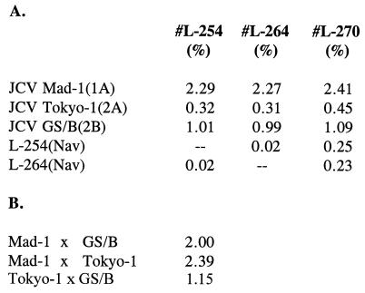
Relationships among three complete JCV genomes from Navahos (#L-254, type 2A; #L-264, type 2A; and #L-270, type 2C) were determined with the gap program of GCG (Fig. 2). These three Navaho JCV strains were also compared with prototypes of the other genotypes. It is noteworthy that #L-254 and #L-264 differed from each other by only a single nucleotide (0.02%). In contrast, they differed from another Navaho strain (#L-270) by 0.23% and 0.25%, respectively, about the same as the difference between #L-270 and a JCV strain (Tokyo-1) isolated from PML brain in Tokyo (0.32%). This indicates that the difference between Navaho type 2 strains can be nearly as great as their difference from Japanese strains. Complete JCV genomes of the Flathead and the Chamorro have not been analyzed.
Figure 2.
Percent difference between the complete genomic coding region (4,854 bp) of JCV strains. (A) Differences between three Navaho type 2 strains and the prototypes type 1 (Mad-1) and types 2A (Tokyo-1) and 2B (GS/B). Navaho strains #L-254 and #L-264 were classified as type 2A in the short typing fragment, whereas #L-270 was a type 2C. #L-270 falls into the type 2A group based on its closer relationship to type 2A prototype Tokyo-1 (0.45% difference), than to prototype type 2B strain (1.09% difference). Specimen #L-254 was obtained from a 26-year-old female, #L-264 from a 78-year-old female, and #L-270 from a 63-year-old male. Comparison of the coding region sequences used the gap program. (B) Differences between prototype strains of JCV.
DISCUSSION
The Navaho and Flathead people in the continental United States and the Chamorro of Guam excrete JCV at a significantly higher frequency than do populations of European origin. The excretion rates in all populations studied to date (40–69%) make it practical to determine the genotype of JCV harbored by ethnic groups in any part of the world.
All three indigenous cohorts excreted predominantly strains of JCV type 2A, the genotype that is characteristic of China and Japan (6, 16). type 2B is a related subtype that comprises a minor group of strains in the United States and that presumably arrived with European settlers. Its actual distribution and prevalence in Europe is still unknown. Among 25 JCV strains obtained in Hungary, all were type 1. We have included the minor variant designated type 2C within the type 2A group. Among the Navaho, the occurrence of JCV type 2A + 2C was 42/46 (91%). Among the Flathead people, where there has been considerable intermarriage with the surrounding Caucasian population (10), the occurrence of JCV type 2A + 2C was 11/16 (69%). This compares to an occurrence of type 2A + 2C in the control population of the United States of only 23%. The strains of JCV in Native Americans appear to be very closely related to those of China and Japan. These Native American strains have changed very little from the presumed parental Northeast Asian strains. The complete genome of the Japanese prototype type 2A strain, termed Tokyo-1, differs from the Navaho type 2A or type 2C strains by only 0.31–0.45%. This is less than the 0.5–1.0% difference that presumptively defines different subtypes of JCV (7). In contrast, the major genotypes of the virus differ by about 1.0–2.5% in their genomic DNA sequence. The fact that these Asian strains have changed so little since ancestors of the Navaho migrated across the Bering land bridge in the late Pleistocene suggests roughly the timing of JCV genotype evolution. The arrival of these first American settlers has been dated by artifacts at prehistoric sites to between 12,000 and 30,000 years ago. The close relationship of Navaho strains to JCV (Tokyo-1) pushes the time of origin of the major JCV genotypes in humans to well before the time of arrival of the first Americans. Because JCV had already differentiated into stable modern genotypes by that time, the origin of JCV in humans probably dates from 50,000 to 100,000 years ago or more. If so, it would appear that JCV may have coevolved with modern humans (Homo sapiens sapiens).
JCV strains from the Chamorro of Guam have not yet been sequenced in their entirety, and it is uncertain whether they represent a distinct subtype of type 2. Nevertheless, they clearly represent a variant of type 2A, in which “A” at position 1805 is consistently replaced by “T” in the Chamorro strains. The existence of this variant on Guam likely reflects the origins of the indigenous Chamorro population, although Spanish and Filipino influence have also occurred in the more recent past. Interestingly, the type 2A variant (“T” at 1805) identified in Guam has also been discovered in the urine of a 43-year-old HIV-positive male Filipino living in Los Angeles (unpublished data). This raises the possibility that JCV strains in Micronesia and the Philippines may have a common ancestor. Further, by this criterion the Navaho strains are more closely related to type 2A from Japan (represented by Tokyo-1) than they are to the Asian Pacific (Chamorro) strains. A final determination awaits analysis of the entire sequence of the Chamorro variant of JCV. It will be of interest to characterize the major JCV genotypes in other Pacific populations (especially Australo-Melanesian and Polynesian groups) in relation to the Asian, Native American, and Micronesian strains.
For a viral marker to be useful for following both early and recent worldwide population movements, it is necessary that it have evolved early, and it is desirable that it be ubiquitous. Moreover, it should be quite stable genetically and should be transmitted largely within the family or the immediate community. JCV is a DNA virus that appears to meet these essential requirements. Like mitochondrial DNA, JCV has evolved relatively rapidly compared with the human genome, yet unlike many highly infectious RNA viruses, its genomic sequence has not changed so rapidly that ancient associations are lost. Other viruses that may have coevolved with humankind and that may serve as markers for early and recent human migrations include retroviruses with unusually high genomic stability such as HTLV-1 (17, 18) and DNA viruses such as the human papillomaviruses (HPV) (19). Despite a superficial morphological similarity between polyomaviruses, such as JCV, and papillomaviruses, the circular double-stranded polyomavirus genome of 5.1 kb that is bi-directionally transcribed is now known to be unrelated by DNA sequence to the circular 8-kb papillomavirus genome with its single coding strand. Interestingly, like JCV, variants of HPV-16 show African, East Asian, and European associations (19). Information about the human diaspora obtained from viruses that coevolved with branches of the human family tree should correlate with that provided by genetic, archeologic, and linguistic data (20).
In summary, the simplicity of the distribution of the three main genotypes of JCV in Europe (type 1), Asia (type 2), and Africa (types 3 and 6) suggests a coincidence of viral evolution with the major lineages of early humans who migrated out of their probable ancestral home in Africa to people the continents of Asia and Europe around 100,000 years ago. The close relationship of JCV found today in Native Americans with that in Northeast Asia, and its distinction from Micronesian strains, is consistent with the migration of Amerind ancestors from Northeast Asia by way of the Bering land bridge. Further detailed study of JCV genomes from indigenous North, Central, and South American populations might identify variant viral genomes that mark successive waves of immigration to the New World.
Acknowledgments
We thank Drs. Elyse J. Singer and Robert W. Baumhefner for control urine samples. The support and encouragement of Dr. Henry deF. Webster is gratefully acknowledged. Urine samples were obtained from the National Neurological Research Specimen Bank, Veterans Administration Medical Center, Los Angeles, which is sponsored by National Institute of Neurological Disorders and Stroke/National Institute of Mental Health, National Multiple Sclerosis Society, Hereditary Disease Foundation, Comprehensive Epilepsy Program, Tourette Syndrome Association, Dystonia Medical Research Foundation, and Veterans Health Service and Research Administration, Department of Veterans Affairs. H.T.A. was supported by the Deutsche Forschungsgemeinschaft, Bonn (Grant No. Ag 19/1-1).
Footnotes
This paper was submitted directly (Track II) to the Proceedings Office.
Abbreviations: JCV, human polyomavirus JC; PML, progressive multifocal leukoencephalopathy; MS, multiple sclerosis; HPV, human papillomavirus.
References
- 1.Åström K E, Mancall E L, Richardson E P., Jr Brain. 1958;81:93–111. doi: 10.1093/brain/81.1.93. [DOI] [PubMed] [Google Scholar]
- 2.Agostini H T, Ryschkewitsch C F, Stoner G L. J Clin Microbiol. 1996;34:159–164. doi: 10.1128/jcm.34.1.159-164.1996. [DOI] [PMC free article] [PubMed] [Google Scholar]
- 3.Yogo Y, Iida T, Taguchi F, Kitamura T, Aso Y. J Clin Microbiol. 1991;29:2130–2138. doi: 10.1128/jcm.29.10.2130-2138.1991. [DOI] [PMC free article] [PubMed] [Google Scholar]
- 4.Ault G S, Stoner G L. J Gen Virol. 1992;73:2669–2678. doi: 10.1099/0022-1317-73-10-2669. [DOI] [PubMed] [Google Scholar]
- 5.Agostini H T, Brubaker G R, Shao J, Levin A, Ryschkewitsch C F, Blattner W A, Stoner G L. Arch Virol. 1995;140:1919–1934. doi: 10.1007/BF01322682. [DOI] [PubMed] [Google Scholar]
- 6.Guo J, Kitamura T, Ebihara H, Sugimoto C, Kunitake T, Takehisa J, Na Y Q, Al-Ahdal M N, Hallin A, Kawabe K, Taguchi F, Yogo Y. J Gen Virol. 1996;77:919–927. doi: 10.1099/0022-1317-77-5-919. [DOI] [PubMed] [Google Scholar]
- 7.Agostini H T, Ryschkewitsch C F, Brubaker G R, Shao J, Stoner G L. Arch Virol. 1997;142:637–655. doi: 10.1007/s007050050108. [DOI] [PubMed] [Google Scholar]
- 8.Loeber G, Dörries K. J Virol. 1988;62:1730–1735. doi: 10.1128/jvi.62.5.1730-1735.1988. [DOI] [PMC free article] [PubMed] [Google Scholar]
- 9.Agostini H T, Ryschkewitsch C F, Singer E J, Stoner G L. J Neurovirol. 1996;2:259–267. doi: 10.3109/13550289609146889. [DOI] [PubMed] [Google Scholar]
- 10.Ronan P. Historical Sketch of the Flathead Indian Nation. Hudson, WI: Ross & Haines; 1991. [Google Scholar]
- 11.Yanagihara R T, Garruto R M, Gajdusek D C. Ann Neurol. 1983;13:79–86. doi: 10.1002/ana.410130117. [DOI] [PubMed] [Google Scholar]
- 12.Agostini H T, Ryschkewitsch C F, Mory R, Singer E J, Stoner G L. J Infect Dis. 1997;176:1–8. doi: 10.1086/514010. [DOI] [PubMed] [Google Scholar]
- 13.Stoner G L, Agostini H T, Ryschkewitsch C F, Baumhefner R W, Tourtellotte W W. Multiple Sclerosis. 1996;1:193–199. [PubMed] [Google Scholar]
- 14.Stoner, G. L., Agostini, H. T., Ryschkewitsch, C. F. & Komoly, S. (1997) Multiple Sclerosis, in press. [DOI] [PubMed]
- 15.Agostini H T, Stoner G L. J Neurovirol. 1995;1:316–320. doi: 10.3109/13550289509114028. [DOI] [PubMed] [Google Scholar]
- 16.Kato A, Kitamura T, Sugimoto C, Ogawa Y, Nakazato K, Nagashima K, Hall W W, Kawabe K, Yogo Y. Arch Virol. 1997;142:875–882. doi: 10.1007/s007050050125. [DOI] [PubMed] [Google Scholar]
- 17.Yanagihara, R., Saitou, N., Nerurkar, V. R., Song, K.-J., Bastian, I., Franchini, G. & Gajdusek, D. C. (1995) Cell. Mol. Biol. 41, Suppl. I, S145–S161. [PubMed]
- 18.Yanagihara R. Adv Virus Res. 1994;43:147–186. doi: 10.1016/s0065-3527(08)60048-2. [DOI] [PubMed] [Google Scholar]
- 19.Chan S-Y, Ho L, Ong C K, Chow V, Drescher B, Durst M, ter Meulen J, Villa L, Luande J, Mgaya H N, Bernard H-U. J Virol. 1992;66:2057–2066. doi: 10.1128/jvi.66.4.2057-2066.1992. [DOI] [PMC free article] [PubMed] [Google Scholar]
- 20.Cavalli-Sforza L, Piazza A, Menozzi P, Mountain J. Proc Natl Acad Sci USA. 1988;85:6002–6006. doi: 10.1073/pnas.85.16.6002. [DOI] [PMC free article] [PubMed] [Google Scholar]


