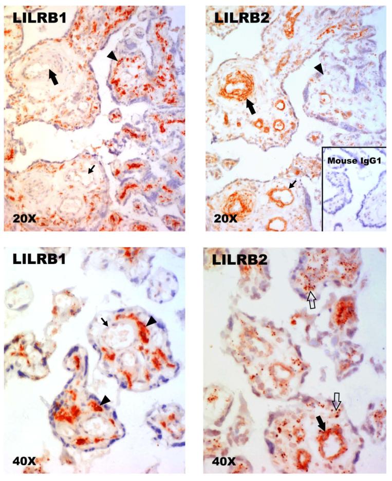
Fig. 1. Identification of LILRB1 and LILRB2 proteins in human term villous placentas by immunohistology.
(Upper left panel) Anti-LILRB1 (Amgen) fails to reveal signals in mesenchymal cells cuffing a small artery (large arrow) and a small vein (small arrow) but is strongly positive with pleiomorphic stromal cells (arrowhead). (Upper right panel) Anti-LILRB2 (Amgen) identifies positive cells cuffing a small artery (large arrow) and a small vein (small arrow) but does not yield readily identifiable signal in stromal cells (arrowhead). (Lower left panel) Anti-LILRB1 (Amgen) fails to reveal signals in cells cuffing a small vessel (large arrow) but is strongly positive with pleiomorphic stromal cells (arrowhead). (Lower right panel) Anti-LILRB2 (Amgen) identifies positive cells cuffing a small vessel (large filled arrow) and identifies diffuse, punctuate staining within the stroma (open arrows). Upper panels, original magnifications ×200; lower panels, original magnifications ×400. Positive stains in these experiments are identified by red signals. The slides were counterstained with Mayer’s hematoxylin.

