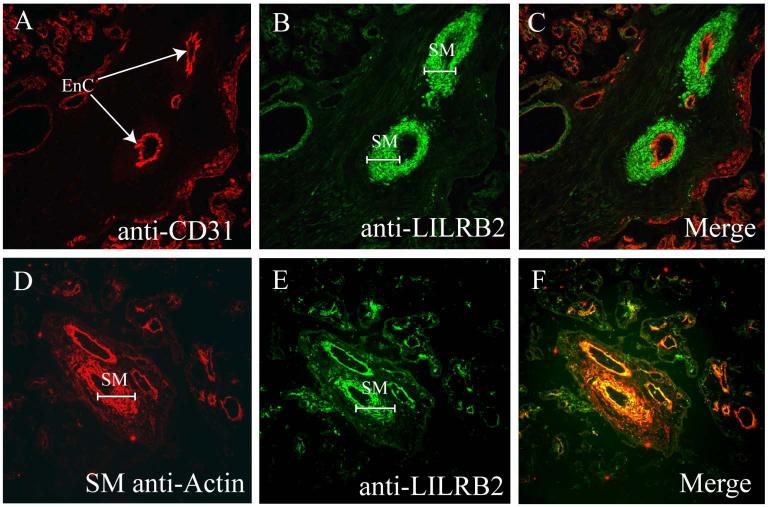
Fig. 2. Identification of LILRB2 positive vascular smooth muscle cells and LILRB2 negative endothelial cells in term villous placenta by double label immunofluorescence.
(A) Anti-CD31. Arrows point to CD31+ arterial endothelial cells (EnC) in a term placental villus. (B) Anti-LILRB2. Brackets mark LILRB2+ smooth muscle (SM) cuffs around small arteries. (C) Merging of frames (A) and (B) reveals no double staining (yellow) in either endothelial cells or smooth muscle. (D) Anti-smooth muscle actin marks smooth muscle (SM) cuffs around villous placental vessels. (E) Anti-LILRB2. As in Panel (B), LILRB2 is prominent in smooth muscle (SM) cuffs around placental vessels. (F) Merging of frames (D) and (E) demonstrates bright yellow staining indicating that the vascular smooth muscle is LILRB2+. Images were captured at magnification ×200.

