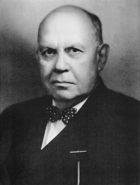
Dr. Charles F. Painter is shown. Figure is ©1947 by the Journal of Bone and Joint Surgery, Inc., and is reprinted with permission from Charles Fairbank Painter 1869–1947. J Bone Joint Surg Am. 1947;29:540–541.
Charles Fairbank Painter was born in Grand Haven, Michigan, the son of a clergyman [1]. He evidently spent one year at Williams College, then went to Johns Hopkins University. He obtained his medical education at Harvard Medical School, from which he graduated in 1895 (in the same class with Harvey Cushing and E. Amory Codman). While a house officer at the Massachusetts General Hospital he became interested in orthopaedic surgery. He taught at both Harvard and Tufts and in 1913 became Dean of the Medical School of Tufts College. As with many prominent orthopaedists of the time, he served in the military during WWI, as the orthopaedic surgeon to the Chelsea Naval Hospital. At various times he practiced at the Robert Breck Brigham Hospital, the House of the Good Samaritan, the Beth Israel Hospital, the Massachusetts’s Women’s Hospital, and the Carney Hospital.
Dr. Painter not only produced a large number of scholarly works (including his contributions as coeditor of Diseases of the Bones and Joints with Drs. Joel E. Goldthwait and Robert B. Osgood in 1910 [2]), but was active in community and national orthopaedic organizations. He was librarian of the Boston Medical Library for 8 years. He served as President of the American Orthopaedic Association in 1916, and in 1943 served as acting editor of The Journal of Bone and Joint Surgery after the death of the editor, Dr. Murray S. Danforth. He published a number of articles, but seemed particularly interested in arthritis and inflammatory and infection conditions. He wrote an early description of myositis ossificans in 1909 (and commented, “It is likely to be a long time before the medical profession will be in a position to offer the sufferer from myositis ossificans any material aid.”) [4].
His earliest publication relates to the topic of this month’s symposium, “Molecular and Clinical Developments in Tendinopathy.” Published three years after his graduation from medical school, “Inflammation of the Post-calcaneal Bursa Associated With Exostosis” [3] reflects the thinking of the time. The concept of inflammation in tendons unassociated with tuberculosis or syphilis or rheumatic condition was not well developed in the late 1800s and early 1900s. However, most of the eight cases he described had long-standing pain and symptoms and signs of inflammation about the Achilles tendon. Perhaps owing to the radiographic appearance of exostoses, Painter directed his attention to that feature. (X-rays had just been discovered by Roentgen in November, 1895 [6]; it is remarkable little more than two years later the use of roentgenograms in clinical practice was widespread.) He stated, “It is possible to eliminate from the etiology influenza, scarlet fever, syphilis, gonorrhœa, acute rheumatism, fibromata, neuromata, and acute suppurative processes.” “Trauma,” he commented, “does not seem to have played any prominent part, unless it be in the third case, that of the bicycle rider. If the trauma of the shoe were an important causative factor there would be more cases of this trouble....However, as has been pointed out by Dr. William Adams and others, the osteo-arthritic lesions are manifested in the course of tendons, in bursal sacs, in the cartilage over joint surfaces, and in the fringes of the synovial membrane within the large joints, especially the knee, shoulder, and elbow” [2].
Painter’s use of the term “osteo-arthritis” cannot be considered analogous to current usage. Our contemporary distinctions of osteoarthritis arose decades later than when Painter was writing. The earliest use of the term I can find is from 1889, in an article by John Kent Spender [8], although the cases he describes seem to have no relationship to what we could currently consider osteoarthritis. Rather, Spender even included “The so-called rheumatoid arthritis” in his title. Spender begins his article, “Few things are so apt to cause a feeling of drowsy despair at a medical meeting as the prospect of an academic discussion on the etiology of osteo-arthritis...one master says one thing, and another says another; so that the self-styled practical man cries out in his confusion, How have all these speculations helped me” [8]. Clearly, there was great confusion at the time, so it is not surprising Painter focused on the obvious: the calcaneal exostoses that result from longstanding inflammation of the Achilles tendon.
The literature uses several words related to the condition we know today as tendinopathy: “tendinopathy,” “tendonopathy,” “tendinosis,” and “tendonosis.” (Interestingly, Stedman’s Medical Dictionary contains none of these words.) The spelling of both of these terms with “i” rather than “o” is clearly preferred in the literature: PubMed lists 5109 (262 in the title) with the tendinopathy and only 27 (11 in the title) with the tendonopathy, 5020 (145 in the title) with tendinosis and only 9 (four in the title) with tendonosis. The earliest reference I could find to any of these is to tendinosis in a thesis from 1831, “De Ulcerum Diagnosi et Aetiologia Nonulla” in the context of abscesses [7].
The concept of tendinopathy as we know it today is relatively recent: The earliest reference in PubMed to any of the four terms I noted is 1950, and Google Scholar lists none earlier. None of these terms appear in an article title until 1969. One of the earliest articles addressing the role of chronic stress in tendinopathy appeared only in 1976 by Perugia, et al. [5]. They recognized three forms: (1) pure peritendinitis; (2) peritendinitis associated with tendinosis; and (3) pure tendinosis, thus describing the elements we recognize today. They further recognized the role of stress in the origin of the condition.
One wonders why a condition that is relatively common would receive such little recognition and clarification. My only explanation is the condition generally does not completely disable people, and the natural history (not well described in the literature that I can find) is one of a condition that waxes and wanes for months or years, typically limiting but not disabling a person until it eventually subsides clinically. Prior to the mid 20th century, many patients in the industrialized world may simply have accepted such conditions as part of life, as likely did their doctors with such little understanding of the condition. Therefore, tendinopathy may simply have not received the attention of more urgent conditions.
References
Charles Fairbank Painter 1869–1947. J Bone Joint Surg Am. 1947;29:540–541.
Goldthwait JE, Painter CF, Osgood RB. Diseases of the Bones and Joints: Clinical Studies. Boston, MA: W. M. Leonard; 1910.
Painter CF. Inflammation of the post-calcaneal bursa associated with exostosis. J Bone Joint Surg Am. 1898;s1–11:169–180.
Painter CF, Clarke JD. Myositis ossificans. J Bone Joint Surg Am. 1909;s2–6:626–651.
Perugia L, Ippolitio E, Postacchini F. A new approach to the pathology, clinical features and treatment of stress tendinopathy of the Achilles tendon. Ital J Orthop Traumatol. 1976;2:5–21.
Röntgen WC. Über eine neue Art von Strahlen. Physic-med Ges Wuertzburg. 1895.
Rust C. De Ulcerum Diagnosi et Aetiologia Nonulla. 1831. Berolini. Available at: http://books.google.com/books?hl=en&lr=&id=pDkAAAAAQAAJ&oi=fnd&pg=PA3&dq=+tendinosis+OR+tendonosis&ots=HsJWbBs2Uo&sig=dFrxuihm-EMGnSRAa81kCx6VT70. Accessed March 31, 2008.
Spender J. On some hitherto undescribed symptoms in the early history of osteo-arthritis (The so-called rheumatoid arthritis). BMJ. 1888:781–783.
During the past eighteen months I have seen in the Orthopedic Clinic at the Boston Dispensary six cases of this affection, and through the kindness of Dr. Goldthwait two other cases in private practice. All of these cases, with one exception, had the exostoses in addition to the bursal inflammation. The frequency of the occurrence of this condition in a series of perhaps two hundred cases of painful affections of the feet of one sort or another suggested the possibility of the condition having been overlooked in certain cases where anti-flat-foot treatment was of no avail.
Certain facts observed in the etiology and certain features of the clinical history have suggested the possibility of a constitutional diathesis being at the bottom of the whole matter. If the conclusions from these facts are correct as to etiology they have an important bearing upon treatment.
Patients suffering from this condition present the following symptoms: They complain of pain at the insertion of the tendo Achillis and along its course for a variable distance. All motion of the feet is painful, especially dorsal flexion, and is more or less restricted. Walking in the bare feet is impossible in some cases, and the gait is that of an extreme valgus. There is sometimes a valgus associated. On either side of the tendon, filling up the lateral fossæ, is a swelling, which may be dense, firm, non-elastic, or it may be soft, but is always tender to pressure. Pain may also be referred to the plantar surface of the os calcis, which may also be tender to pressure. The swollen area is usually slightly hyperæmic, and the surface temperature is increased.
In this situation a process giving rise to such signs and symptoms could be located in the tendon sheaths, in the bursal sac, or in the bone. Schüler and Rössler have established the existence of this post-calcaneal bursa as a normal anatomical structure from an investigation on some three hundred cadavers. There is a similar though smaller one situated beneath the os calcis, separating it from the plantar muscles. The walls of the bursæ are normally quite thick, and the serous surfaces are thrown into rugæ. Heinecke was the first to describe these structures anatomically. Nancrede has shown that cartilage is normally present in the anterior wall of the post-calcaneal bursa.
A study of the literature brings out a number of cases of this sort, or analogous to them. Rössler reports nine cases, presenting symptoms like those just mentioned, in which he believes that long-continued pressure had caused an inflammation and consequent thickening of the periosteum over the os calcis.
Frankë believes the process to be primary in the periosteum; he also reports cases following influenza.
Rosenthal found a neuroma in a patient with these symptoms which was being pressed upon by the tendo Achillis. Raynard has reported a few cases of acute suppuration in the sheath of the tendo Achillis following certain general infections, showing that such tissues are vulnerable to infection.
Kirmisson also reports two cases of peritendinous inflammation about the Achillis tendon. One case was suffering from gonorrhœal synovitis and the other from multiple fibromata, one of which was beneath the tendo Achillis. The first one had an excess of uric acid in the urine and nodosities at the metacarpo-phalangeal joints.
Da Costa reports eight cases of inflammation of this bursa. The bursal sacs contained osteophytes, some of them tipped with cartilage. They occurred in both the anterior and posterior walls of the bursa.
In the above cases the causes assigned have been trauma, gonorrhœa, syphilis, rheumatism, influenza, sciatica, tuberculosis of os calcis, scarlet fever, uric-acid diathesis, etc.
A condition comparable to this, in a way, is that described by P. Busquet, in which he calls attention to the painful exostoses on the dorsal surfaces of the second, third, and fourth metatarsals, occurring in men whose duties require them to walk long distances. He divides the cases etiologically into three groups: one where the trauma from walking is direct, one where it is indirect, and one where it is dependent upon a diathesis.
Pathology. In three of the cases which I am about to report the bone removed from beneath the tendo Achillis has been examined histologically, with the following results: The marrow spaces are filled with a young granulation tissue in which are numerous plasma and lymphoid cells, and this is plentifully supplied with new-formed bloodvessels. The cartilage over this is the normal hyaline variety. The trabeculæ are surrounded by osteoblasts. The subungual exostoses present the same appearance under the microscope. This would seem to indicate an inflammatory process. Grossly, at the time of operation, a profuse capillary hemorrhage has been noted, as though the tissue were engorged with blood, and this would also suggest the venous stasis of inflammatory changes.
Case I. The patient (C. H. H.) was twenty-seven years of age, a hotel clerk by occupation. He has had trouble with his feet for four or five years, complaining of pain along the tendo Achillis. He was wearing bronze flat-foot plates, applied three years ago. There was no appreciable valgus. He had not been at all relieved by the use of the plates. There was no rheumatic, syphilitic, or gonorrhœal history obtainable. There was marked thickening, which was tender to pressure on either side of the posterior portion of the os calcis, suggesting a bursitis or an exostosis. The x-rays revealed nothing. He was admitted to the Carney Hospital, and was operated upon June 14, 1897. Through a two-and-a-half-inch incision, parallel with, but a little in front of, the tendo Achillis, a bursal sac was dissected out which was thickened; this was opened, and no fluid found within, but in place of it a small amount of viscid, myxomatous-looking tissue jutted out from the walls of the sac anterior to this, and projecting from the posterior surface of the os calcis was a rough line of bony spicules. These were curetted off and the wound closed. On June 18th he was discharged from the hospital, and since that time has been under observation. For a time he experienced some relief from the operation, but this has not been permanent. He was not sufficiently encouraged to submit to operation upon the other foot; he also complains now of pain on the plantar surface of both heels. This patient was seen walking upon the street recently, and, so far as the gait was an indication, he was apparently well.
Case II. This patient (G. R.) was twenty-one years of age, employed in the print works as a dyer, where he has been obliged to be exposed to changes of temperature in going from a hot, moist atmosphere into cold surroundings. For ten years he has had pain in his heels to such an extent that he has lost a considerable amount of time from his work. Three months ago he was obliged to give up entirely. He is a large, heavy man, who walks with a marked limp in both feet, using two canes. The arches of his feet are strong and apparently efficient. The calves of his legs are small in proportion to his muscular development elsewhere. On either side of both tendo Achilles are marked thickenings, which are very tender to pressure; he is also tender beneath the point of the heel. The x-ray was not employed. He cannot walk a step in his bare feet without the use of his canes. He was operated upon through incisions similar to the ones employed in the previous case, except that the incisions were made on either side of the tendon in order to get at the bony thickening more readily. There was considerable fluid in this bursal sac, the interior of which was thrown into folds, but there were no osteophytes seen; beneath these was a diffuse bony projection, with two or three sharp spines coming from it. This was removed readily with a heavy chisel. Fourteen days later he was allowed to get out of bed, and could walk in his bare feet without pain, except for a trifling soreness, without cane or crutch. Two days later he was discharged. Since the operation he has been able to return to his work gradually, having had two or three attacks of considerable soreness about his heels, but by means of massage, bathing, and moderate exercise he has now, May 1st, so far recovered that he can walk four or five miles without discomfort, and is able to return to his old employment. Examination with the fluoroscope does not show any exostoses.
Case III. This patient (P. L.) was twenty years of age, a loom-tender. He complains of pain under the point of the heel and along the tendo Achillis, which followed the use of a bicycle, the pedals of which were too long; consequently he was obliged to use toe-clips to keep his foot upon the pedal, always riding with his foot plantar flexed. He always rode in high shoes. This would bring the counter of the shoe in contact with the skin over the insertion of the tendo Achillis throughout the whole circuit of the pedal. He has had this trouble for twenty months. He states that his father had chronic rheumatism which laid him up for two years before his death, which apparently resulted from cancer of the throat. He also states that he has been obliged himself to work where his feet were wet a good share of the time. For this condition, which had resulted in such a degree of plantar flexion of the foot as to compel him to walk on his toes, he had a tenotomy of his tendo Achillis performed in August, 1897. At the same time the bursal sac beneath the tendo Achillis was opened, and kept open, being allowed to close four months later. He was then permitted to get up, but, the condition for which he had his tenotomy performed having returned, he had a second tenotomy in November; he was then fitted with flat-foot plates. These operations and the plates gave him no relief whatever. He never met with any injury, and was never ill in his life, except for the grippe five years ago. Fluoroscope showed marked exostoses beneath tendo Achillis. He was operated upon two months ago in a manner similar to that already described. Large exostoses were removed. Pain about the tendo Achillis was relieved, but he still complains of pain on plantar surface of the heels. A report from him May 10th says that he was benefited a little for a time, but that now he is as bad as ever.
Case IV. This was the case of a man, aged twenty-eight years, a city laborer, who came to the dispensary about two years ago because of pain in the foot, referred to the instep. He had a very rigid foot, held in moderate pronation. Under ether, an ineffectual attempt was made to break up the adhesions and correct the position. After this, with exercises and a Thomas sole, he got along fairly well, and was lost sight of until three or four months ago, when he returned, with his pain now referred to the insertion of his tendo Achillis. There was tenderness to pressure at this point, and a prominent spur was evident in the fluoroscope, projecting from the posterior and upper surface of the os calcis. The fulness on the sides of the tendo Achillis was not very marked. There was nothing in his history which had any bearing on the causation of the trouble.
Case V. This patient is a man, aged twenty-nine years, a sailor. For nine years he has been utterly unable to do any work on account of pain in his feet. This pain was referred to both tendo Achilles as well as to the plantar surface of the heel. There was nothing in his family or personal history which was at all suggestive. During these nine years he has tried all sorts of anti-rheumatic treatment, has been at various baths in this country and abroad, but is still unable to get about without great difficulty and pain. Uses canes; walking in the bare feet was impossible. On physical examination there were very marked thickenings on either side of the tendo Achillis, filling up the normal fossæ at these points; tender to pressure; hard and not fluctuating. No x-ray photograph was taken. There are also some thickenings at the metacarpo-phalangeal joints, particularly in the thumb, and he uses the hands awkwardly, as patients do with these painful joints, though in his case these are not painful, though they had been. Through incisions similar to the one employed in the last case the bony thickenings were chiselled off, together with the bursa, which was also thickened. At the end of two weeks he was discharged, being able to walk in his bare feet the first time he tried. For some time this improvement continued; but, like the previous case, he has had several setbacks which have not responded as readily to treatment as in the case of the preceding patient. His pain, however, has left the region of the insertion of the tendo Achillis, and is now confined to the plantar surface of the heel. In spite of these setbacks, he regards his condition as much better than before. Now, on physical examination, there is still considerable thickening at the seat of operation—more than should come, it seems to me, following a first-intention wound, as all these have been.
Case VI. M. E. S., aged thirty-three years, a lawyer. Five years ago he had an attack of rheumatism in the left knee which lasted three or four weeks. After that he had no trouble until the fall of 1896. He got his feet and lower part of his body wet in a heavy rain-storm, and the next day both feet were sore. Five days later he refereed a foot-ball game in low shoes, and was exposed for several hours in deep mud and slush. That evening his feet were very sore. Ever since they have troubled him a great deal, being fairly comfortable for a few days, and then bad again. Always much worse stormy days.
The right foot is swollen behind the heel; pain and tenderness. Motion in all directions painful, especially on flexion. Walking without shoes almost impossible. In the left foot there is no swelling behind the heel, and the pain is on plantar surface of the heel. The left foot is pronated. His treatment consisted in being confined to his bed, the application of dry heat, and bandages.
The necessity for his going to bed was brought on by his getting his feet very wet.
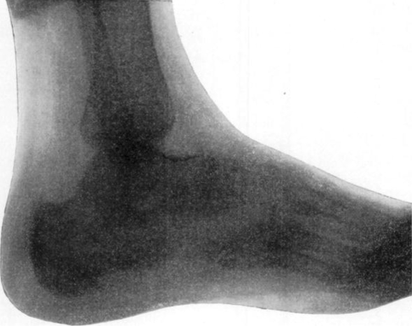
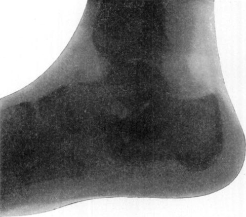
The x-ray photographs (Figs. 1 and 2) show the bony growth behind the tendo Achillis and also in its plantar surface.
Fig. 1.
Case VI. Shows bony spurs on upper and lower surfaces of the os calcis.
Fig. 2.
Case VII. H. F. F. before operation, showing bony thickening on the upper surface of os calcis, which was removed.
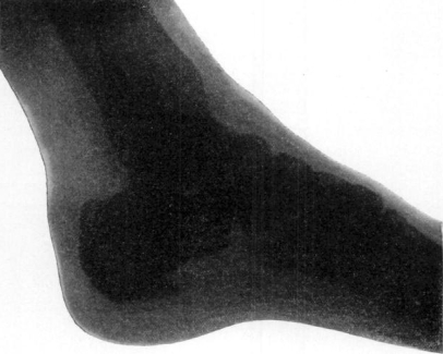
Case VII. This patient (H. F. F.), a man, aged about twenty years, came to the Carney Hospital in November, 1895, on account of painful feet. He had not walked for over a year. Figure 2 shows the bony outgrowths beneath the tendo Achillis before operation, which were removed in the spring of 1897, with a very satisfactory immediate result. His symptoms recurred, however, in a short time, and Fig. 3 shows his condition one year after the exostoses shown in Fig. 2 had been chiselled away, showing that there has been a recurrence of the bony outgrowth. He has been very much benefited by the use of dry heat at high temperatures.
Fig. 3.
Case VII. H. F. F. one year after operation, showing prominent spur on plantar surface of os calcis.
Case VIII. This patient (B. M.) was twenty-seven years of age. Family history of phthisis; mother died of it. He had pain, swelling, and tenderness, with local heat and redness over the sheath of the tendon. This has turned out to be a case of tuberculosis of the calcaneal bursa and of the os calcis.
In addition to these cases of which I have details, I have seen three other cases which presented precisely similar clinical symptoms and physical signs.
In this series of cases it is possible to eliminate from the etiology influenza, scarlet fever, syphilis, gonorrhœa, acute rheumatism, fibromata, neuromata, and acute suppurative processes. Trauma does not seem to have played any prominent part, unless it be in the third case, that of the bicycle rider. If the trauma of the shoe were an important causative factor there would be more cases of this trouble.
I cannot rule out the uric-acid diathesis mentioned by some observers, because I did not test the urine. In the eighth case tuberculosis, primary either in the os calcis, which is probable, or in the bursal sac or the sheath of the tendon, accounts for one of the eight. Of the other seven, one, the fifth, presents arthritic changes in other joints, which suggest an osteo-arthritic process; Ortho Soc 12 three give no history of any probable cause, and the other three give a history of being exposed to cold, wet, and sudden changes of temperature. In the x-ray photographs the osteophytes show very clearly in Cases VI. and VII. Cases I., III., and IV. revealed the same condition through the fluoroscope, and Case VIII., the tuberculous case, did not show any definite osseous changes. The only case not examined by the x-ray before operation—viz., Case II.—did not show any osteophytes five months after operation; this is also the only case which can be considered well which has been operated upon. Case VII. has photographs before operation and one year later also. The last picture shows that there was a recurrence of the bony growth. In both the sixth and seventh cases the osteophytes appear on the plantar surface of the os calcis as well as beneath the tendo Achillis.
We have, then, a condition to deal with which is not sufficiently accounted for by traumatic influences alone; which is not dependent upon any of the acute infective processes, certainly not in this series; which runs an essentially chronic course, none of the cases being of less than two years’ standing; which presents the histological characteristics of a subacute or chronic osseous inflammation, and, lastly, which does not respond to the treatment which we should expect to give permanent relief to a simple exostosis, the only case operated upon which has remained relieved being Case II., in which case the duration of the disease before operation had been nine years. All the other cases have been immediately relieved, only to be as bad as before within six months or less. Such characteristics must indicate, of course, an inflammatory process of some sort, dependent probably upon a constitutional diathesis. From the clinical stand-point, osteoarthritis would seem to fit the condition better than any other. Unfortunately, we do not get the opportunity to study histologically the joints of osteo-arthritic patients while they are in the acute stage, and in the late chronic stage, after acute symptoms have subsided, the lesions presented are not distinctive of any special disease. However, as has been pointed out by Dr. William Adams and others, the osteo-arthritic lesions are manifested in the course of tendons, in bursal sacs, in the cartilage over joint surfaces, and in the fringes of synovial membrane within the large joints, especially the knee, shoulder, and elbow.
He also calls attention to the fact that trauma and exposure to cold and wet are prominent factors in the etiology of that disease, and cites the unusual prevalence of osteo-arthritis in the north of Ireland, where all three of these factors are present.
In conclusion, then, though an absolutely clear case cannot be established for the osteo-arthritic hypothesis as to the cause of these osteophytes about the os calcis and metatarsus, still it would seem to afford a better explanation than can be given on any other theory.
The question of treatment, in the light of the cases here cited, must be decided on the theory that this is an osteo-arthritic process. It is well recognized that surgical interference in that disease during its acute or subacute stage is contraindicated, and such interference must be deferred until the quiescent stage is reached. The treatment in the acute stage must consist of rest, the application of dry heat and fixation bandages.
I wish to thank Dr. Goldthwait for the use of two cases in the above series, and for suggestions as to the etiology and treatment.
Conclusions: Exostoses beneath the post-calcaneal bursa are most commonly manifestations of an osteo-arthritic process. The exciting causes of this are exposure to cold and wet and trauma. The treatment should be operative only after the acute stage has subsided. The condition is one to be carefully looked for where persistent pain in the foot is present not yielding to the ordinary treatment.
Footnotes
This article is ©1898 by the Journal of Bone and Joint Surgery, Inc. and is reprinted with permission from Painter CF. Inflammation of the post-calcaneal bursa associated with exostosis. J Bone Joint Surg Am. 1898;s1-11:169–180.
Richard A. Brand MD ✉ Clinical Orthopaedics and Related Research, 1600 Spruce Street, Philadelphia, PA 19103, USA e-mail: dick.brand@clinorthop.org
References
- Albert, E. “Achillodyne.” Wiener Medical Presse, 1893, No. 2.
- Adams, William. “Chronic Rheumatic Arthritis.” British Medical Journal, 1886, vol. ii. 915–919.
- Baker, Morrant. Trans. Chir. Society of London, 1885, vol. xviii. p. 62.
- Brackett, E. G. Boston Medical and Surgical Journal, 1896.
- Busquet, P. “De 1’Osteo Periostite ossifiante des Metatarsiens.” Revue de Chirurgie, 1897, No. 12.
- Byers. “Exostosis Bursata.” Mont. Medical Journal, 1895–96, vol. xxiv. 967.
- Da Costa. “Achillodynia.” Phila. Medical Journal, March 12, 1898.
- Dent, E. G. “Arthritis Deformans.” Edinborough Medical Journal, 1897, vol. ii.
- Finotti, E. “Ueber Tuberculose des Calcaneus.” Deutsche Zeitschrift für klin. Chirurgie, vol. xl.
- Franke, F. “Ueber die Erkrankungen der Knochen-Celenke and Bănder bei der Influenza.” Langenbeck’s Archive für klin. Chir., vol. xlix.
- Goldthwait, J. E. Boston Medical and Surgical Journal, 1897.
- Heinecke. “Anatomic and Pathologie der Schleimbeutel und Sehnenscheiden.” Erlangen, 1868.
- Hirsch. “Das Verholten der Achillessehnen bel Kontraction der Wadenmuskenlatur.” Centralblatt für Chir., January 15, 1898, No. 2.
- Lloyd, S. “Achilles Bursitis Anterior.” St. Louis Medical Review, May 15, 1897.
- Mackenzie, S. “On the Various Forms of Rheumatism, Especially in Reference to Age and Sex.” Edinburgh Medical Journal, 1897, Nos. 1 and 2.
- Michalovisez. Lehrbuch der descriptiven und topographischen Anatomie (Ungarlach), 1888.
- Parker. “On the Diagnosis of Certain So-called Rheumatic Diseases from Each Other and from Rheumatism.” Lancet, June 26, 1897.
- Raynal. “Cellulite peritendineuse du tendon d’Achille.” Archives Générales de Médecine, 1883.
- Rona, S. “Casuistischer Beitrag zu der Entzundung der Sehnenscheiden Schleimbeutel Muskeln und peripherer Nerven in Verlaufe der Gonorrhoe.” Archives für Derm. und Syph., 1892, Heft 2.
- Rosenthal, L. “Bermerkung zur Achillodynie.” Wiener medical Presse, 1893, No. 10.
- Scülller. “Bemerkung zur Achillodynie.” Wiener medical Presse, 1893, No. 7.
- “The Relation of Rheumatic Arthritis to Diseases of the Nervous System.” “Tuberculosis and Rheumatism.” British Medical Journal, 1897, vol. ii. 1225–29.


