Abstract
For patients with refractory retrocalcaneal bursitis (Haglund’s syndrome), the most effective surgical approach has not been defined. We asked whether patients undergoing the tendon-splitting approach and the lateral approach would have comparably effective relief of pain for both types of calcaneal ostectomies. We retrospectively reviewed 30 patients (31 feet) who underwent the tendon-splitting approach and compared their results with 32 previous patients (35 feet) who had a lateral incision. Minimum followup was 12 months (mean, 16 months; range, 12–23 months) for the tendon-splitting group and 15 months (mean, 51 months; range, 15–109 months) for the lateral group. The mean American Orthopaedic Foot and Ankle Society score improved from 43 points preoperatively to 81 points (range, 8–100 points) postoperatively in the tendon-splitting group and from 54 points to 86 points (range, 55–100 points) in the lateral group. The mean physical component score of the Short Form-36, version 2, at followup was 52 (range, 22–61) in the tendon-splitting group and 49 (range, 34–63) in the lateral group. The median return to normal function was 4.1 months (range, 3–13 months) in the tendon-splitting group and 6.4 months (range, 4–20 months) in the lateral group. Both approaches to calcaneal ostectomy provided symptomatic pain relief. However, patients in the tendon-splitting group returned to normal function quicker than patients in the lateral group.
Level of Evidence: Level III, retrospective comparative study. See the Guidelines for Authors for a complete description of levels of evidence.
Introduction
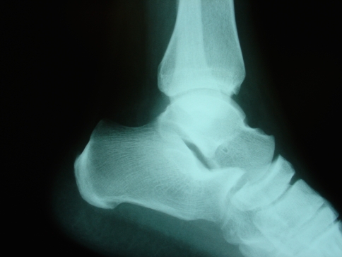
Retrocalcaneal bursitis (Haglund’s deformity), may be difficult to treat effectively by nonoperative measures alone. It originally was described as a prominence of the posterior superolateral calcaneus (Fig. 1) affecting the superoanterior bursa and the Achilles tendon [5]. Nonoperative measures, including analgesia and modified shoe wear, may provide relief [3, 6, 12]. Although McGarvey et al. reported 89% of their patients with insertional Achilles tendinosis improved with nonoperative treatment, they and others believe surgery was a reasonable option for patients not responding to nonoperative treatment [13, 14, 17].
Fig. 1.
A lateral view radiograph shows the prominent posterior calcaneus. (Reprinted with permission from Brunner J, Anderson J, O’Malley M, Bohne W, Deland J, Kennedy J. Physician and patient based outcomes following surgical resection of Haglund’s deformity. Acta Orthop Belg. 2005;71:718–723.)
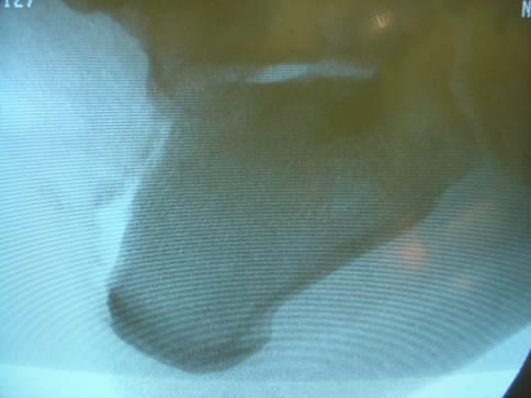
For patients with Haglund’s deformity who do not respond adequately to nonoperative therapy, there are numerous surgical options available, including calcaneal ostectomy (Fig. 2) with or without Achilles tendon débridement, excision of the retrocalcaneal bursa, and calcaneal osteotomy [1, 2, 4, 9, 16]. However, the results of these procedures, and the outcome measures used, have not been consistent [7, 15, 21]. Adequate relief of symptoms is predicated on removing any osseous irritation to the Achilles insertion. In patients treated with a lateral incision, access to the medial aspect of the calcaneal tuber may be restricted. This may lead to inadequate bone resection and recurrence of symptoms. Furthermore, the anterior aspect of the Achilles tendon may be difficult to see and subsequently débride with a limited lateral or medial incision. By contrast, the Achilles tendon-splitting approach provides excellent exposure of the tendon, facilitating adequate débridement of the tendon and bursa. Concerns regarding the tendon-splitting approach include compromise of the integrity of the tendon with slow return to full function and scar irritation about the heel counter. The most effective approach to this surgery has not been defined.
Fig. 2.
A lateral view obtained after the calcaneal ostectomy is shown. (Reprinted with permission from Brunner J, Anderson J, O’Malley M, Bohne W, Deland J, Kennedy J. Physician and patient based outcomes following surgical resection of Haglund’s deformity. Acta Orthop Belg. 2005;71:718–723.)
We asked whether the two surgical procedures for recalcitrant retrocalcaneal bursitis would provide effective relief of pain, good outcome scores, and few complications.
Materials and Methods
We retrospectively compared the effectiveness of two approaches to calcaneal ostectomies for recalcitrant Haglund’s deformity. The first group underwent 35 (32 patients; 23 females and nine males) consecutive calcaneal ostectomies using a lateral approach performed by one surgeon (JGK). After completion of this group, a second group underwent 31 (30 patients; 17 females and 13 males) consecutive calcaneal ostectomies using a central tendon-splitting approach, also performed by the same surgeon (JGK). Outcome measures using the American Orthopaedic Foot and Ankle Society (AOFAS) score and Short Form-36 version 2 (SF-36v2) scores were calculated and compared between groups.
All patients had symptomatic Haglund’s deformity refractory to nonoperative measures, such as modified shoe wear. Two patients who had calcaneal ostectomies performed other than via central tendon-splitting or lateral approaches and two patients with other concomitant surgery were excluded. It was important that the number of patients in the Achilles-splitting group was similar to the number of patients who had the lateral approach, and also that all patients were followed up for at least 12 months. All patients’ charts were reviewed, and all patients except two (one in each group) were available for followup. The two patients unavailable for followup were not included in the study. The mean age of the patients in the central tendon-splitting group was similar to that of the lateral incision group (50 years, range, 28–82 years versus 51 years, range, 19–81 years, respectively). The mean times from onset of symptoms to surgery also were similar in the tendon-splitting group and lateral incision group (18 months, range, 2–72 months versus 13 months, range, 4–22 months, respectively). The minimum followups were 12 months (mean, 16 months; range, 12–23 months) for the tendon-splitting group and 15 months (mean, 51 months; range, 15–109 months) for the lateral group.
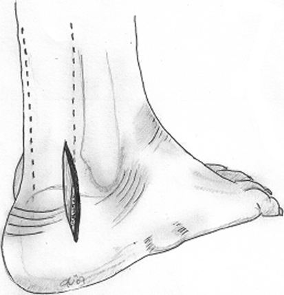
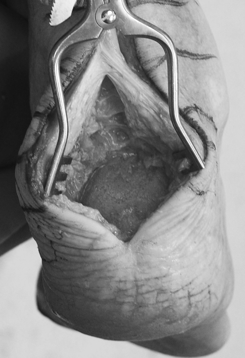
Patients undergoing calcaneal ostectomy were placed in the prone position. We performed a lateral approach in one group of patients (Fig. 3) and a central tendon-splitting approach in the other group (Fig. 4). Patients undergoing a central splitting approach had an incision centered over the Achilles tendon, approximately 10 cm proximal to the Achilles tendon insertion and extending distally to the glabrous skin. An incision was made directly down to the paratenon to avoid unnecessary dissection planes. One central split then was placed in the tendon and a Weitlaner retractor was used to facilitate exposure. The tendon then was resected from the most posterior aspect of the calcaneus. As much as 50% of this can be resected before strength is compromised [14]. All areas of fibrous degeneration and calcification then were removed from the tendon with sharp dissection. The retrocalcaneal bursa was resected to expose the superior aspect of the calcaneus and the posterior aspect of the subtalar joint. A ½-inch curved osteotome was used to make a stress-relieving corticotomy 1 cm proximal to the subtalar joint. The osteotome was used to resect the dorsal calcar tuber first and then the posterior aspect of the Achilles attachment. A rasp was used to smooth all edges. Mitek suture anchors (Raynham, MA) were used to anchor the Achilles to the calcar if greater than 50% of the Achilles tendon insertion had been resected. As the medial and lateral edges of the Achilles tendon were intact, the central part of the tendon could be tensioned accurately before suture anchoring. The tendon then was closed with a No. 2 Ethibond (Smith & Nephew, Memphis, TN) suture.
Fig. 3.
The diagram shows the lateral approach for Haglund’s procedure.
Fig. 4.
The tendon-splitting approach for Haglund’s procedure is shown.
Patients who had a lateral approach had a 6- to 8-cm lateral incision along the lateral border of the Achilles tendon insertion. Once again, full-thickness skin flaps were made to the tendon. The insertion of the Achilles tendon was identified and resected along the lateral border, exposing the prominent calcar tuber. Using a ½-inch curved osteotome, this was resected and the edges smoothed with a rongeur and rasp. The Achilles tendon also was débrided of any visible fibrosis or calcification.
In all cases, the dorsal ostectomy was performed initially, allowing exposure of the remaining posterior component, which subsequently was resected and rasped to remove sharp edges. We also removed any peritendinitis or calcifications in the Achilles tendon.
Both groups of patients wore a below-knee cast for 4 weeks followed by a CAM boot for an additional 4 to 6 weeks. The patients began range of motion exercises at 4 weeks and touchdown-weightbearing at 6 to 8 weeks under the supervision of a physical therapist. Thereafter, walking without the use of walking aids was encouraged. Supervised physical therapy included gastrocnemius and soleus muscle strengthening, and stretching exercises twice weekly for 4 weeks.
We (JK, POL, JA) evaluated patients at 6 weeks, 3 months, 6 months, and 12 months after surgery. The AOFAS ankle-hindfoot scale [11] and the SF-36v2 [22] were used to evaluate patients before and, at a minimum of 12 months, after surgery. The AOFAS ankle-hindfoot score evaluates pain (40 points), function (50 points), and alignment (10 points). The SF-36v2 is a general health survey with physical and mental components and has been validated internally and externally in foot and ankle hindfoot surgery [10, 22].
We used the nonparametric Mann-Whitney test to compare differences in outcome scores between the two groups.
Results
The majority of patients in both groups reported alleviation of pain. At 1 year followup, one patient in the tendon-splitting group had ongoing mild to moderate pain, whereas in the lateral group, one patient had mild to moderate pain and three had moderate to severe pain.
The mean AOFAS scores preoperatively were similar in both groups and similarly improved in both groups (from means of 43 points preoperatively to 81 points postoperatively in the tendon-splitting group and 54 points preoperatively to 86 points postoperatively in the lateral incision group) (Table 1).
Table 1.
Outcomes [2]
| Variable | Tendon-splitting group | Lateral group | p Value |
|---|---|---|---|
| Mean preoperative AOFAS score (points) | 43 | 54 | > 0.05 |
| Range | (10–67) | (10–72) | |
| Mean postoperative AOFAS score (points) | 81 | 86 | > 0.05 |
| Range | (10–100) | (10–100) | |
| Mean SF-36 Version 2 physical score (points) | 52 | 49 | > 0.05 |
| Range | (22–61) | (20–59) | |
| Mean SF-36 Version 2 mental score (points) | 54 | 54 | > 0.05 |
| Range | (27–61) | (22–61) | |
| Return to normal function (months) | 4.1 | 6.4 | 0.02* |
| Range | (3–13) | (4–20) | |
| Return to sporting activity (months) | 5.4 | 6.5 | > 0.05 |
| Range | (4–21) | (4–27) |
AOFAS = American Orthopaedic Foot and Ankle Society; *statistically significant.
The mean physical and mental component scores of the SF-36v2 at followup were similar between groups (Table 1).
The median time to return to normal function was greater (p = 0.02) in the lateral incision group than in the tendon-splitting group. The median time for return to sporting activity was the same between groups (Table 1).
Two patients in each group had superficial wound infections that promptly responded to antibiotic therapy. In the tendon-splitting group, one patient had a hypertrophic scar over the heel incision, which necessitated restriction in certain shoe wear. In this same group, another patient had traumatic rupture of the Achilles tendon 3 months after the procedure when he fell down an airplane stairway. This patient subsequently had an uneventful recovery.
Discussion
Patients with recalcitrant Haglund’s deformity may benefit from calcaneal ostectomy. However, the most effective approach to this procedure has not been well defined. We therefore asked whether the tendon-splitting and lateral approaches for a calcaneal ostectomy would result in comparable effective relief of pain, outcome scores, and complications.
Our study was not a randomized, controlled trial and rather reflected retrospectively collected cohorts. However, all patients were seen before, during, and after surgery by the same surgeon (JGK). Furthermore, all patients received the same outcome methods before and at a minimum of 1 year after surgery. The two groups had similar demographics, time of onset of symptoms, and preoperative outcome scores. While not designed as specific scores for the hindfoot, the AOFAS hindfoot and the SF-36 scores have been used previously for this purpose [10].
Our data suggest tendon-splitting and lateral approaches to calcaneal ostectomy for refractory Haglund’s syndrome produced effective relief of pain with reasonable outcomes and few complications. The AOFAS and SF-32v2 scores suggested substantial improvement postoperatively, supporting the use of surgical resection in patients with refractory Haglund’s syndrome.
Reasonable outcomes were reported for 36 patients who underwent calcaneal ostectomies using a lateral or medial incision that did not routinely involve detachment of the Achilles tendon [2]. Our results are similar to those reported by Sella et al., using the AOFAS score [20], and Sammarco and Taylor, using the Maryland foot score [17] (Table 2). We observed no differences between groups in terms of AOFAS or SF-36v2 scores.
Table 2.
Studies with acceptable results after surgery for Haglund’s deformity [2]
| Authors | Number of patients (feet) | Surgical approach | Outcome system | Preoperative score (points) | Postoperative score (points) |
|---|---|---|---|---|---|
| Current study | 30 (31) | Tendon-splitting | AOFAS | 43/100 | 81/100 |
| ankle-hindfoot scale | |||||
| Brunner et al. [2] | 35 (32) | 31 lateral | AOFAS | 54/100 | 86/100 |
| 4 medial | ankle-hindfoot scale | ||||
| Sella et al. [20] | 13 (16) | Lateral | AOFAS | 13 Good | |
| outcome study committee | 3 Failures | ||||
| Sammarco and Taylor [17] | 65 (53) | Medial | Maryland foot score | 67/100 | 92/100 |
Poor results in patients with Haglund’s deformity undergoing calcaneal ostectomies may have been the result of inconsistent surgical approaches and methods of evaluation. Nesse and Finsen pooled the results of 13 surgeons operating on 23 patients and used a visual analog scale only to determine outcome [15]. Schneider et al. performed concurrent bilateral lateral approaches in nine of the 36 patients, and it took these patients longer to resume full sporting activities than it did for patients who had the unilateral procedure [19].
The time needed by patients for return to normal activity after surgery for Haglund’s deformity has been reported. Saxena’s study of 11 track athletes who underwent Haglund’s procedure using various lateral and curvilinear approaches resulted in a mean return to activity of 15 weeks [18]. McGarvey et al. used a central tendon-splitting approach that resulted in 20 of the 21 patients being able to resume routine activities by 3 months [14]. Similarly, Johnson et al. used a central tendon-splitting approach on 22 patients and the patients’ ability to work full-time increased from 45% preoperatively to 91% after surgery [8]. In our study, patients returned to normal function considerably earlier in the tendon-splitting group (4 months) when compared with the lateral approach group (6 months). There was no difference between groups in the time required for patients to return to sporting activities.
Adequate bony resection is required for good clinical outcome. Sella et al. highlighted the importance of enough bone being resected to allow decompression of the tendon and the retrocalcaneal bursa [20]. Also, for long-term pain relief, they recommend a two-step ostectomy to remove the dorsal and posterior prominences. A tendon-splitting approach allows good observation, and therefore adequate resection, of the periosteum on the medial and lateral sides of the calcaneus and of the tendon. Conversely, a unilateral approach may not allow direct access to these structures.
When Haglund’s deformity does not respond to nonoperative treatment, we believe surgery is warranted. Our data suggest tendon-splitting and lateral approaches to a calcaneal ostectomy produce effective pain relief, high functional scores, and return to activity. However, patients in the tendon-splitting group returned to normal function more quickly than patients who had the lateral approach.
Acknowledgments
We thank Walther B. Bohne, MD, and Inga Zhygalo for their valuable contributions.
Footnotes
Each author certifies that he has no commercial associations (eg, consultancies, stock ownership, equity interest, patent/licensing arrangements, etc) that might pose a conflict of interest in connection with the submitted article.
Each author certifies that his or her institution has approved the human protocol for this investigation, that all investigations were conducted in conformity with ethical principles of research, and that informed consent was obtained.
References
- 1.Angermann P. Chronic retrocalcaneal bursitis treated by resection of the calcaneus. Foot Ankle. 1990;10:285–287. [DOI] [PubMed]
- 2.Brunner J, Anderson JA, O’Malley M, Bohne W, Deland J, Kennedy J. Physician and patient based outcomes following surgical resection of Haglund’s deformity. Acta Orthop Belg. 2005;71:718–723. [PubMed]
- 3.Fiamengo SA, Warren RF, Marshall JL, Vigorita VT, Hersh A. Posterior heel pain associated with a calcaneal step and Achilles tendon calcification. Clin Orthop Relat Res. 1982;167:203–211. [PubMed]
- 4.Green AH, Hass MI, Tubridy SP, Goldberg MM, Perry JB. Calcaneal osteotomy for retrocalcaneal exostosis. Clin Podiatr Med Surg. 1991;8:659–665. [PubMed]
- 5.Haglund P. Beitrag zur Klinik der Achillessehne. Z Orthop Chir. 1927;49:49–58.
- 6.Heneghan MA, Pavlov H. The Haglund painful heel syndrome experimental investigation of cause and therapeutic implications. Clin Orthop Relat Res. 1984;187:228–234. [PubMed]
- 7.Huber HM. Prominence of the calcaneus: late results of bone resection. J Bone Joint Surg Br. 1992;74:315–316. [DOI] [PubMed]
- 8.Johnson KW, Zalavras C, Thordarson DB. Surgical management of insertional calcific Achilles tendinosis with a central tendon splitting approach. Foot Ankle Int. 2006;27:245–250. [DOI] [PubMed]
- 9.Jones DC, James SL. Partial calcaneal osteotomy for retrocalcaneal bursitis. Am J Sports Med. 1984;12:72–73. [DOI] [PubMed]
- 10.Kennedy JG, Harty JA, Casey K, Jan W, Quinlan WB. Outcome after single technique ankle arthodesis in patients with rheumatoid arthritis. Clin Orthop Relat Res. 2003;412:131–138. [DOI] [PubMed]
- 11.Kitaoka HB, Alexander IJ, Adelaar RS, Nunley JA, Myerson MS, Sanders M. Clinical rating systems for the ankle-hindfoot, midfoot, hallux, and lesser toes. Foot Ankle Int. 1994;15:349–353. [DOI] [PubMed]
- 12.Kolodziej P, Glisson RR, Nunley JA. Risk of avulsion of the Achilles tendon after partial excision for treatment of insertional tendonitis and Haglund’s deformity: a biomechanical study. Foot Ankle Int. 1999;20:433–437. [DOI] [PubMed]
- 13.Lohrer H, Nauck T, Dorn NV, Konerding MA. Comparison of endoscopic and open resection for Haglund tuberosity in a cadaver study. Foot Ankle Int. 2006;27:445–450. [DOI] [PubMed]
- 14.McGarvey WC, Palumbo RC, Baxter DE, Leibman BD. Insertional Achilles tendinosis: surgical treatment through a central tendon splitting approach. Foot Ankle Int. 2002;23:19–25. [DOI] [PubMed]
- 15.Nesse E, Finsen V. Poor results after resection for Haglund’s heel: analysis of 35 heels in 23 patients after 3 years. Acta Orthop Scand. 1994;65:107–109. [DOI] [PubMed]
- 16.Pauker M, Katz K, Yosipovitch Z. Calcaneal ostectomy for Haglund disease. J Foot Surg. 1992;31:588–589. [PubMed]
- 17.Sammarco GJ, Taylor AL. Operative management of Haglund’s deformity in the nonathlete: a retrospective study. Foot Ankle Int. 1998;19:724–729. [DOI] [PubMed]
- 18.Saxena A. Results of chronic Achilles tendinopathy surgery on elite and nonelite track athletes. Foot Ankle Int. 2003;24:712–720. [DOI] [PubMed]
- 19.Schneider W, Niehaus W, Knahr K. Haglund’s syndrome: disappointing results following surgery: a clinical and radiographic analysis. Foot Ankle Int. 2000;21:26–30. [DOI] [PubMed]
- 20.Sella EJ, Caminear DS, McLarney EA. Haglund’s syndrome. J Foot Ankle Surg. 1998;37:110–114; discussion 173. [DOI] [PubMed]
- 21.Taylor GJ. Prominence of the calcaneus: is operation justified? J Bone Joint Surg Br. 1986;68:467–470. [DOI] [PubMed]
- 22.Ware JE Jr, Sherbourne CD. The MOS 36-item short-form health survey (SF-36). I. Conceptual framework and item selection. Med Care. 1992;30:473–483. [DOI] [PubMed]


